Copyright
©The Author(s) 2024.
World J Hepatol. Dec 27, 2024; 16(12): 1505-1514
Published online Dec 27, 2024. doi: 10.4254/wjh.v16.i12.1505
Published online Dec 27, 2024. doi: 10.4254/wjh.v16.i12.1505
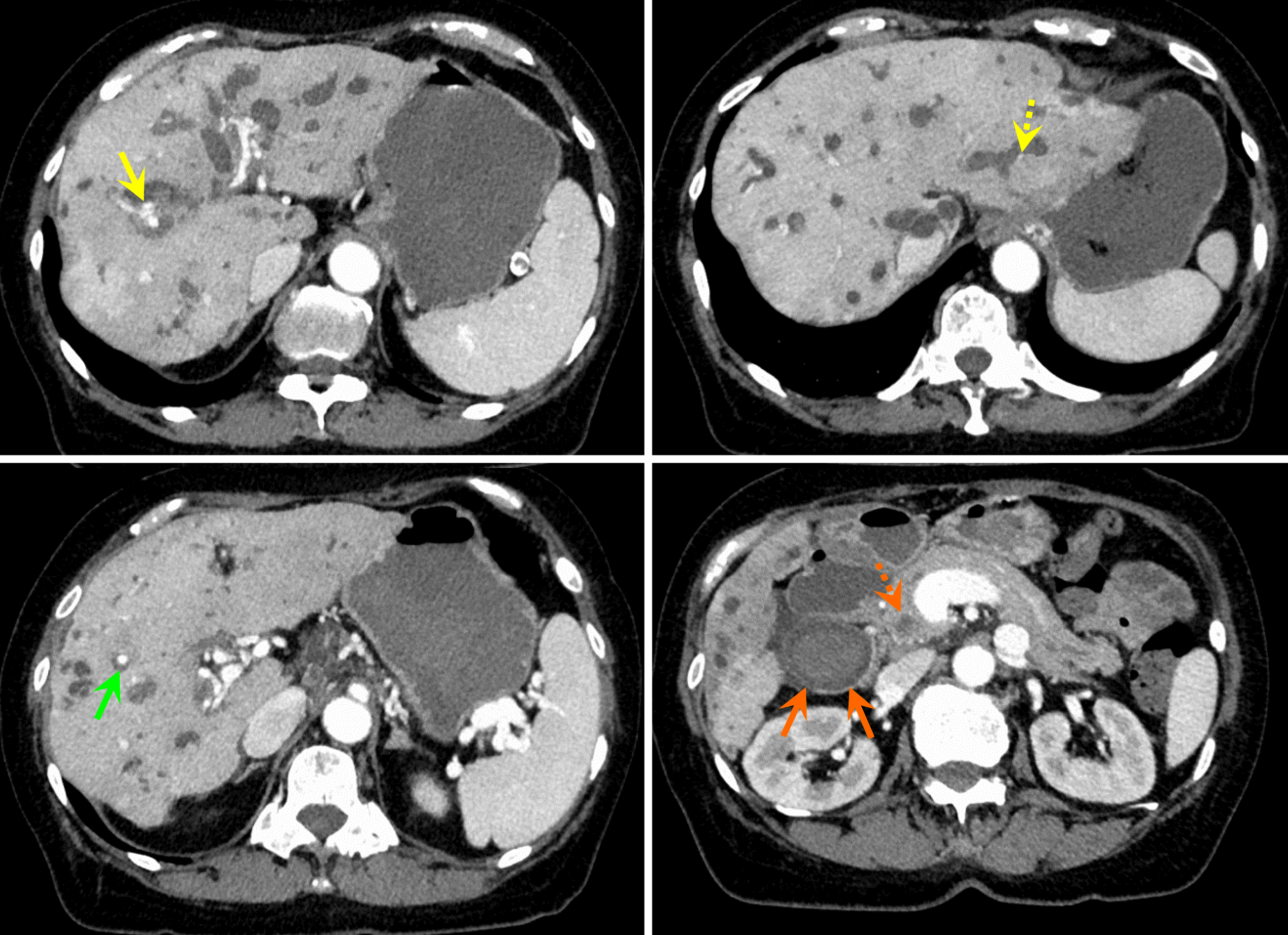
Figure 1 Emergency contrast-enhanced computed tomography scan of the abdomen at the initial visit.
Multiple intrahepatic bile duct dilations and nodular contrast-enhanced areas were observed within the bile duct (yellow, green, and dotted yellow arrows). In addition, biliary hemorrhage was suspected because of the high absorption from the gallbladder (orange arrows) into the common bile duct (dotted orange arrow).
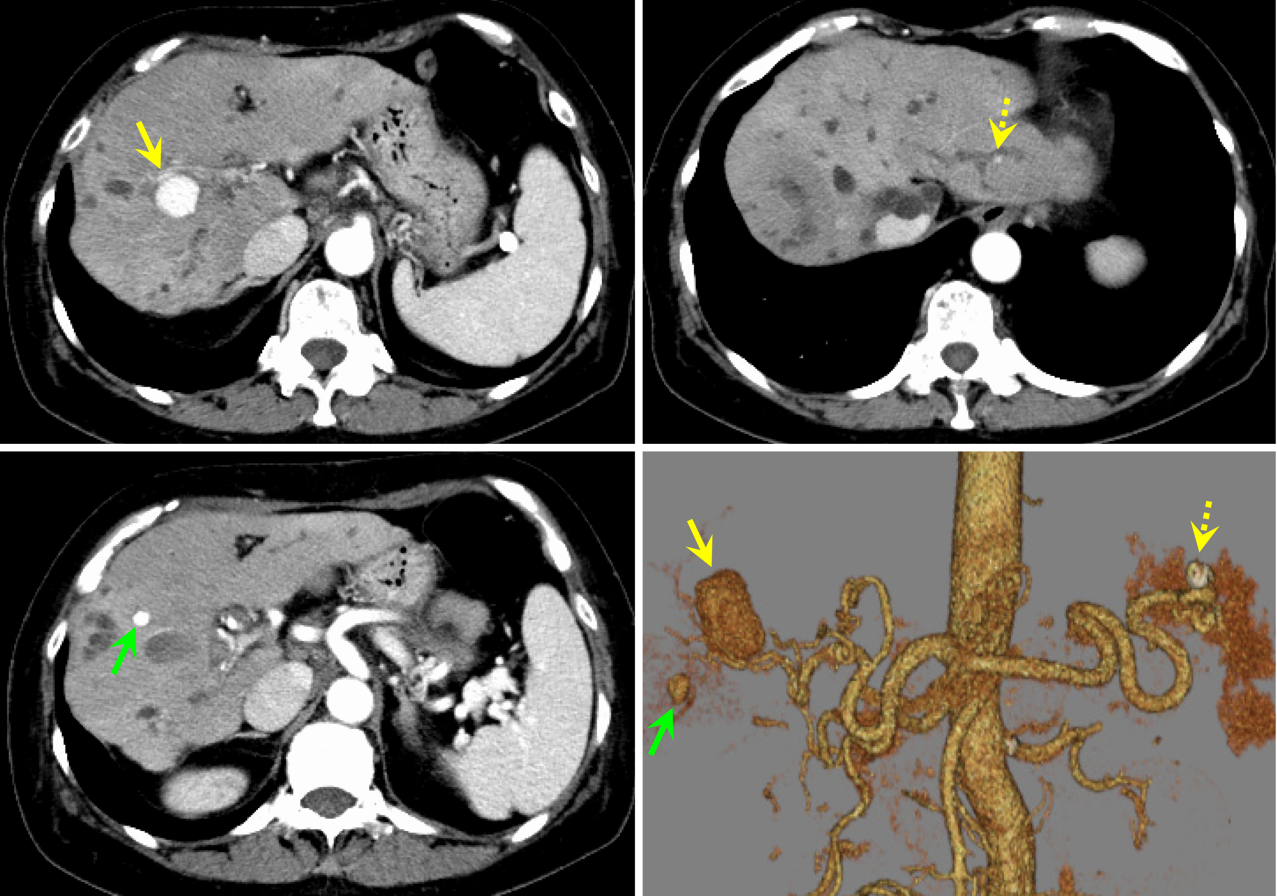
Figure 2 Contrast-enhanced computed tomography scan of the abdomen at diagnosis.
Intrahepatic artery aneurysms were suspected as nodular contrast-enhanced areas approximately 20 mm in size appeared in S 7/8 (yellow arrow), 7 mm in size in S 5 (green arrow), and 4 mm in size in the lateral segment (dotted yellow arrow).
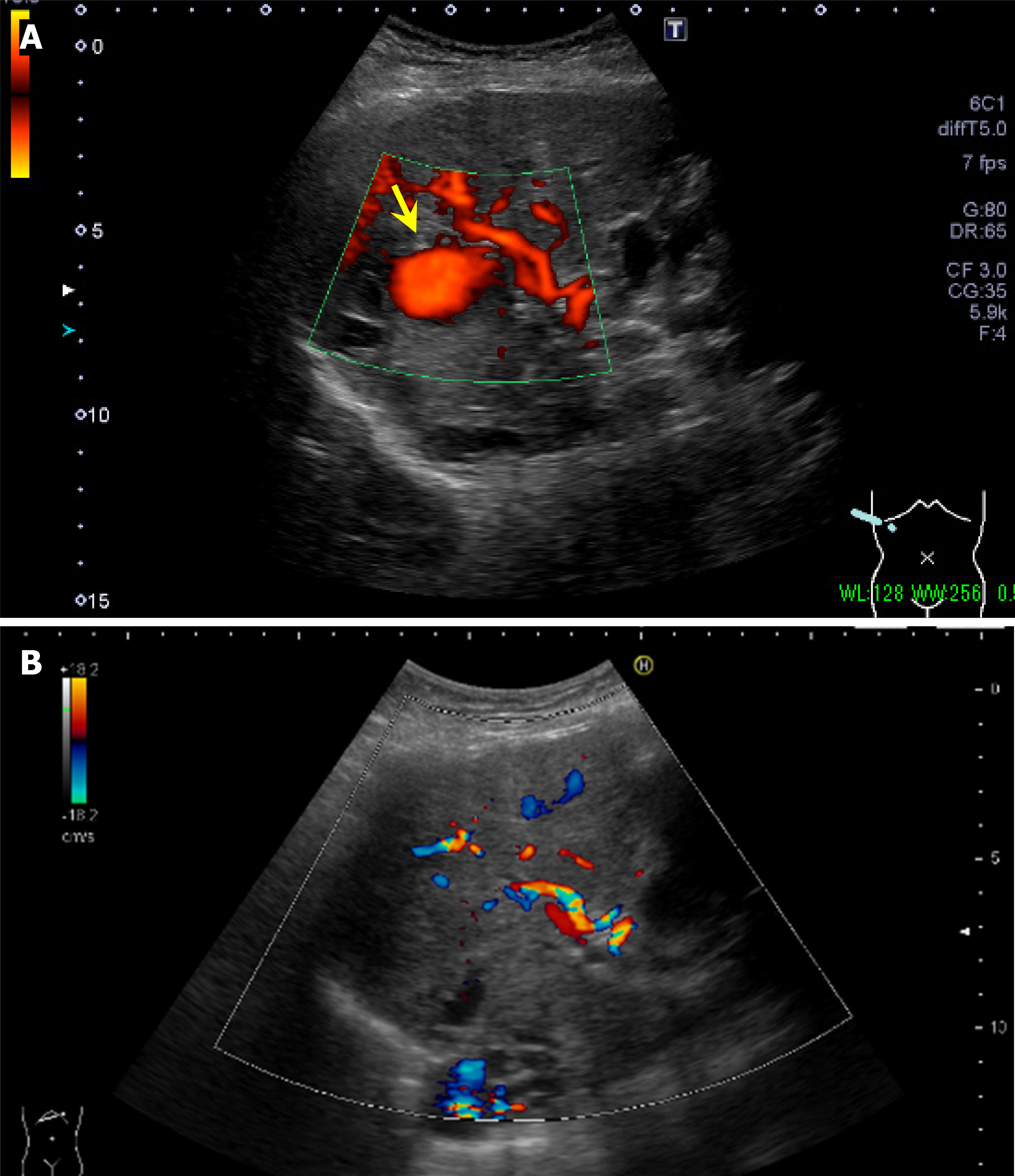
Figure 3 Abdominal ultrasonography.
A: Abdominal ultrasonography before aneurysm embolization. A mass lesion at hepatic S 7/8 showing pulsating blood flow (yellow arrow) was diagnosed as a hepatic artery aneurysm; B: Abdominal ultrasonography after aneurysm embolization. The intrahepatic artery aneurysm seen before aneurysm embolization had disappeared.
Figure 4 Aneurysm embolization.
A and B: Arterial aneurysms are located at A 7 in the right lobe (yellow and green arrows) and at A 2 in the left lobe (dotted yellow arrow); C: After injecting the gel-embolizing agent peripherally, the central part was embolized using microcoils (blue arrows).
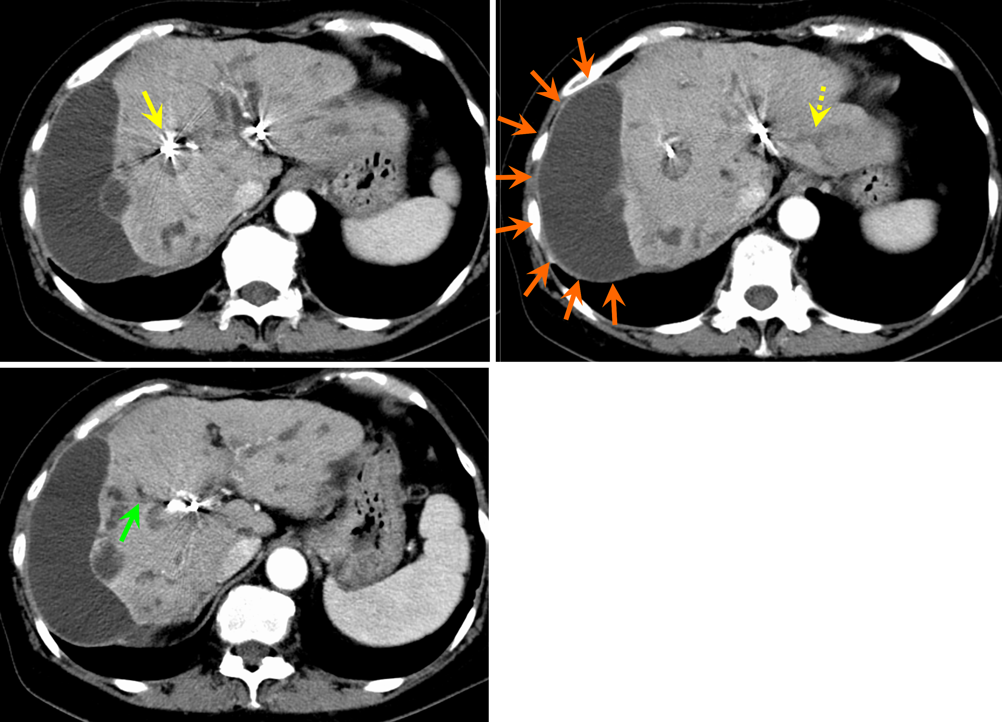
Figure 5 Contrast-enhanced computed tomography scan of the abdomen after aneurysm embolization.
Diffuse atrophy was observed in the liver. The hepatic artery aneurysms in S 7/8 (yellow arrow), S 5 (green arrow), and the lateral segment (dotted yellow arrow) disappeared. The fluid collection on the surface of the right lobe of the liver (orange arrows) was thought to be a biloma caused by the rupture of a peripheral bile duct.
- Citation: Tamura H, Ozono Y, Uchida K, Uchiyama N, Hatada H, Ogawa S, Iwakiri H, Kawakami H. Multiple intrahepatic artery aneurysms during the treatment for IgG4-related sclerosing cholangitis: A case report. World J Hepatol 2024; 16(12): 1505-1514
- URL: https://www.wjgnet.com/1948-5182/full/v16/i12/1505.htm
- DOI: https://dx.doi.org/10.4254/wjh.v16.i12.1505


