Copyright
©The Author(s) 2023.
World J Hepatol. Jun 27, 2023; 15(6): 755-774
Published online Jun 27, 2023. doi: 10.4254/wjh.v15.i6.755
Published online Jun 27, 2023. doi: 10.4254/wjh.v15.i6.755
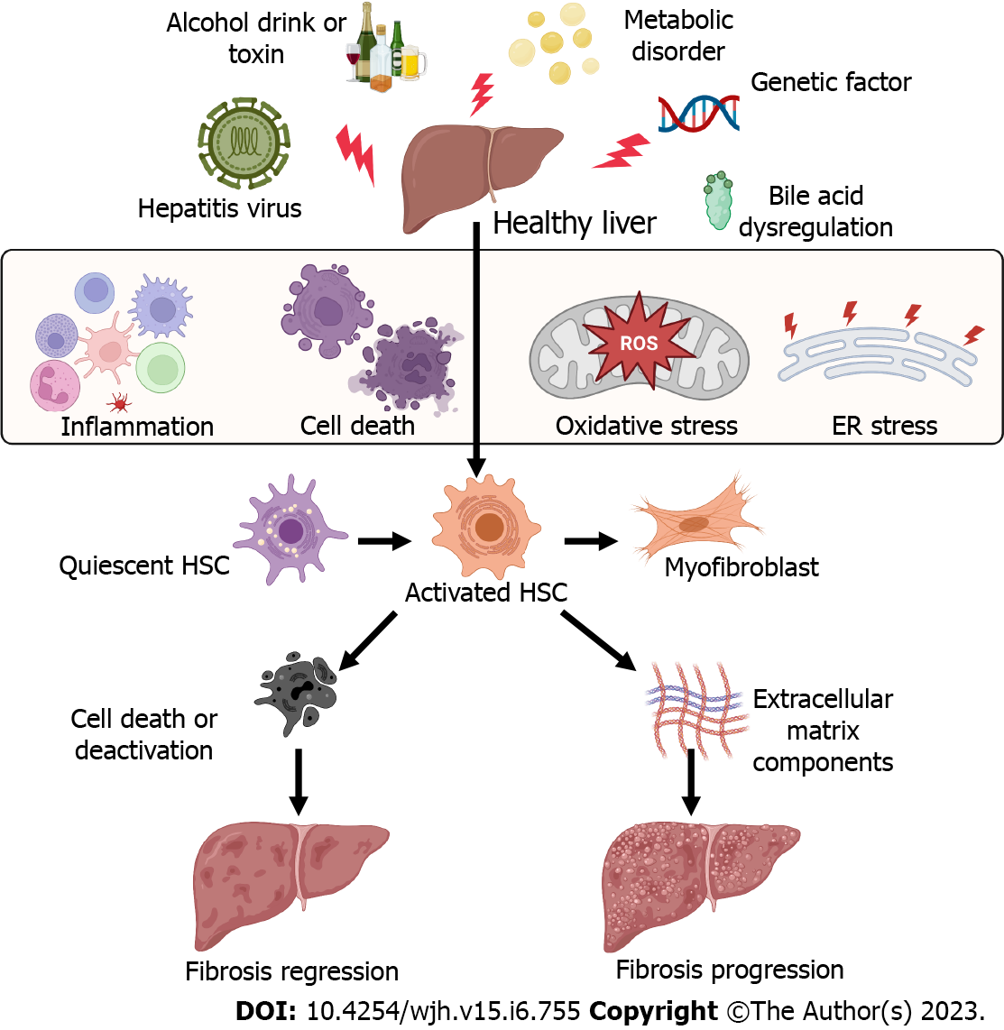
Figure 1 Factors causing the activation of hepatic stellate cells and liver fibrosis.
Many factors can cause liver injury and hepatic cell death and inflammation, including hepatitis viral infection, alcohol consumption, metabolic liver disease, abnormal bile acid products, and genetic factors. These pathogenic factors cause immune cell inflammation, hepatocyte death, oxidative stress, and endoplasmic reticulum stress, resulting in hepatic stellate cell activation and differentiation to myofibroblasts to lead to liver fibrosis. All cartoons in this figure were prepared using Biorender (https://biorender.com). ER: Endoplasmic reticulum; HSC: Hepatic stellate cell; ROS: Reactive oxygen species.
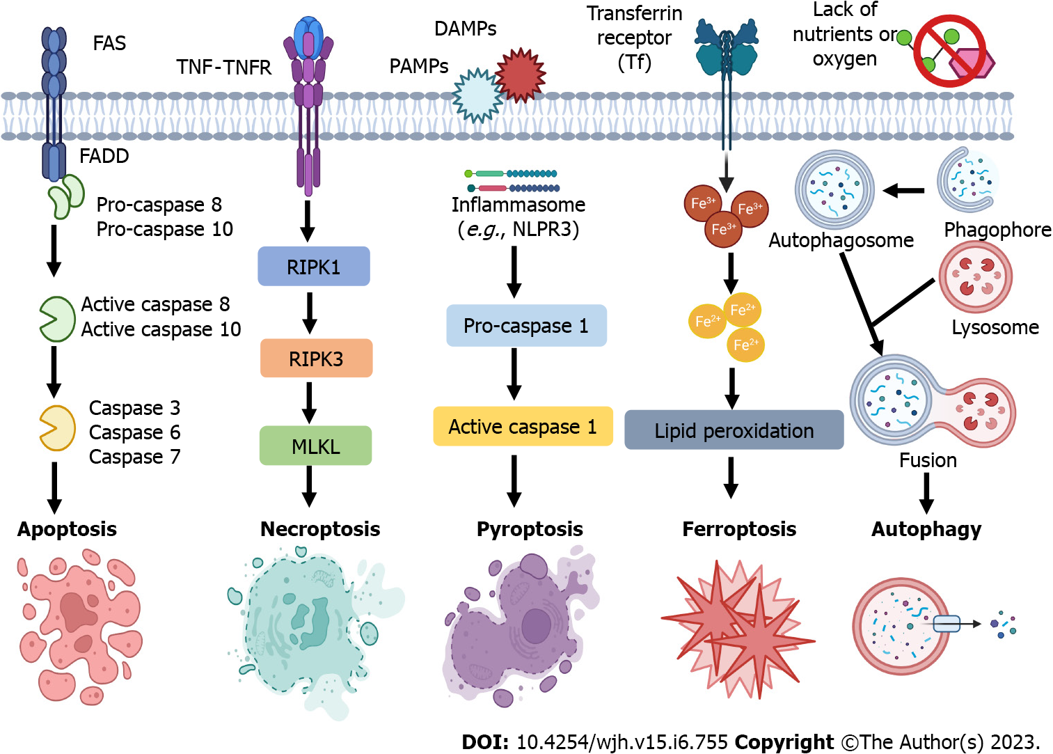
Figure 2 Programmed cell death subtypes of hepatic cells, including apoptosis, necroptosis, pyroptosis, ferroptosis, and autophagy-mediated cell death.
Apoptosis, necroptosis, pyroptosis, and ferroptosis are programmed forms of cell death, while necrosis is unprogrammed cell death. Autophagy-mediated cell death should be defined when autophagic flux is raised without the involvement of other types of programmed cell death, and pharmacological or genetic inhibition of autophagy blocks cell death. DAMPs: Danger-associated molecular patterns; FADD: Fas-associated protein with a death domain; FAS: Fas cell surface death receptor; MLKL: Mixed lineage kinase domain-like; NLRP3: Nod-like receptor family, pyrin domain containing 3; PAMPs: Pathogen-associated molecular patterns; RIPK1/3: receptor-interacting protein kinase 1/3; TNF: Tumor necrosis factor; TNFR: Tumor necrosis factor receptor. All cartoons in this figure were prepared using Biorender
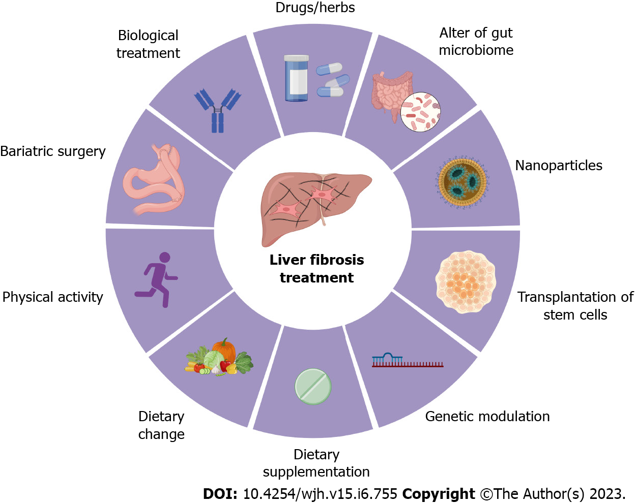
Figure 3 Treatment options for liver fibrosis.
Currently, the preventive and therapeutic treatments for liver fibrosis include physical activity (e.g., running), dietary change (e.g., avoid of high-fat and high-sugar diet), dietary supplementation (e.g., vitamin C), biological treatment (e.g., simtuzumab), bariatric surgery (e.g., Roux-en-Y-gastric procedure), drugs and herb medicines (e.g., pegbelfermin), change of gut microbiota (e.g., probiotics), nanoparticles (e.g., BMS-986263), genetic regulation (e.g., non-coding RNAs), and transplantation of stem cells (e.g., hematopoietic stem cells). All cartoons in this figure were prepared using Biorender (https://biorender.com) .
- Citation: Zhang CY, Liu S, Yang M. Treatment of liver fibrosis: Past, current, and future. World J Hepatol 2023; 15(6): 755-774
- URL: https://www.wjgnet.com/1948-5182/full/v15/i6/755.htm
- DOI: https://dx.doi.org/10.4254/wjh.v15.i6.755


