Copyright
©The Author(s) 2022.
World J Hepatol. Aug 27, 2022; 14(8): 1678-1686
Published online Aug 27, 2022. doi: 10.4254/wjh.v14.i8.1678
Published online Aug 27, 2022. doi: 10.4254/wjh.v14.i8.1678
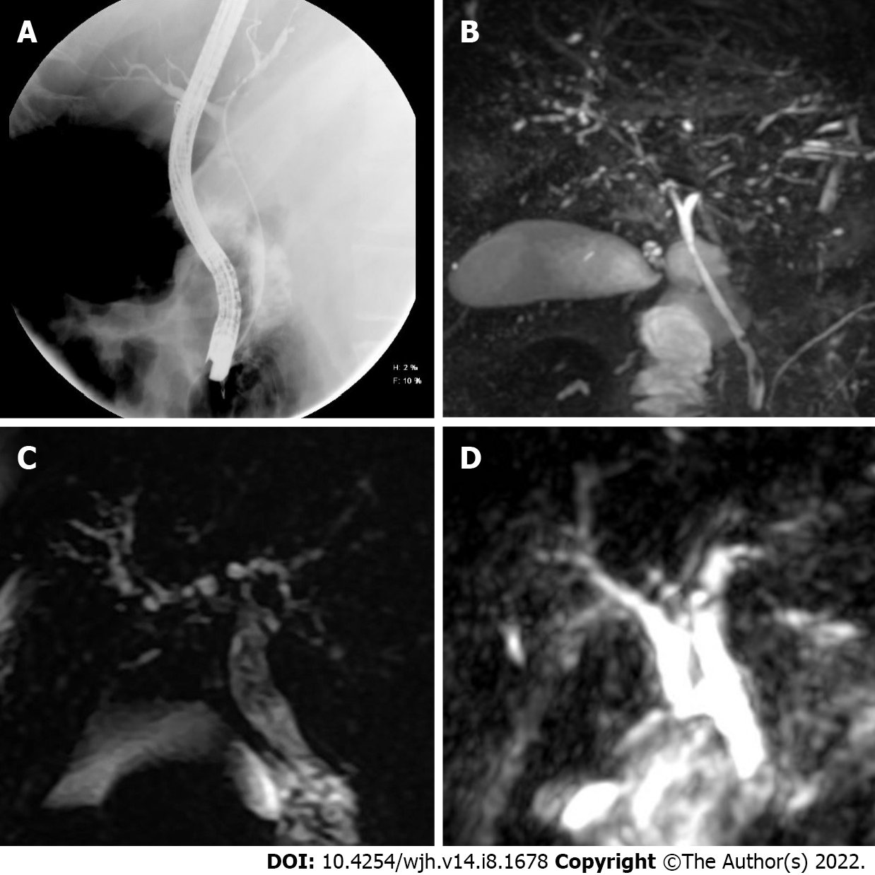
Figure 1 Sclerosing cholangitis imaging findings.
A: Cholangiogram showed dilatation of the common bile duct and its intrahepatic branches in the first case; B: Magnetic resonance imaging (MRI) demonstrated multiple short stenosis of the intrahepatic bile ducts; C and D: In the second and third case, the MRI showed multiple stenosis of the intrahepatic bile duct.
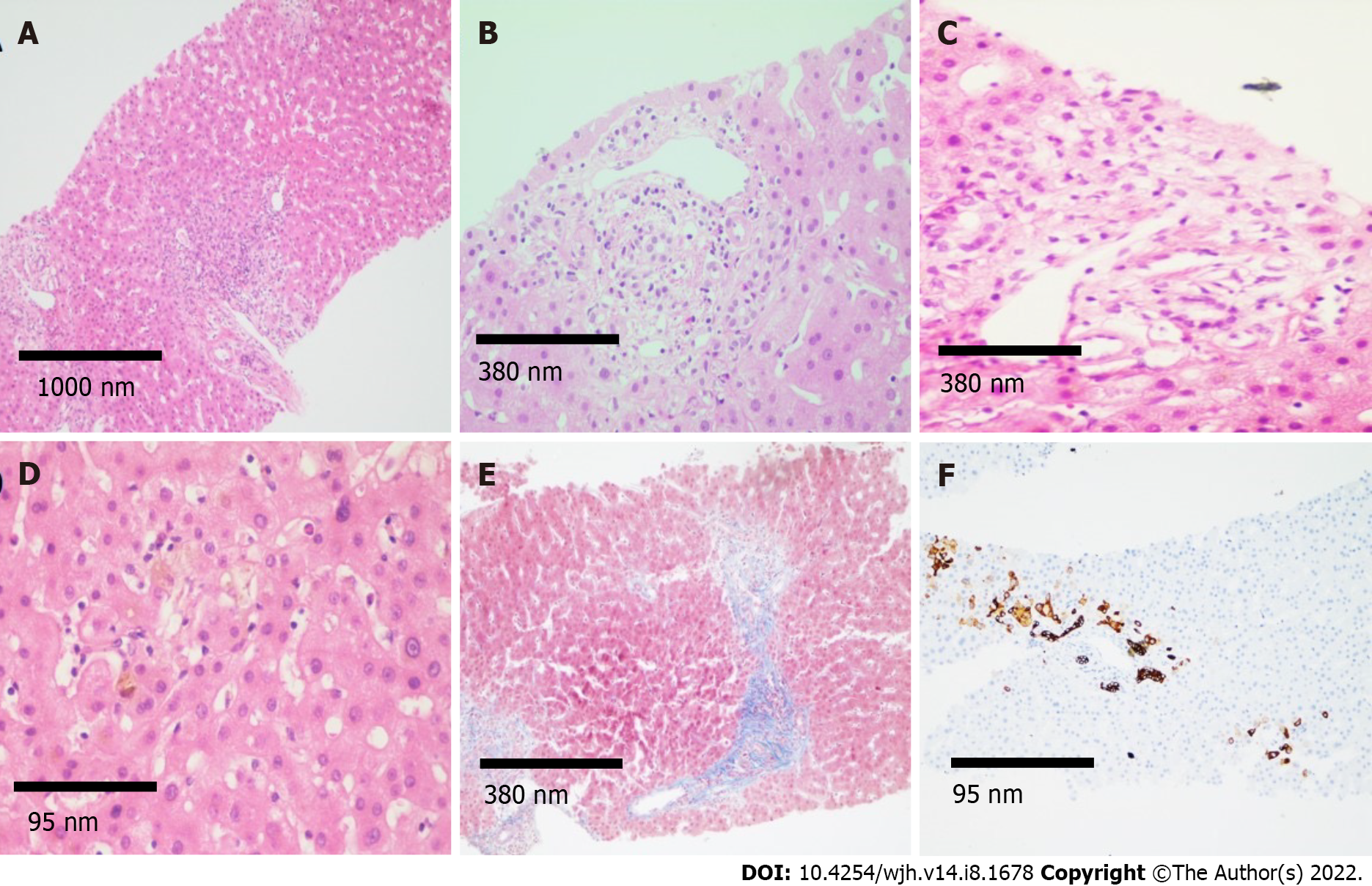
Figure 2 Histological findings.
A: Histological sections of the liver (magnification 4 ×) stained with hematoxylin and eosin (H&E) showing mixed inflammatory infiltrate in portal spaces; B and C: H&E (magnification 10 ×) showing regenerative changes and swelling of cholangiocytes, as well as presence of inflammatory infiltrate in the portal space vein and hepatic artery; D and E: Intracanalicular and cytoplasmic cholestasis is observed predominantly in space 3, fibrosis in portal and periportal space (magnification 40 × and 10 ×, respectively); F: Immunohistochemistry for cytokeratin 7 (CK7) demonstrating CK7 metaplasia in hepatocytes and ductular reaction (magnification 40 ×).
- Citation: Mayorquín-Aguilar JM, Lara-Reyes A, Revuelta-Rodríguez LA, Flores-García NC, Ruiz-Margáin A, Jiménez-Ferreira MA, Macías-Rodríguez RU. Secondary sclerosing cholangitis after critical COVID-19: Three case reports. World J Hepatol 2022; 14(8): 1678-1686
- URL: https://www.wjgnet.com/1948-5182/full/v14/i8/1678.htm
- DOI: https://dx.doi.org/10.4254/wjh.v14.i8.1678


