Copyright
©The Author(s) 2022.
World J Hepatol. May 27, 2022; 14(5): 911-922
Published online May 27, 2022. doi: 10.4254/wjh.v14.i5.911
Published online May 27, 2022. doi: 10.4254/wjh.v14.i5.911
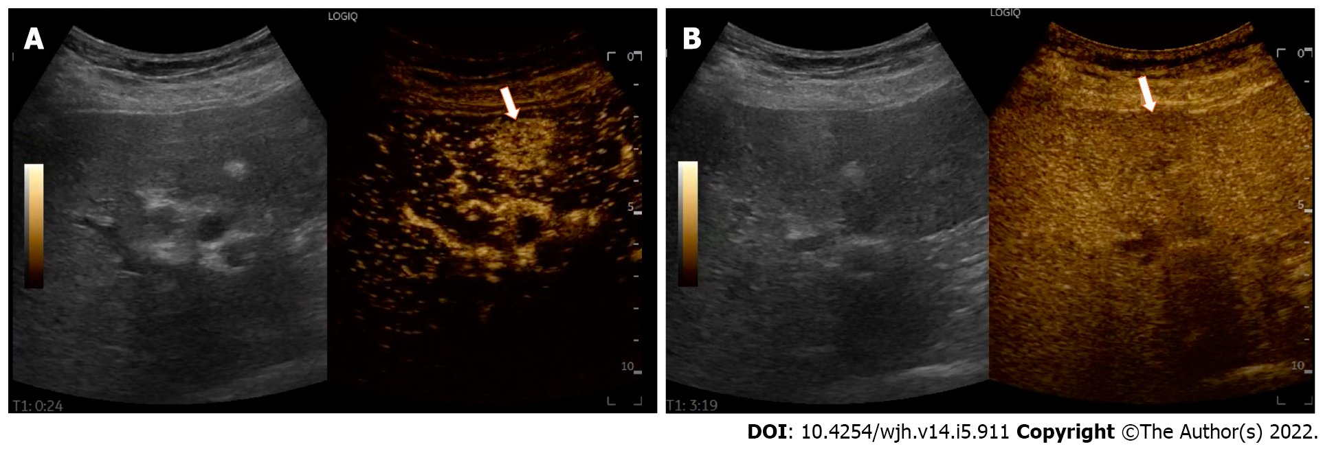
Figure 1 Typical contrast-enhancement pattern of a hepatocellular carcinoma in a 72-year-old woman with hepatitis C virus related cirrhosis.
A: In the arterial phase (24 s after the injection of a microbubble-based contrast agent) contrast enhanced ultrasound depicts a hypervascular tumor sized 2 cm in the IV liver segment (white arrow); B: In the late phase, 199 s after the injection, the lesion shows a mild wash-out (white arrow).
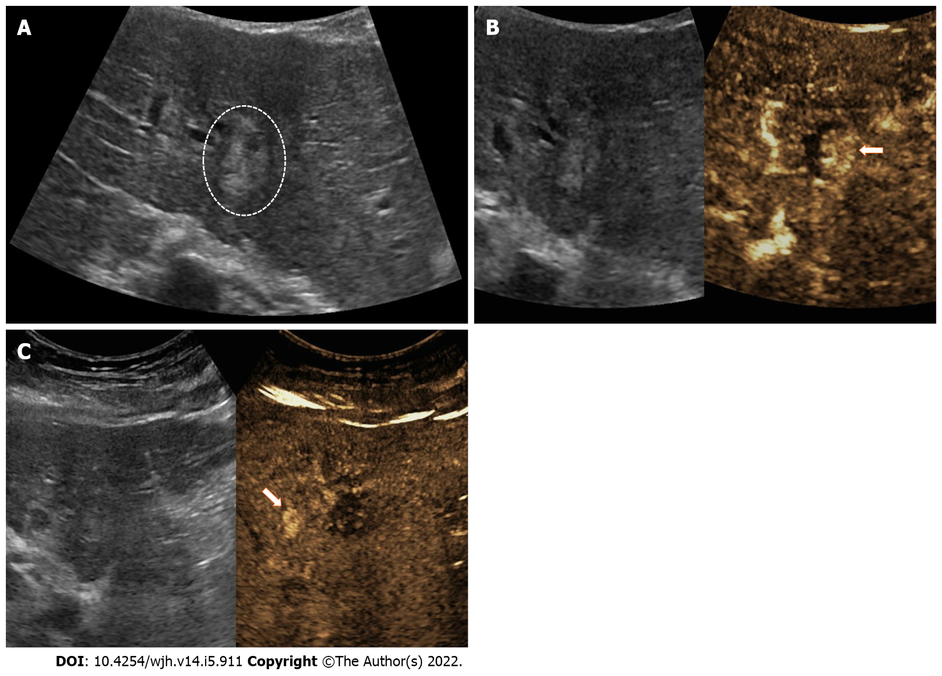
Figure 2 Intrazonal recurrence after radiofrequency ablation in a 79-year-old woman with hepatitis C virus-related cirrhosis.
A: Gray-scale ultrasound image achieved 1 yr after ablation shows a heterogeneous hyperechoic nodule (circle); B: Contrast enhanced ultrasound (CEUS) imaging achieved 15 s after injection shows a nodular token of arterial phase hyperenhancement (white arrow); C: CEUS image obtained at 2 min shows a wash-out. The patient underwent a new radiofrequency ablation in the same session. Simultaneously CEUS shows small hemangioma in the same segment (white arrow).
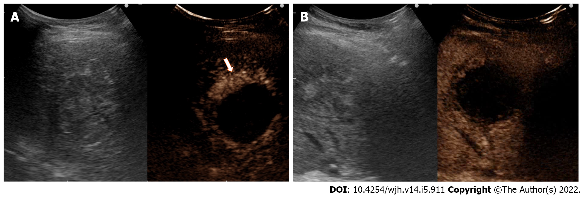
Figure 3 Extrazonal recurrence.
Follow-up of hepatocellular carcinoma treated with microwave ablation in a 79-year-old man with hepatitis C virus and ethanol alcohol-related cirrhosis. A: Contrast enhanced ultrasound image achieved 12 mo after treatment shows a peripheral arterial enhancement 20 s after injection (white arrow); B: Subsequent wash-out is evident at 3 min after injection.
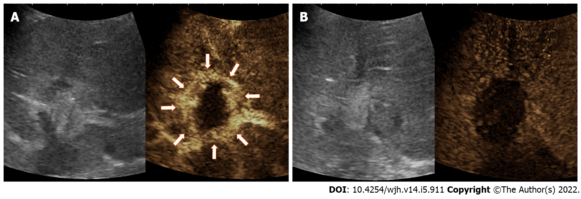
Figure 4 Post procedural contrast enhanced ultrasound (20 min after microwave ablation) in a 72-year-old man with chronic hepatitis C virus-related hepatocellular carcinoma.
Reactive hyperemia achieved immediately after the procedure (22 s). A: Thick and regular rim of arterial phase hyperenhancement surrounds the treatment area (arrows); B: Absence of wash-out in the late phase (4 min) confirms the reactive significance of hyperemia.
- Citation: Inzerillo A, Meloni MF, Taibbi A, Bartolotta TV. Loco-regional treatment of hepatocellular carcinoma: Role of contrast-enhanced ultrasonography. World J Hepatol 2022; 14(5): 911-922
- URL: https://www.wjgnet.com/1948-5182/full/v14/i5/911.htm
- DOI: https://dx.doi.org/10.4254/wjh.v14.i5.911


