Copyright
©The Author(s) 2022.
World J Hepatol. Apr 27, 2022; 14(4): 854-859
Published online Apr 27, 2022. doi: 10.4254/wjh.v14.i4.854
Published online Apr 27, 2022. doi: 10.4254/wjh.v14.i4.854
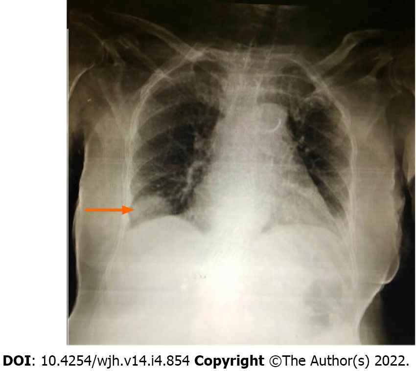
Figure 1
Chest radiograph demonstrates a well-defined soft tissue mass noted just above the right hemi-diaphragm making an obtuse costophrenic angle suggesting pleural or extra-pleural mass.
Figure 2 Chest computed tomography images.
A: Axial view showing herniated part of the liver through focal defect in the right hemi-diaphragm (arrow) mimicking a pleural/pulmonary mass; B: Coronal view shows extension of liver parenchyma into the thoracic cavity with hepatic artery within herniated liver (arrow); C: Sagittal view shows nubbin of liver parenchyma herniated through diaphragmatic defect posteriorly (arrow).
- Citation: Tebha SS, Zaidi ZA, Sethar S, Virk MAA, Yousaf MN. Angiotensin converting enzyme inhibitor associated spontaneous herniation of liver mimicking a pleural mass: A case report. World J Hepatol 2022; 14(4): 854-859
- URL: https://www.wjgnet.com/1948-5182/full/v14/i4/854.htm
- DOI: https://dx.doi.org/10.4254/wjh.v14.i4.854


