Copyright
©The Author(s) 2022.
World J Hepatol. Apr 27, 2022; 14(4): 846-853
Published online Apr 27, 2022. doi: 10.4254/wjh.v14.i4.846
Published online Apr 27, 2022. doi: 10.4254/wjh.v14.i4.846
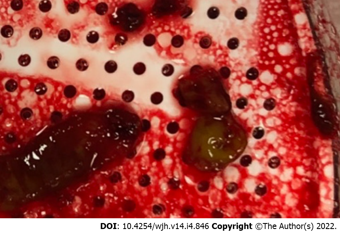
Figure 1 Transjugular intrahepatic portosystemic shunt thrombectomy.
Thrombus extracted during first transjugular intrahepatic portosystemic shunt thrombectomy was notable for size and infected appearance.
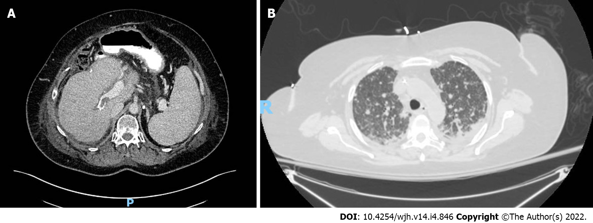
Figure 2 Computed tomography images.
A: Computed tomography of upper abdomen. An axial-contrast enhanced computed tomography of the upper abdomen showing an occluded (hypodense area) transjugular intrahepatic portosystemic shunt; B: Computed tomography of chest. An axial non-enhanced computed tomography of the chest showing diffuse bilateral ground-glass and interstitial opacities with innumerable bilateral pulmonary nodules and small bilateral pleural effusions.
- Citation: Perez IC, Haskal ZJ, Hogan JI, Argo CK. Late polymicrobial transjugular intrahepatic portosystemic shunt infection in a liver transplant patient: A case report. World J Hepatol 2022; 14(4): 846-853
- URL: https://www.wjgnet.com/1948-5182/full/v14/i4/846.htm
- DOI: https://dx.doi.org/10.4254/wjh.v14.i4.846


