Copyright
©The Author(s) 2022.
World J Hepatol. Apr 27, 2022; 14(4): 719-728
Published online Apr 27, 2022. doi: 10.4254/wjh.v14.i4.719
Published online Apr 27, 2022. doi: 10.4254/wjh.v14.i4.719
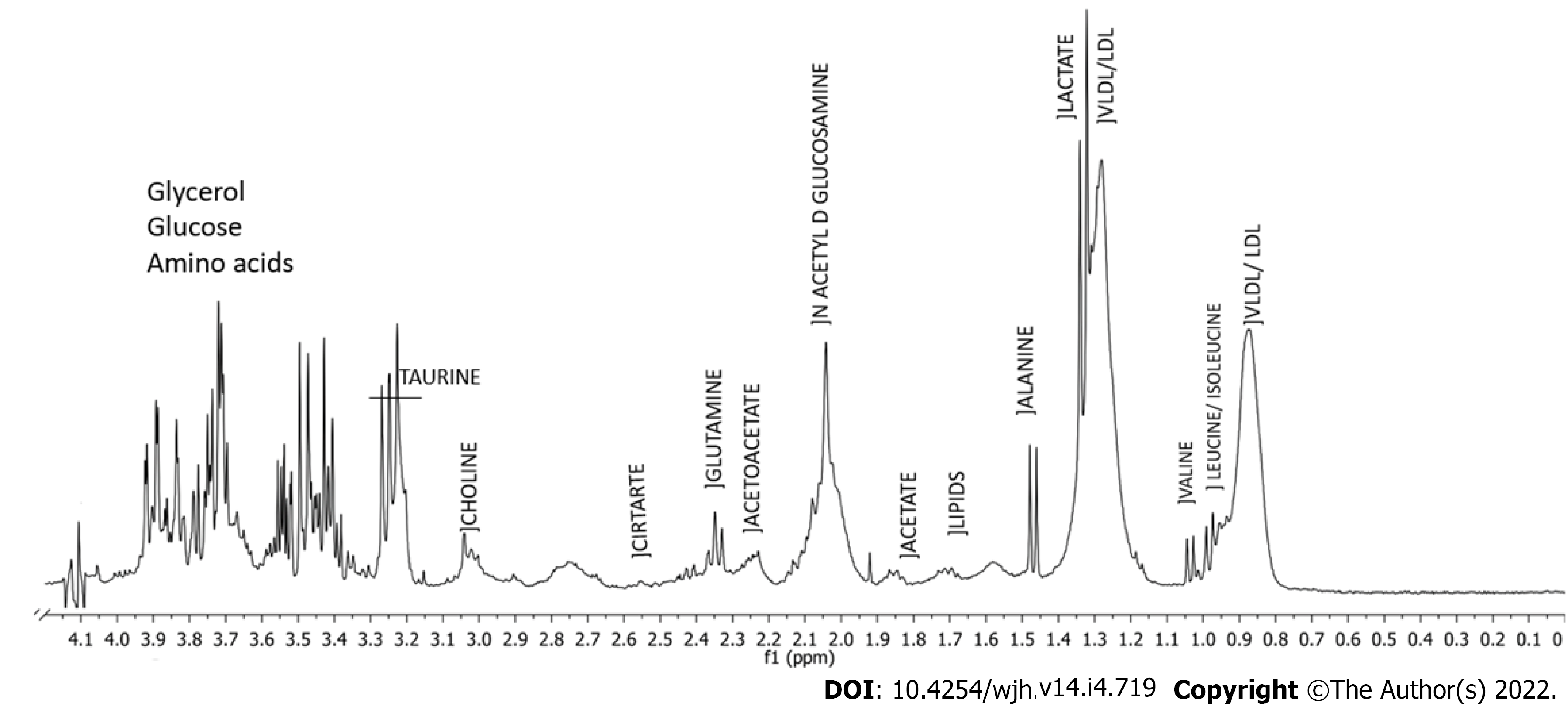
Figure 1 Typical 1H-nuclear magnetic resonance spectrum (400 MHz, D2O, presaturation-Carr-Purcell-Meiboom-Gill) of serum from a patient with Schistosomiasis mansoni, Pernambuco, Brazil, 2020.
Integration areas under the signal are associated with the concentration of metabolites weighted by the number of hydrogen nuclei in each chemical environment. Some assignments are presented in the spectrum.
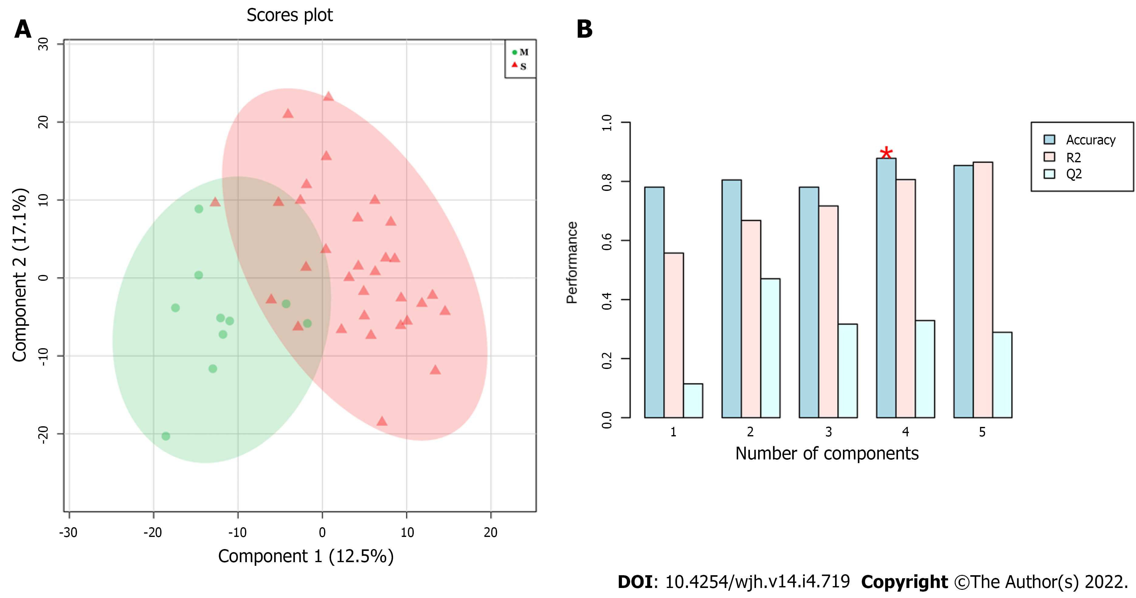
Figure 2 Results of partial least squares-discriminant analysis modelling using 41 samples of patients with Schistosomiasis mansoni, Pernambuco, Brazil, 2020.
A: Score plot–significant (red) and mild (green) PPF patterns; B: Performance of metabonomics models (Red star: Best number of components for modelling).
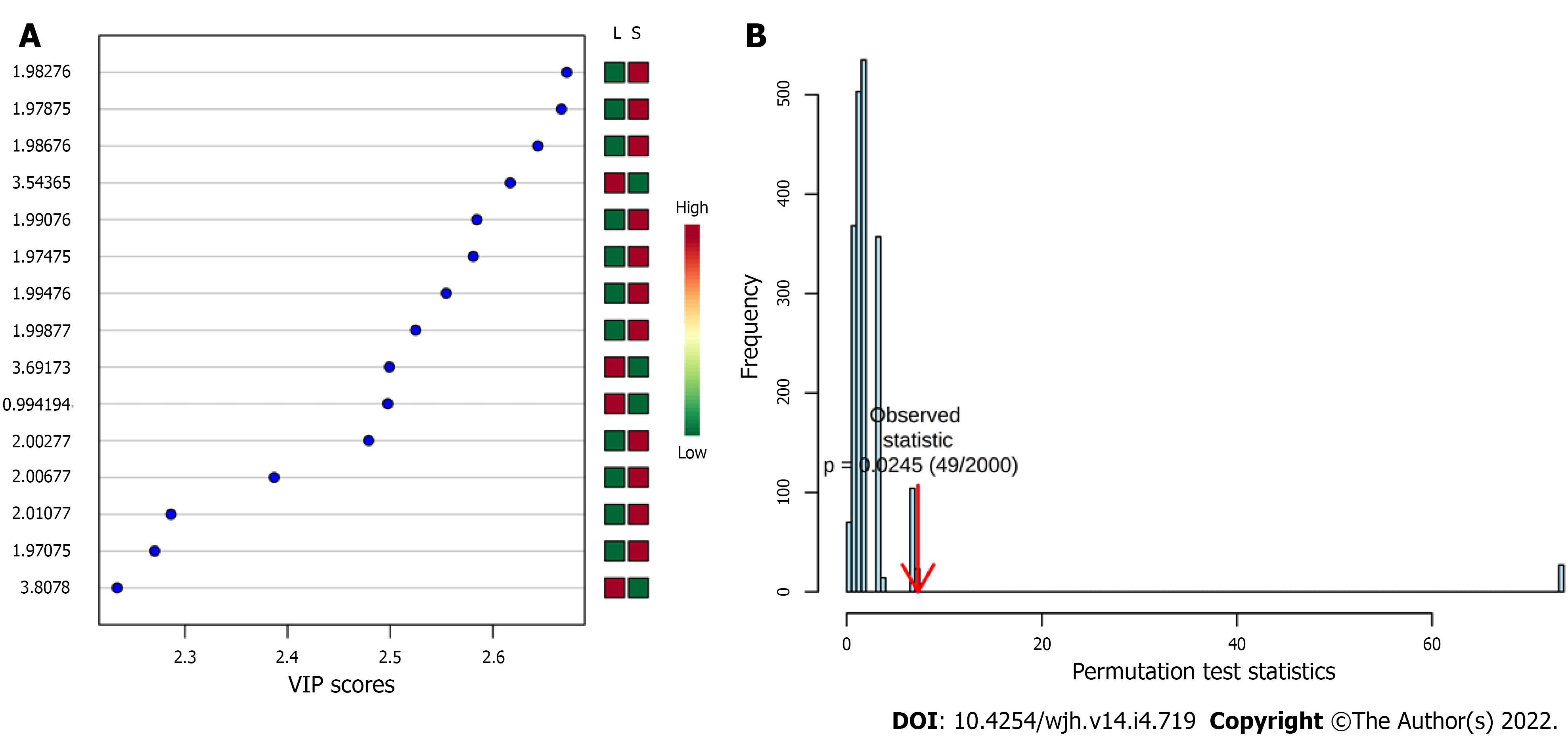
Figure 3 Results of partial least squares-discriminant analysis modelling using 41 samples of patients with Schistosomiasis mansoni, Pernambuco, Brazil, 2020.
A: Variable importance in the projection score plot; B: Permutation test statistic at 2000 permutations with observed statistic of the model prediction accuracy with P value = 0.0245.
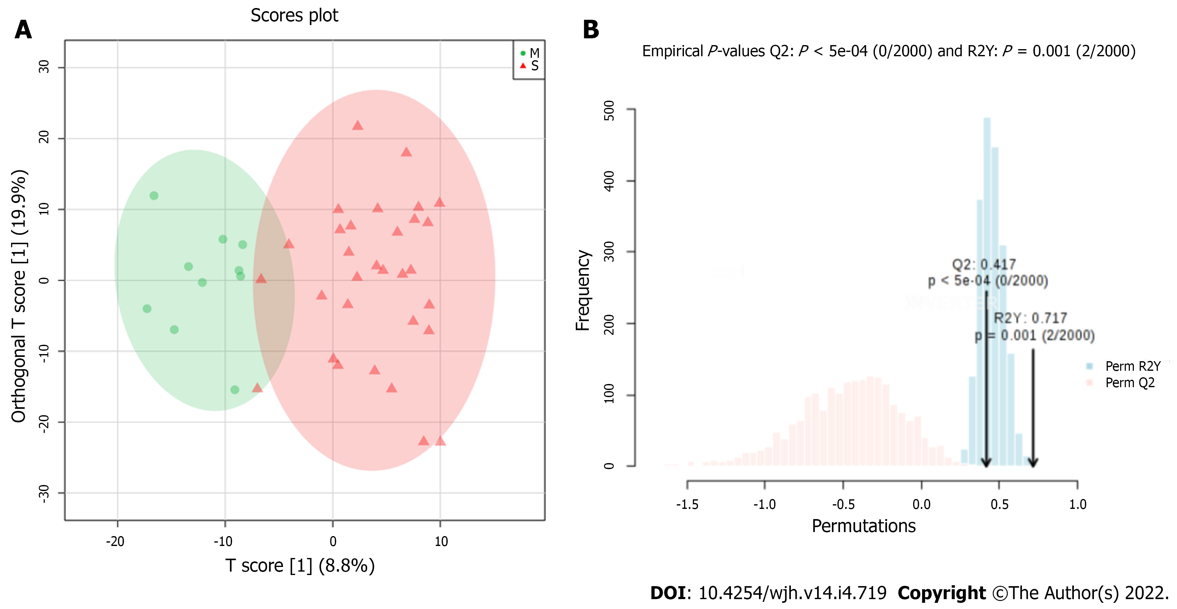
Figure 4 Results of orthogonal projections to latent structures discriminant analysis modelling using 41 samples of patients with Schistosomiasis mansoni, Pernambuco, Brazil, 2020.
A: Score plot–Significant (red) and Mild (green) PPF patterns; B: Permutation test statistic at 2000 permutations with observed statistic of the model prediction accuracy with P value = 0.001.
- Citation: Rodrigues ML, da Luz TPSR, Pereira CLD, Batista AD, Domingues ALC, Silva RO, Lopes EP. Assessment of periportal fibrosis in Schistosomiasis mansoni patients by proton nuclear magnetic resonance-based metabonomics models. World J Hepatol 2022; 14(4): 719-728
- URL: https://www.wjgnet.com/1948-5182/full/v14/i4/719.htm
- DOI: https://dx.doi.org/10.4254/wjh.v14.i4.719


