Copyright
©The Author(s) 2021.
World J Hepatol. Feb 27, 2022; 14(2): 464-470
Published online Feb 27, 2022. doi: 10.4254/wjh.v14.i2.464
Published online Feb 27, 2022. doi: 10.4254/wjh.v14.i2.464
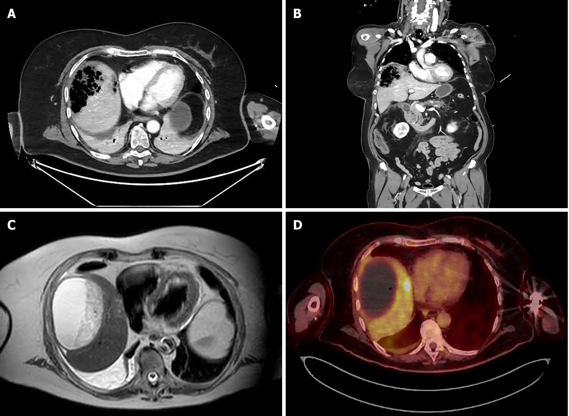
Figure 1 Imaging of emphysematous hepatitis.
A and B: Axial (A) and coronal (B) computed tomography scans on admission, showing a 9 cm air-filled cavity in the right liver lobe; C: Magnetic resonance imaging, showing a 10 cm fluid-filled collection in the right liver lobe with heterogeneous content at 6 wk after initial presentation; D: Positron emission tomography performed at 9 wk after initial presentation, showing no metabolic activity in the large collection in the right liver lobe and a 2-cm nodule with positive metabolic activity in segment VIII.
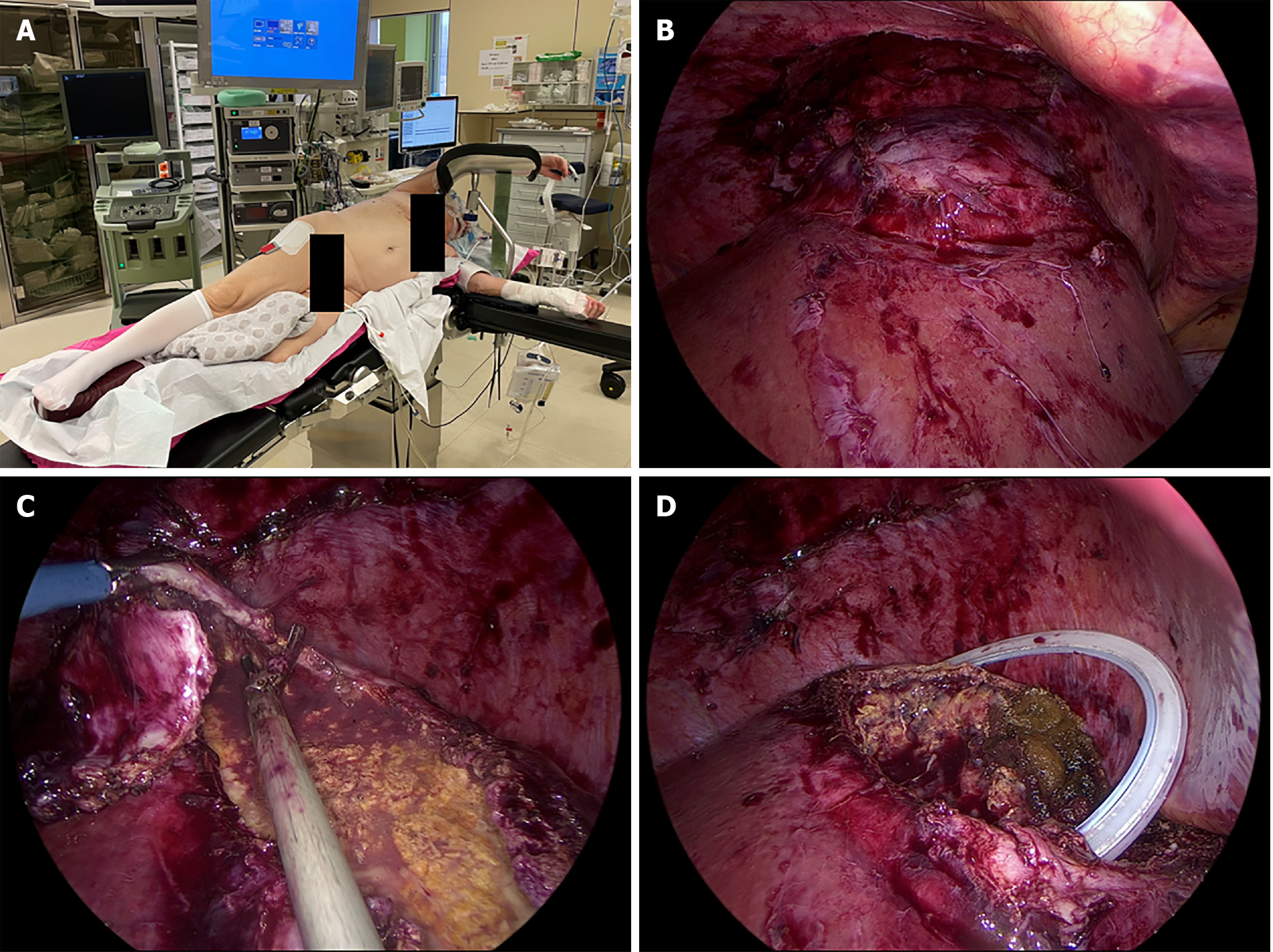
Figure 2 Laparoscopic treatment of emphysematous hepatitis.
A: Semi-pone positioning of the patient; B: Laparoscopic view of liver segments VII and VIII after mobilization of the right liver lobe; C: Laparoscopic deroofing of the liver capsule of segments VII and VIII; D: Partial hepatectomy of segment VIII and placement of a surgical drain in the remaining cavity.
- Citation: Francois S, Aerts M, Reynaert H, Van Lancker R, Van Laethem J, Kunda R, Messaoudi N. Step-up approach in emphysematous hepatitis: A case report. World J Hepatol 2022; 14(2): 464-470
- URL: https://www.wjgnet.com/1948-5182/full/v14/i2/464.htm
- DOI: https://dx.doi.org/10.4254/wjh.v14.i2.464


