Copyright
©The Author(s) 2021.
World J Hepatol. Aug 27, 2021; 13(8): 887-895
Published online Aug 27, 2021. doi: 10.4254/wjh.v13.i8.887
Published online Aug 27, 2021. doi: 10.4254/wjh.v13.i8.887
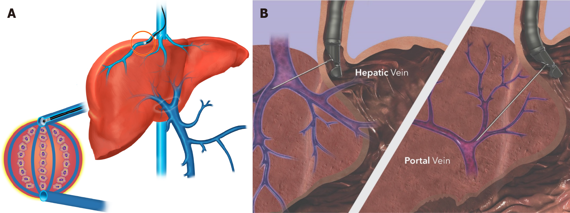
Figure 1 Comparison of modalities for measuring portal hypertension.
A: Methods for obtaining hepatic venous pressure gradient measurement via the transjugular approach. Placement of catheter into right hepatic vein for measurement of free hepatic venous pressure, followed by balloon or “wedged” occlusion (inset) to measure wedged hepatic venous pressure, indirectly measuring the portal vein pressure via the sinusoids; B: Portal pressure gradient measurement via endoscopic ultrasound. The hepatic vein (left panel) and portal vein (right panel) are both directly accessed with transgastric needle puncture. Permission for use granted by Cook Medical, Bloomington, Indiana.
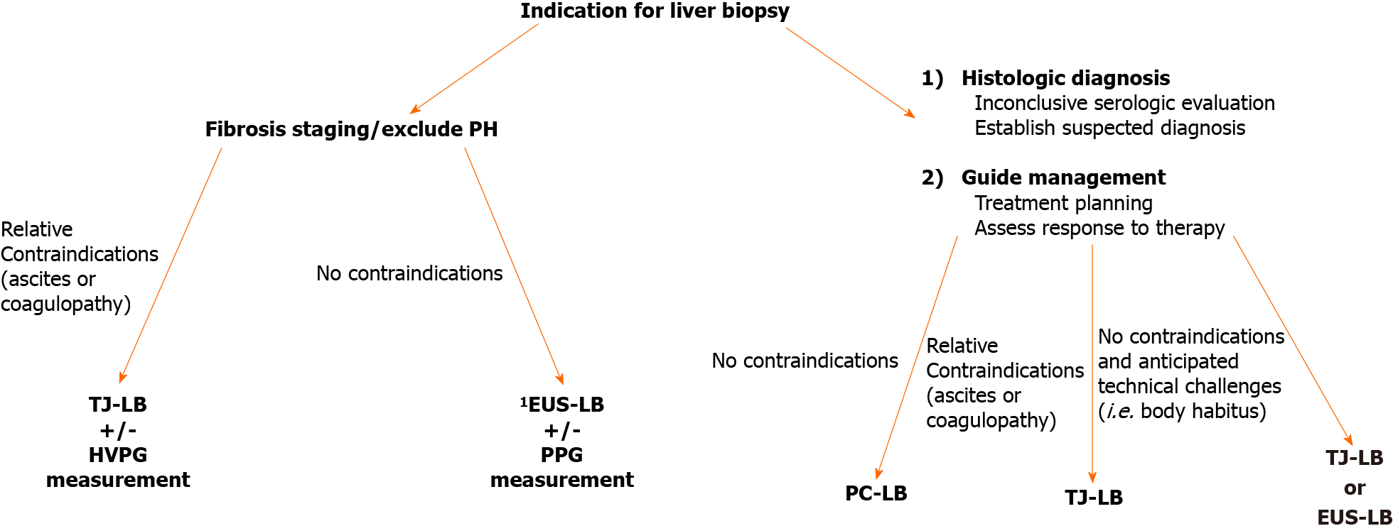
Figure 2 Proposed algorithm for choosing suitable modality for liver biopsy.
1Allows endoscopic exam for evidence of portal hypertension (i.e., varices/PHG), high-resolution endoscopic ultrasound images of liver contours/parenchyma, endoscopic “palpation” of the liver, bi-lobar biopsies, and direct measure of portal pressure gradient. PH: Portal hypertension; TJ-LB: Transjugular liver biopsy; EUS-LB: endoscopic ultrasound-guided liver biopsy; PPG: Portal pressure gradient; HVPG: Hepatic venous pressure gradient; PC-LB: Percutaneous liver biopsy.
- Citation: Rudnick SR, Conway JD, Russo MW. Current state of endohepatology: Diagnosis and treatment of portal hypertension and its complications with endoscopic ultrasound. World J Hepatol 2021; 13(8): 887-895
- URL: https://www.wjgnet.com/1948-5182/full/v13/i8/887.htm
- DOI: https://dx.doi.org/10.4254/wjh.v13.i8.887


