Copyright
©The Author(s) 2021.
World J Hepatol. Feb 27, 2021; 13(2): 166-186
Published online Feb 27, 2021. doi: 10.4254/wjh.v13.i2.166
Published online Feb 27, 2021. doi: 10.4254/wjh.v13.i2.166
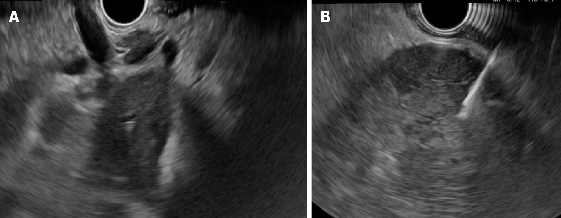
Figure 1 Endoscopic ultrasound for liver evaluation.
A: Hilium view of the liver tumoral mass; B: Fine needle aspiration guided endoscopic ultrasound for left lobe intrahepatic cholangiocarcinoma.
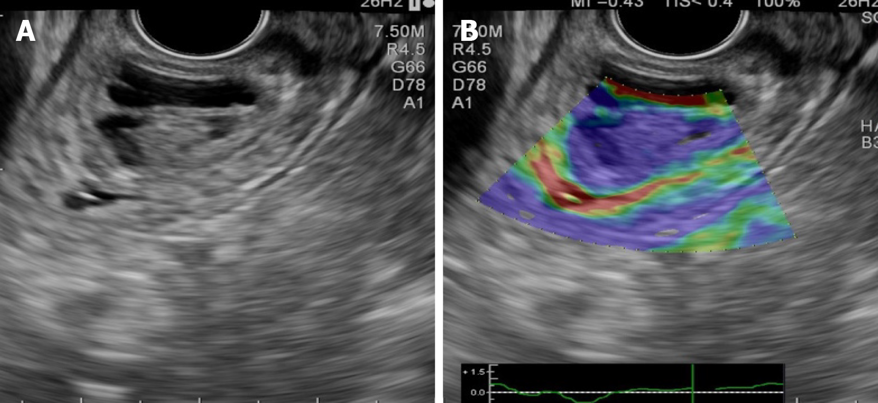
Figure 2 Endoscopic ultrasound of the distal common bile duct.
A: Small non-invasive tumoral mass (distal cholangiocarcinoma); B: Elastography: Blue color of the tumor (hard stiffness).
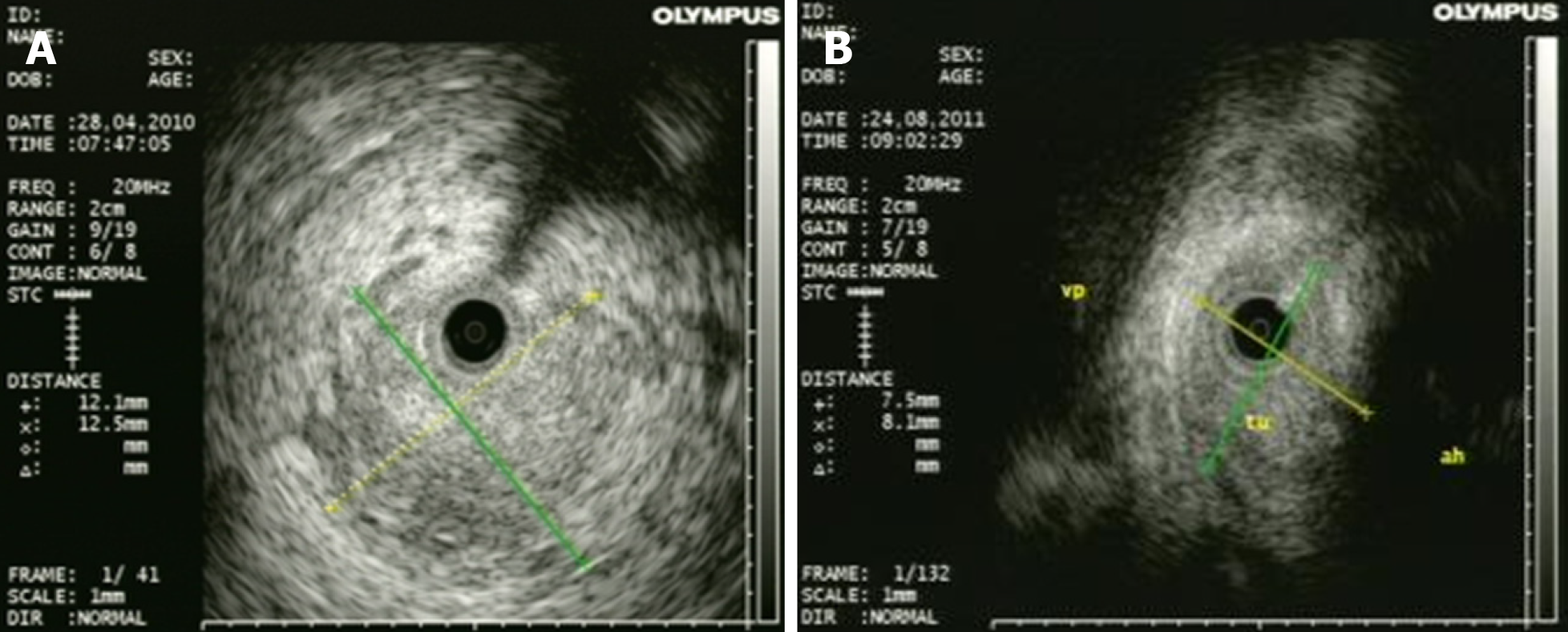
Figure 3 Intraductal ultrasonography for diagnosis of cholangiocarcinoma.
A: Large tumoral mass with invasion of surrounding tissue; B: Infiltrative cholangiocarcinoma in proximity of the hepatic artery.
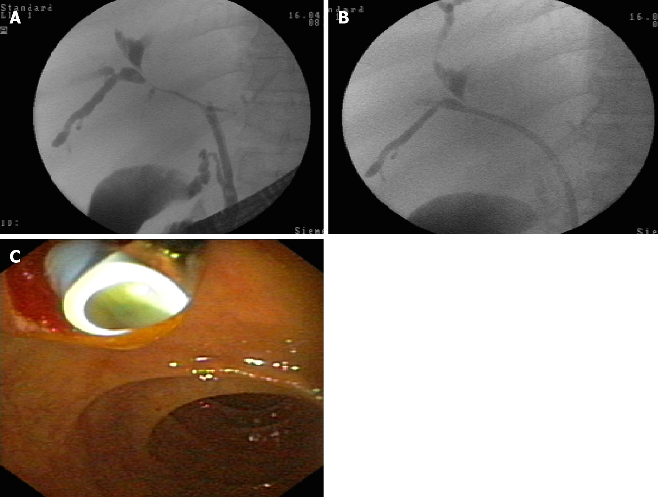
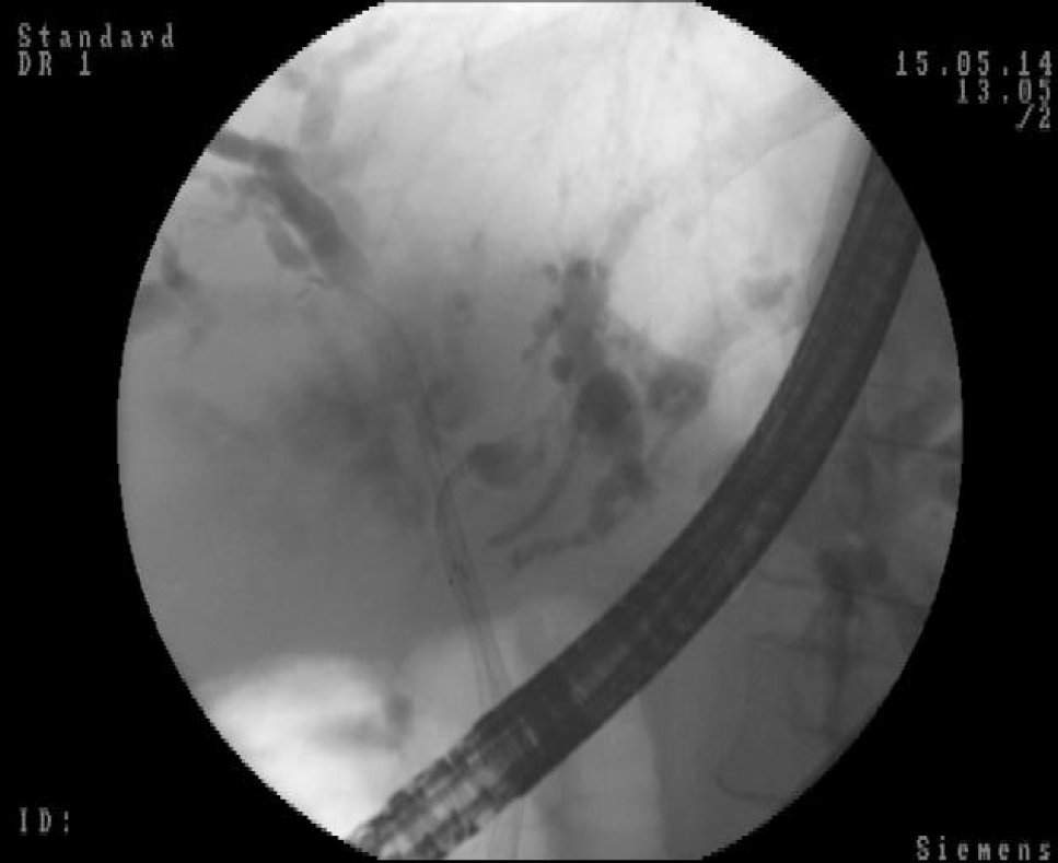
Figure 4 Endoscopic retrograde cholangiopancreatography.
Radiologic and endoscopic view. A: Bismuth IV cholangiocarcinoma of the hilum; B: Endoscopic stenting with plastic stent in place; C: Endoscopic view of plastic stent at the level of the papilla.
Figure 5 Endoscopic retrograde cholangiopancreatography.
Radiologic and endoscopic view. A: Bismuth IV cholangiocarcinoma of the hilum; B: Endoscopic stenting with metallic stent in place; C: Endoscopic view of metallic stent at the level of the papilla.
Figure 6 Endoscopic retrograde cholangiopancreatography.
Radiologic view. Side-by-side technique (metallic stents in both intrahepatic ducts).
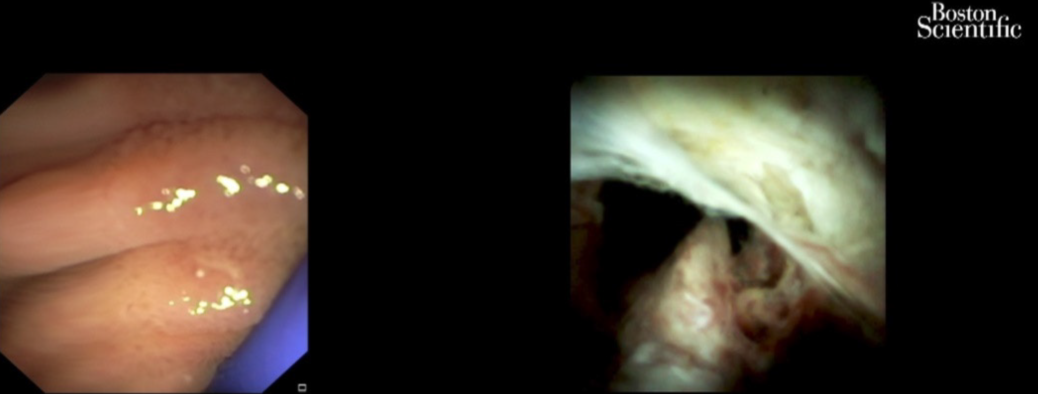
Figure 7 Cholangioscopy: Hilum malignant obstruction.
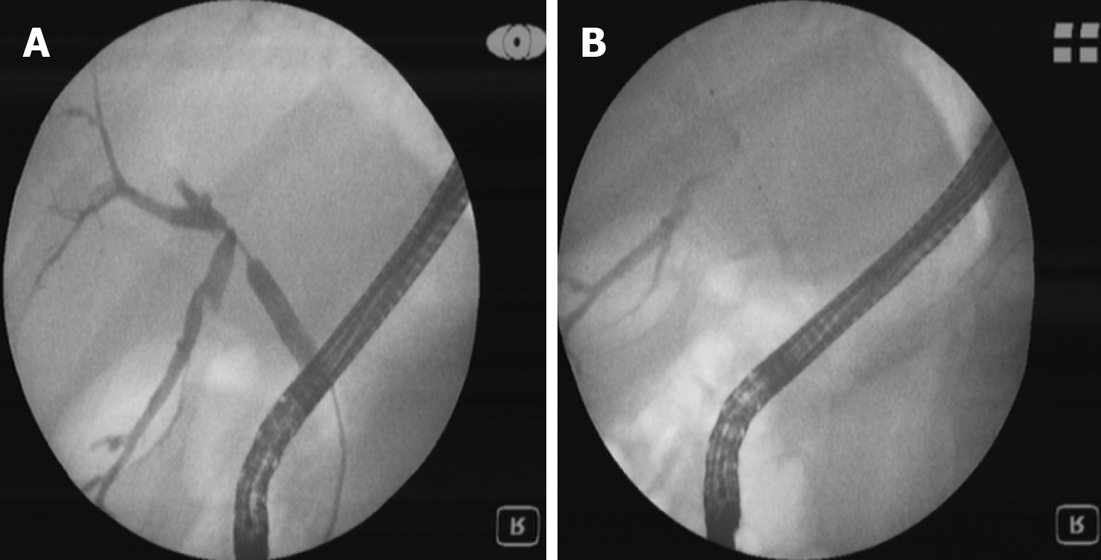
Figure 8 Endoscopic retrograde cholangiopancreatography.
Radiologic view. A: Bismuth III cholangiocarcinoma (guidewire is passing through malign stenosis); B: Photodynamic therapy. The laser fiber at the level of stenosis can be seen.
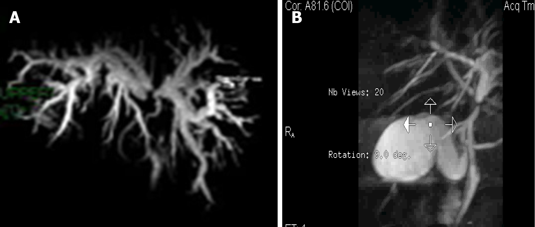
Figure 9 CholangioIRM.
A: Before photodynamic therapy: Bismuth III cholangiocarcinoma (large dilatation of intrahepatic ducts can be seen); B: After photodynamic therapy: The stenosis at the level of hilium and intrahepatic dilatation have been reduced.
- Citation: Tantau AI, Mandrutiu A, Pop A, Zaharie RD, Crisan D, Preda CM, Tantau M, Mercea V. Extrahepatic cholangiocarcinoma: Current status of endoscopic approach and additional therapies. World J Hepatol 2021; 13(2): 166-186
- URL: https://www.wjgnet.com/1948-5182/full/v13/i2/166.htm
- DOI: https://dx.doi.org/10.4254/wjh.v13.i2.166


