Copyright
©The Author(s) 2021.
World J Hepatol. Dec 27, 2021; 13(12): 2168-2178
Published online Dec 27, 2021. doi: 10.4254/wjh.v13.i12.2168
Published online Dec 27, 2021. doi: 10.4254/wjh.v13.i12.2168
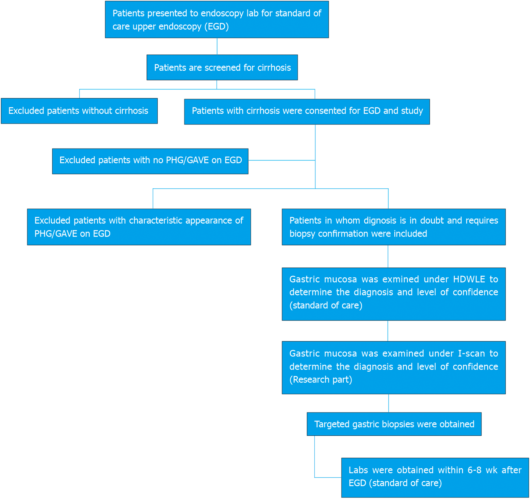
Figure 1 Study design.
PHG: Portal hypertensive gastropathy; GAVE: Gastric antral vascular ectasia; HDWL: High definition white light endoscopy.
Figure 2 Portal hypertensive gastropathy.
A: I-scan with pit edema/capillary engorgement; B: Dilated collecting venules under magnification; C: Intramucosal hemorrhage under magnification; D: Gastric antral vascular ectasia on I-scan defined as presence of capillary ectasia.
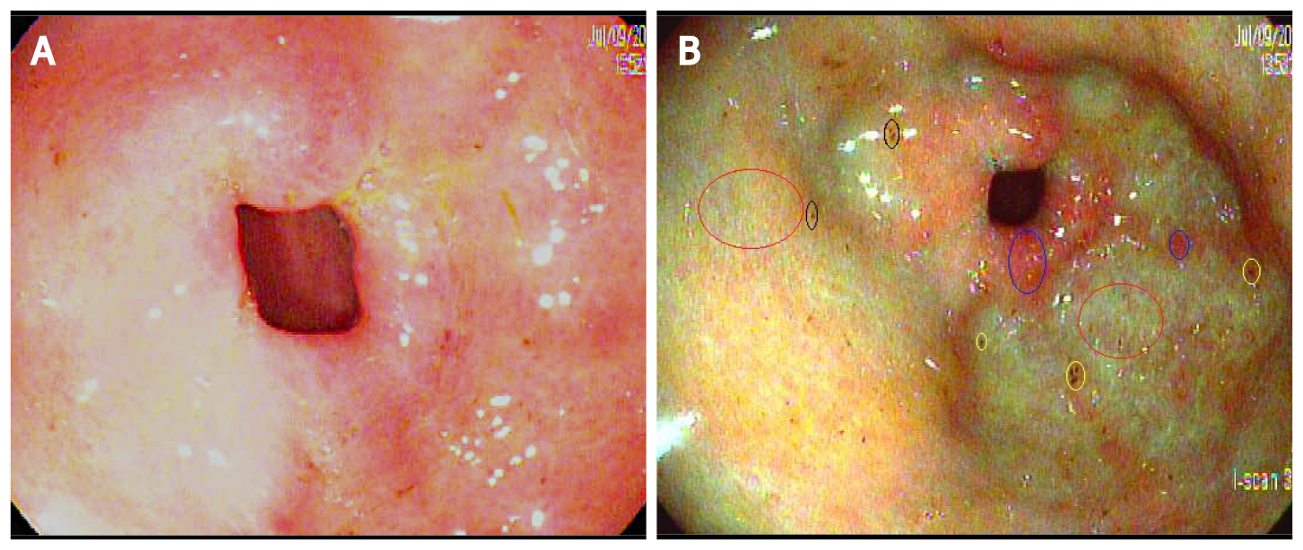
Figure 3 Portal hypertensive gastropathy under high definition white light endoscopy and I-scan Pit edema (red circles), intramucosal hemorrhage (yellow circles), capillary congestion (blue circles), and dilated venules (black circle).
A: High definition white light endoscopy; B: I-scan.
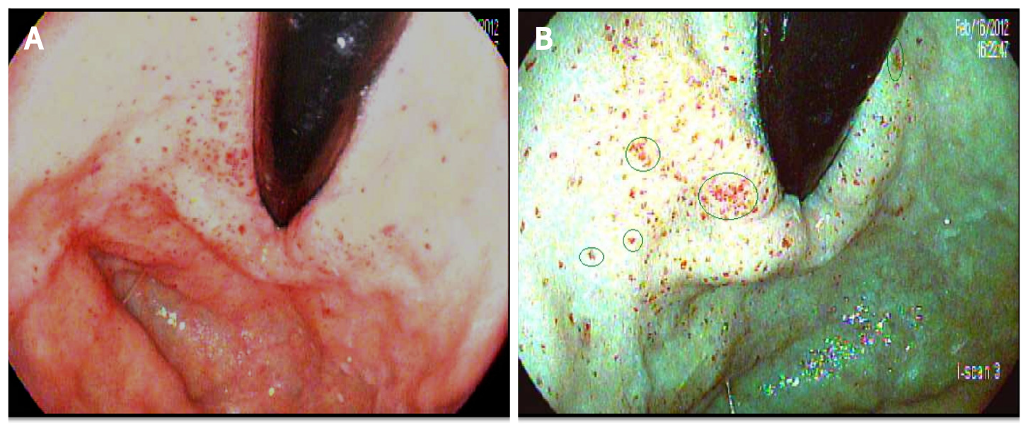
Figure 4 Gastric antral vascular ectasia under high-definition white light and I-scan dilated capillaries (green circles).
A: High-definition white light; B: I-scan.
- Citation: Al-Taee AM, Cubillan MP, Hinton A, Sobotka LA, Befeler AS, Hachem CY, Hussan H. Accuracy of virtual chromoendoscopy in differentiating gastric antral vascular ectasia from portal hypertensive gastropathy: A proof of concept study. World J Hepatol 2021; 13(12): 2168-2178
- URL: https://www.wjgnet.com/1948-5182/full/v13/i12/2168.htm
- DOI: https://dx.doi.org/10.4254/wjh.v13.i12.2168


