Copyright
©The Author(s) 2021.
World J Hepatol. Nov 27, 2021; 13(11): 1707-1726
Published online Nov 27, 2021. doi: 10.4254/wjh.v13.i11.1707
Published online Nov 27, 2021. doi: 10.4254/wjh.v13.i11.1707
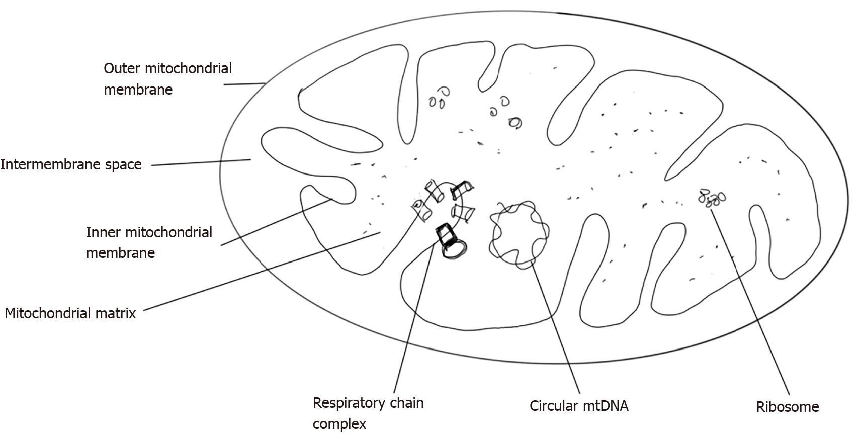
Figure 1 Diagrammatic representation of structure of mitochondria.
mtDNA: Mitochondrial DNA.
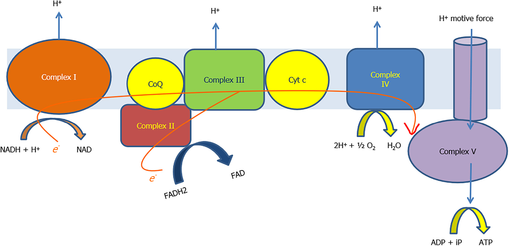
Figure 2 The electron transport chain formed by the respiratory chain complexes and process of oxidative phosphorylation.
ADP: Adenosine diphosphate; ATP: Adenosine triphosphate; CoQ: Coenzyme Q; Cyt c: Cytochrome c; FAD: Flavin adenine dinucleotide; FADH2: Reduced form of FAD; NAD: Nicotinamide adenine dinucleotide; NADH: Reduced form of NAD.
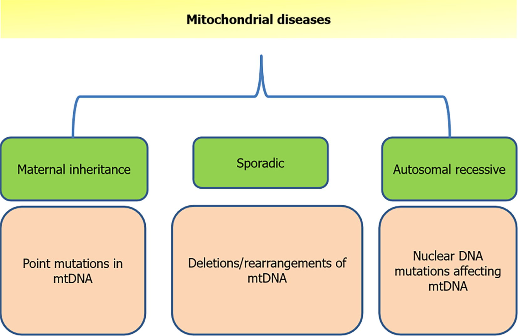
Figure 3 Various modes of inheritance of mitochondrial disease.
mtDNA: Mitochondrial DNA.
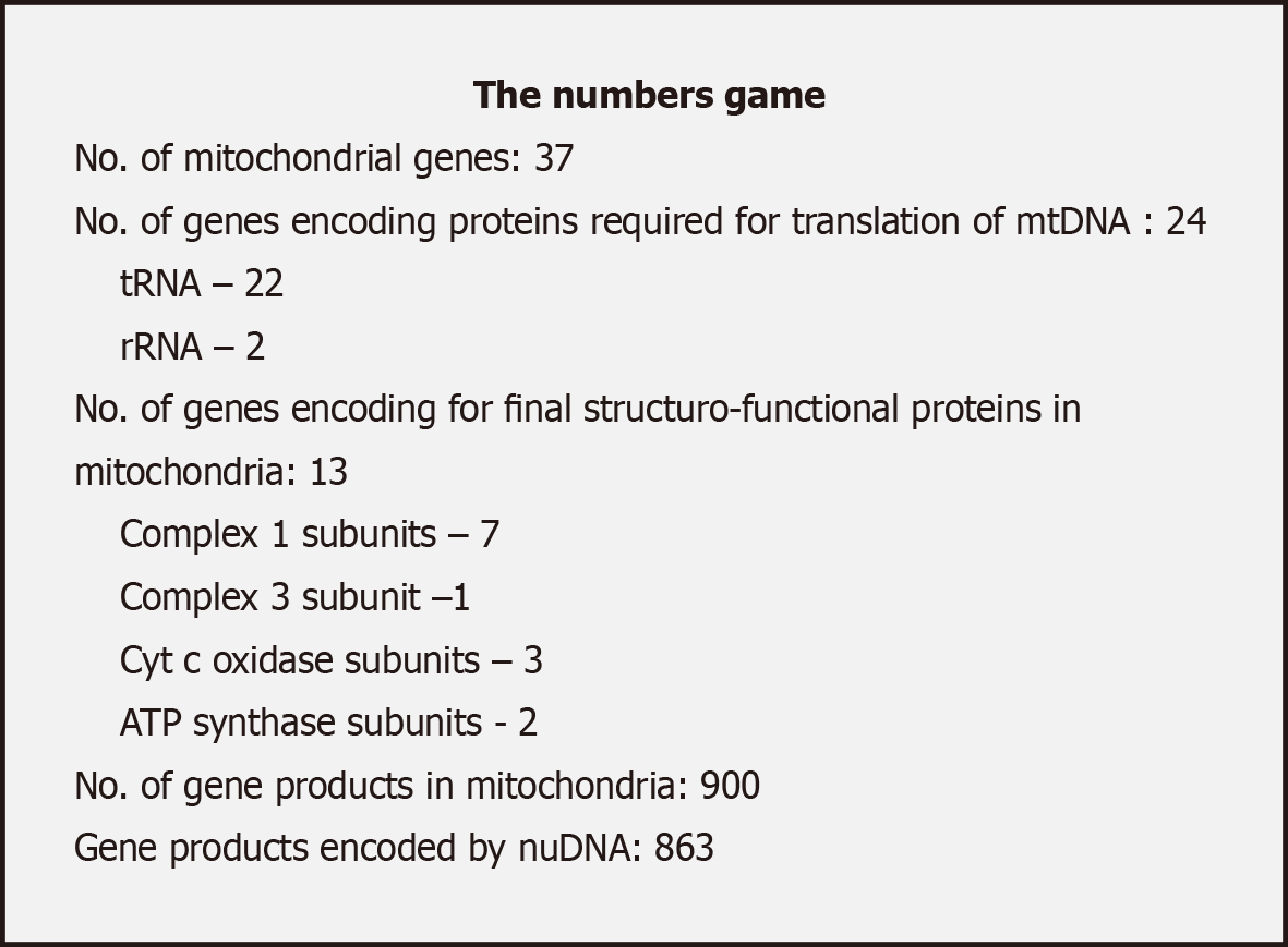
Figure 4 Comparison of mitochondrial and nuclear DNA influence in genetics of mitochondria.
mtDNA: Mitochondrial DNA; rRNA: Ribosomal RNA; tRNA: Transfer RNA.
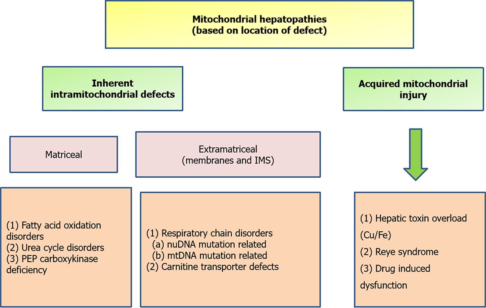
Figure 5 Simplified way of classification of mitochondrial hepatopathies based on location of defect.
IMS: Intermembrane space; mtDNA: Mitochondrial DNA; PEP: Phosphoenolpyruvate.
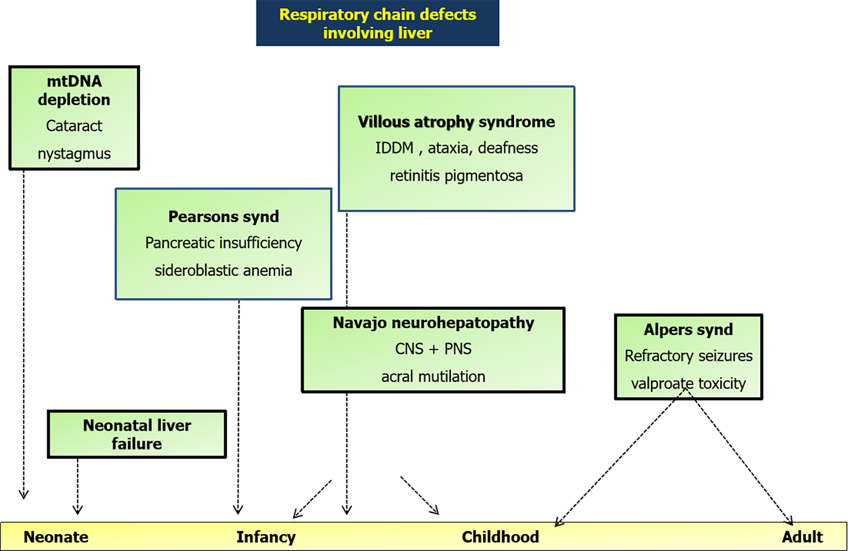
Figure 6 Graphical summary of various respiratory chain disorders involving liver on a timeline with key features.
CNS: Central nervous system; IDDM: Insulin dependent diabetes mellitus; mtDNA: Mitochondrial DNA; PNS: Peripheral nervous system.
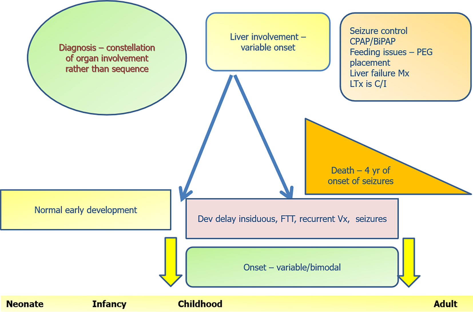
Figure 7 Graphical summary of Alpers Huttenlocher syndrome and its natural history.
BiPAP: Bilevel positive airway pressure; C/I: Contraindicated; CPAP: Continuous positive airway pressure; FTT: Failure to thrive; LTx: Liver transplantation; Mx: Management; PEG: Percutaneous endoscopic gastrostomy; Vx: Vomiting.
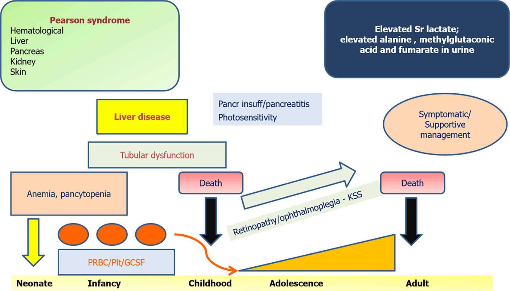
Figure 8 Graphical summary of Pearson marrow pancreas syndrome and its natural history.
GCSF: Granulocyte colony stimulation factor; insuff: Insufficiency; KSS: Kearns Sayre syndrome; Pancr: Pancreatic; Plt: Platelets; PRBC: Packed red blood cells.
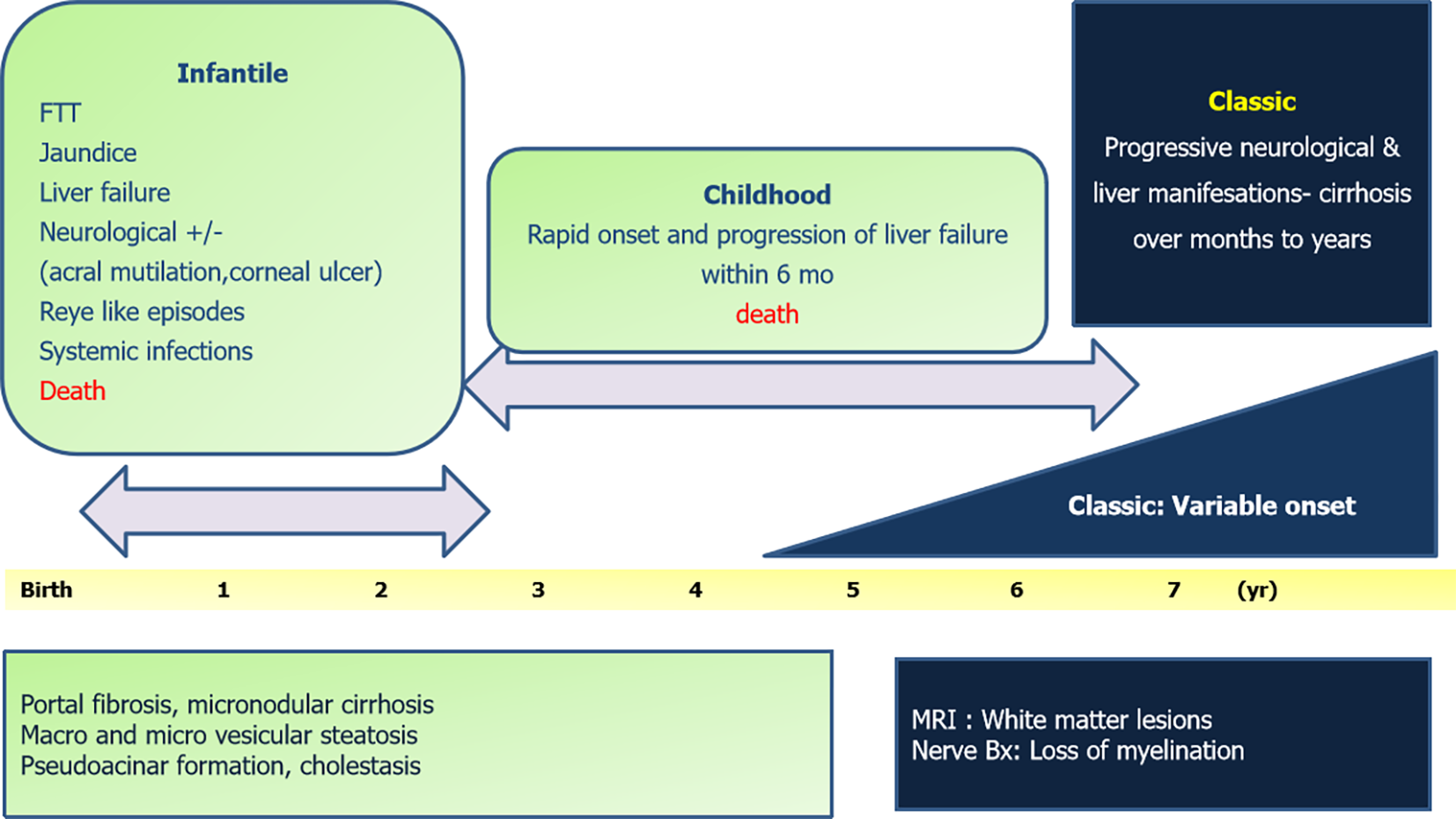
Figure 9 Graphical summary of Navajo neurohepatopathy.
Bx: Biopsy; FTT: Failure to thrive.
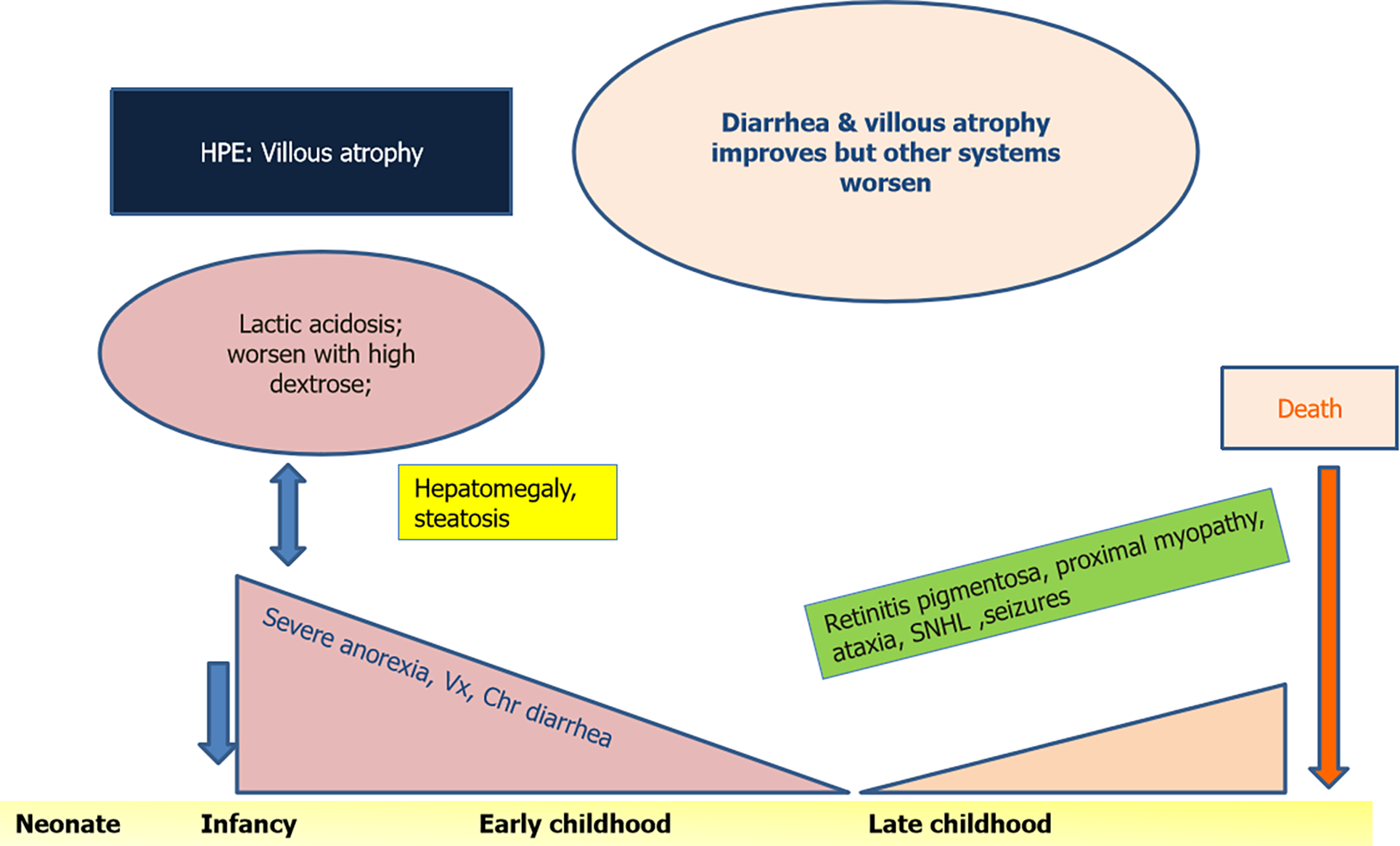
Figure 10 Graphical summary of villous atrophy syndrome and its natural history.
HPE: Histopathological examination; SNHL: Sensorineural hearing loss; Vx: Vomiting.
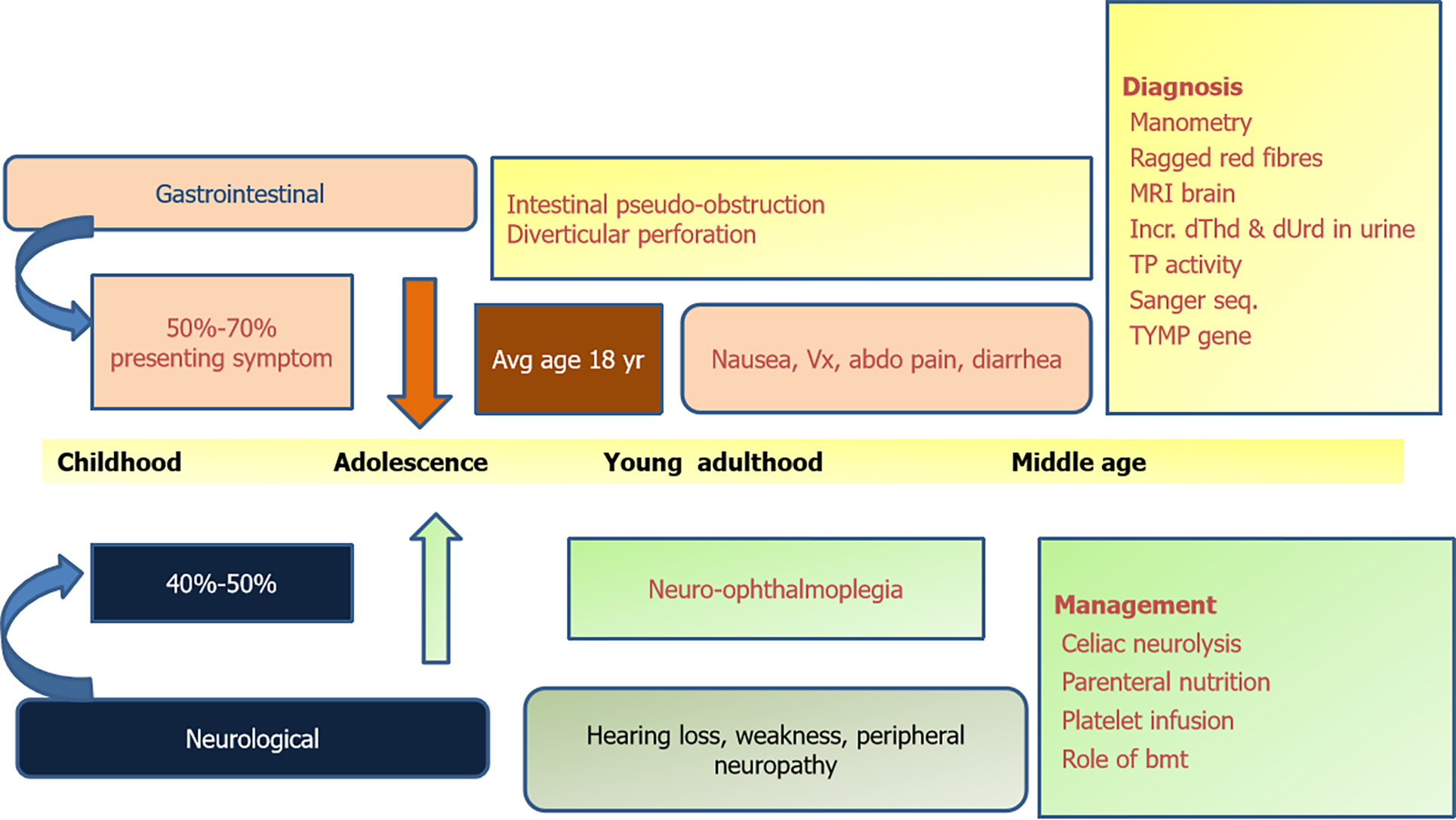
Figure 11 Graphical summary of mitochondrial neurogastrointestinal encephalomyopathy.
BMT: Bone marrow transplantation; dThd: Thymidine; dUrd: Deoxy uridine levels; MRI: Magnetic resonance imaging; TP: Thymidine phosphorylase. Vx: Vomiting.
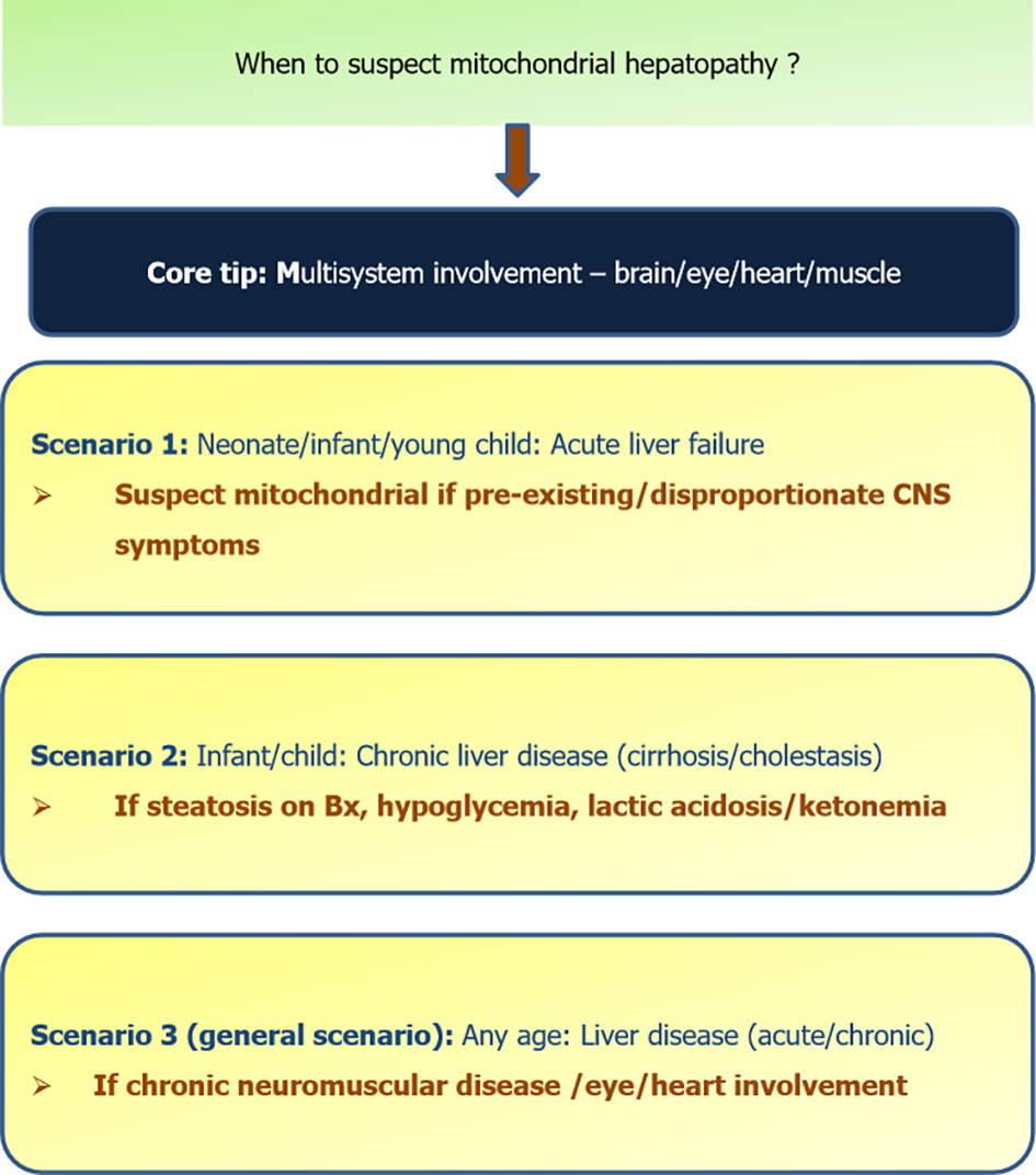
Figure 12 Scenarios when to suspect mitochondrial hepatopathy.
Bx: Biopsy; CNS: Central nervous system.
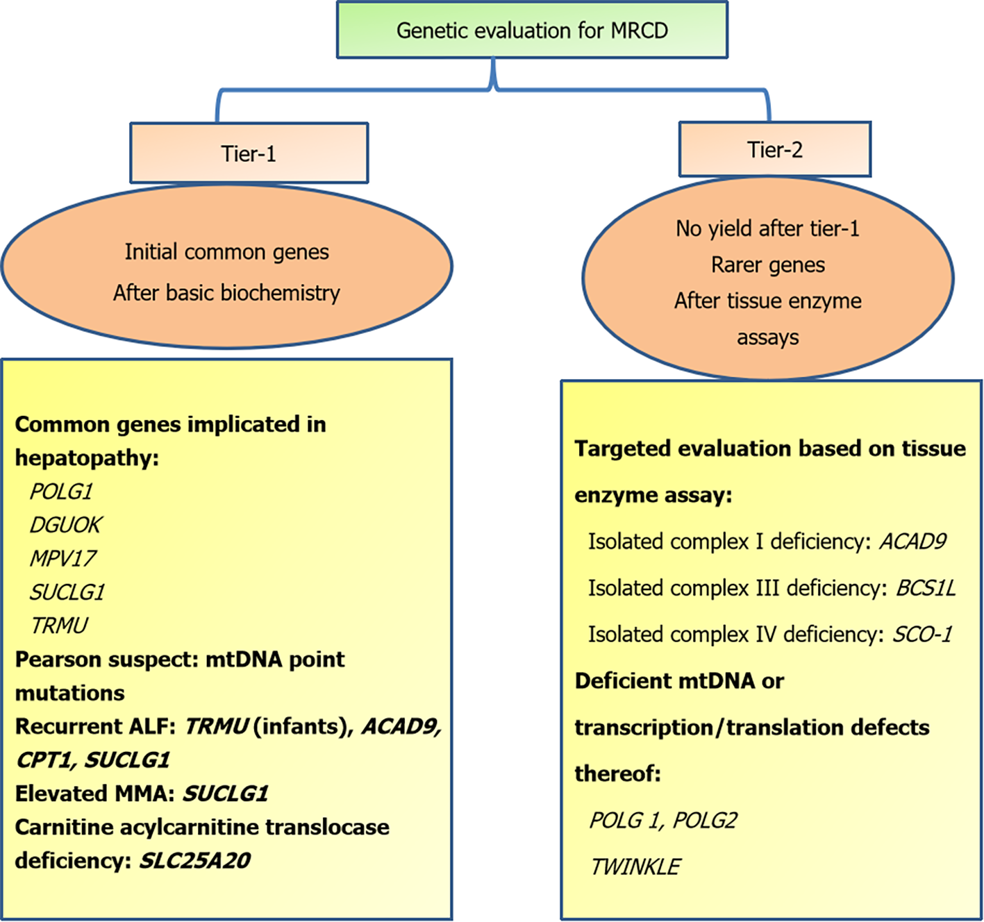
Figure 13 Step-wise strategy of genetic evaluation in mitochondrial respiratory chain defects.
MRCD: Mitochondrial respiratory chain disorders; ALF: Acute liver failure; MMA: Methylmalonic acid.
- Citation: Gopan A, Sarma MS. Mitochondrial hepatopathy: Respiratory chain disorders- ‘breathing in and out of the liver’. World J Hepatol 2021; 13(11): 1707-1726
- URL: https://www.wjgnet.com/1948-5182/full/v13/i11/1707.htm
- DOI: https://dx.doi.org/10.4254/wjh.v13.i11.1707


