Copyright
©The Author(s) 2021.
World J Hepatol. Nov 27, 2021; 13(11): 1459-1483
Published online Nov 27, 2021. doi: 10.4254/wjh.v13.i11.1459
Published online Nov 27, 2021. doi: 10.4254/wjh.v13.i11.1459
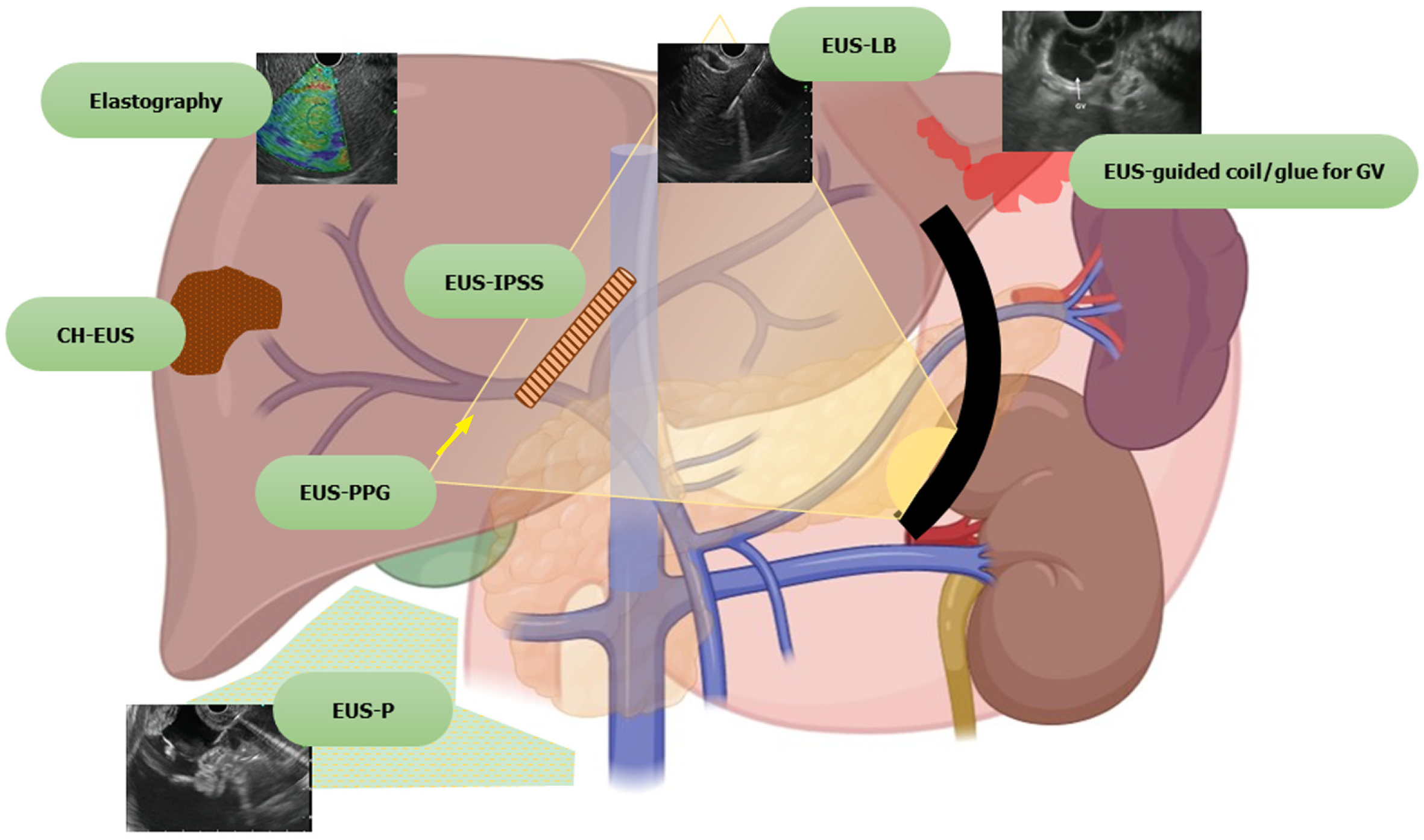
Figure 1 Spectrum of endoscopic ultrasound in hepatology.
EUS: Endoscopic ultrasound; CH-EUS: Contrast harmonic endoscopic ultrasound; EUS-IPSS: Endoscopic ultrasound guided intrahepatic portosystemic shunt; EUS-LB: Endoscopic ultrasound guided liver biopsy; EUS-PPG: Endoscopic ultrasound guided portal pressure gradient; EUS-P: Endoscopic ultrasound guided paracentesis; GV: Gastric varices.
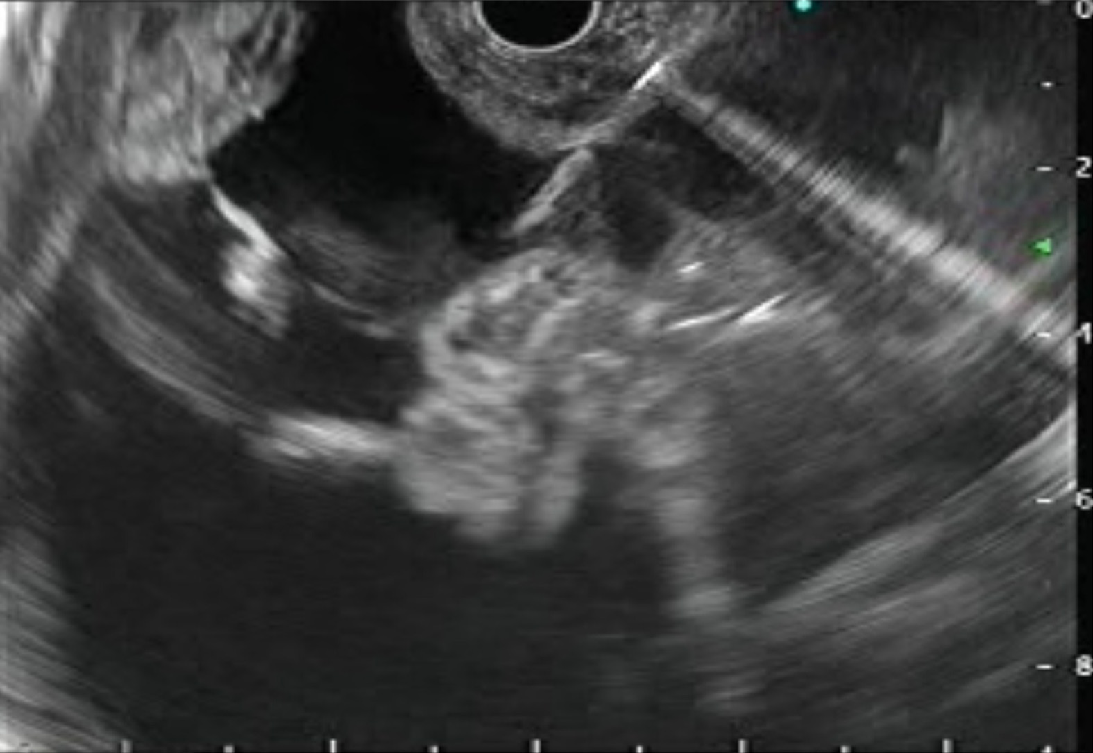
Figure 2 Endoscopic ultrasound guided paracentesis.
Needle is visualized in the ascitic fluid.
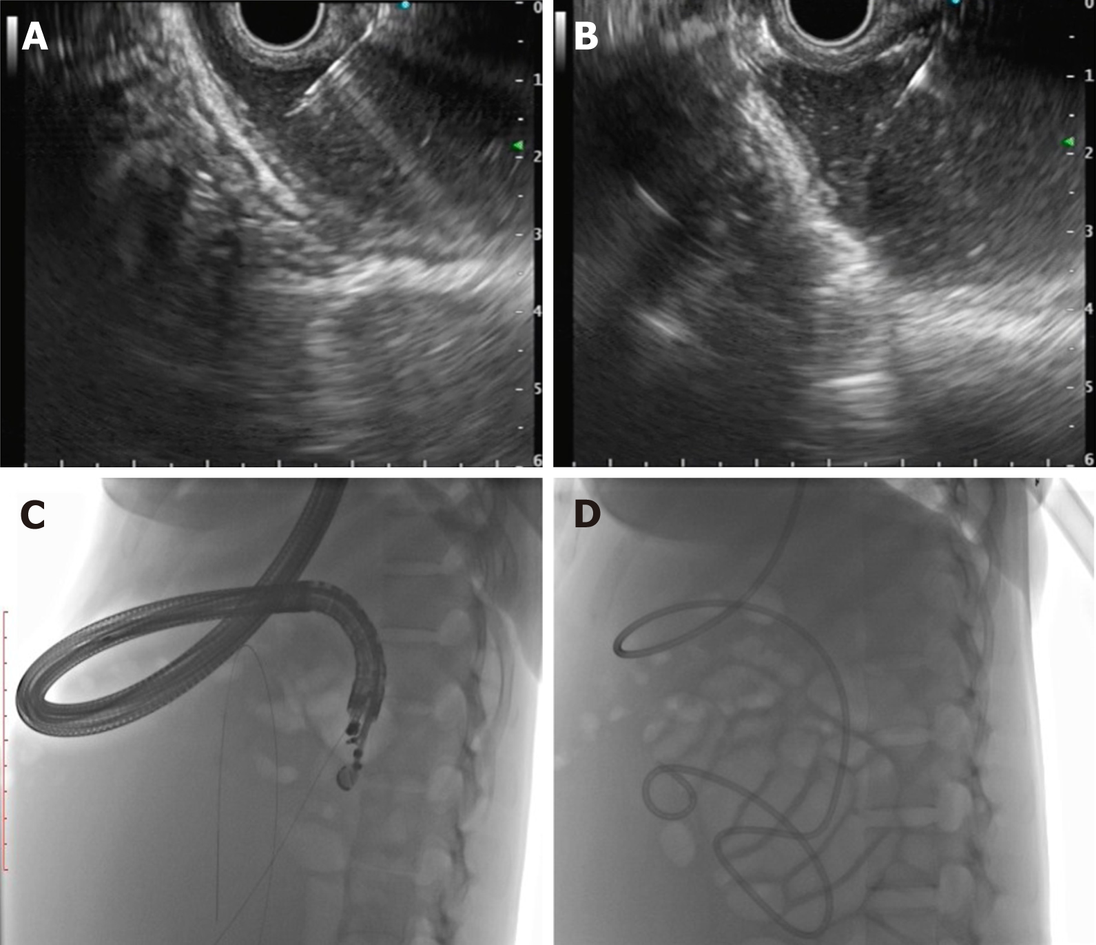
Figure 3 Endoscopic ultrasound-guided internal drainage of loculated ascites.
A: Puncture of the loculated ascites with 19-G aspiration needle; B: Guidewire negotiated across as visualized on endoscopic ultrasound; C: Fluoroscopic view of guidewire coiled inside the loculated ascites; D: Naso-cystic drain placed inside the loculated ascites.
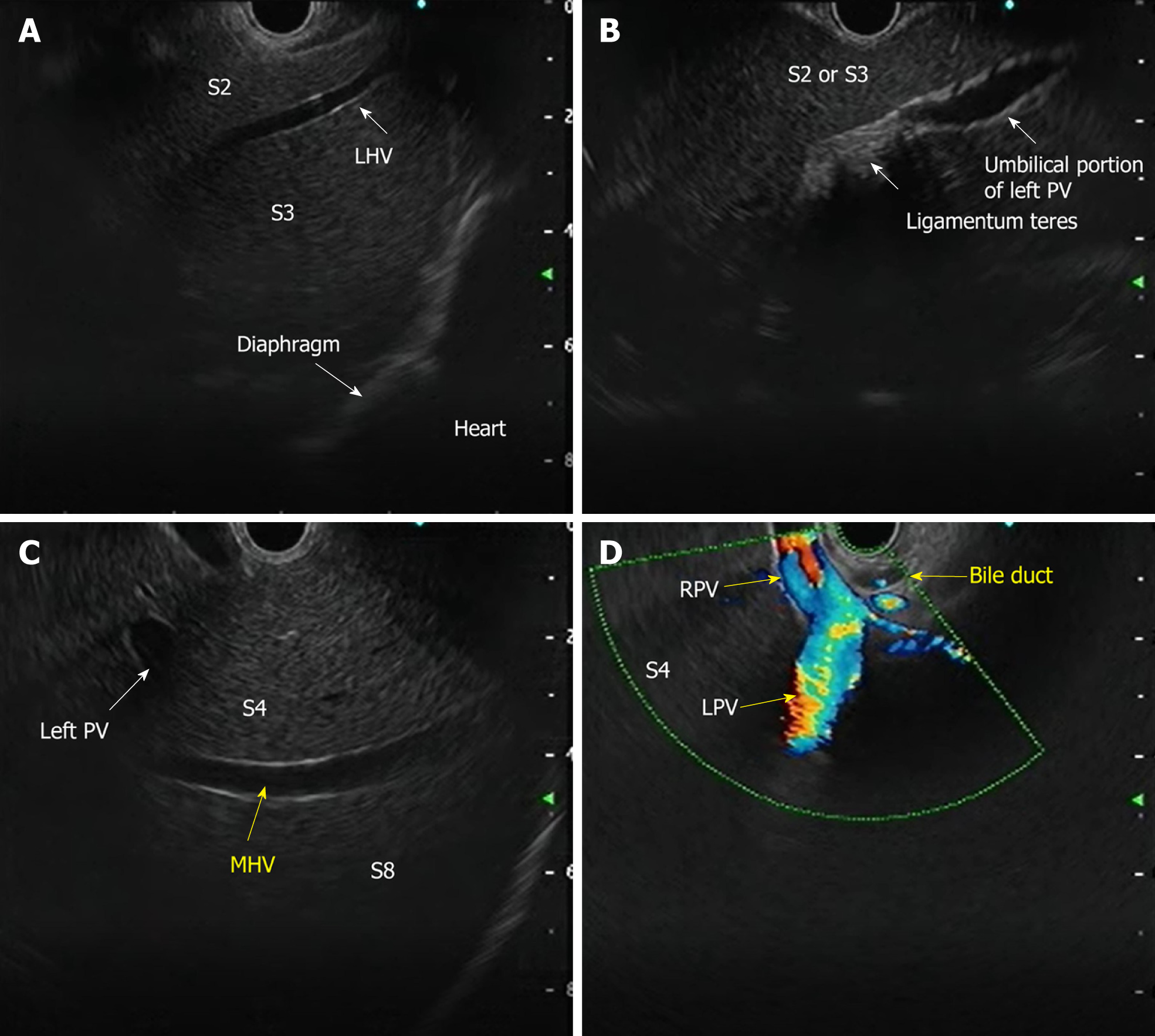
Figure 4 Endoscopic ultrasound anatomy of liver segments.
A: Anatomy of the left lobe with S2 and S3 segments; B: Ligamentum teres with umbilical portion of the left portal vein; C: Middle hepatic vein with segments of the liver; D: Anatomy of the bifurcation of portal vein from the duodenal bulb. PV: Portal vein; MHV: Middle hepatic vein; LHV: Left hepatic vein; RPV: Right portal vein; LPV: Left portal vein.
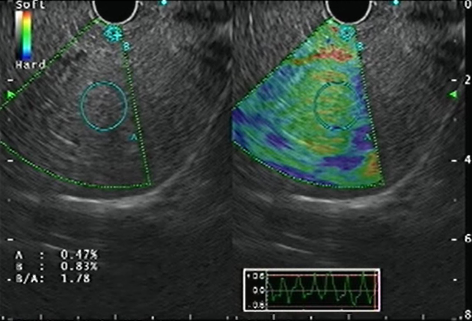
Figure 5 Endoscopic ultrasound elastography of the liver parenchyma.
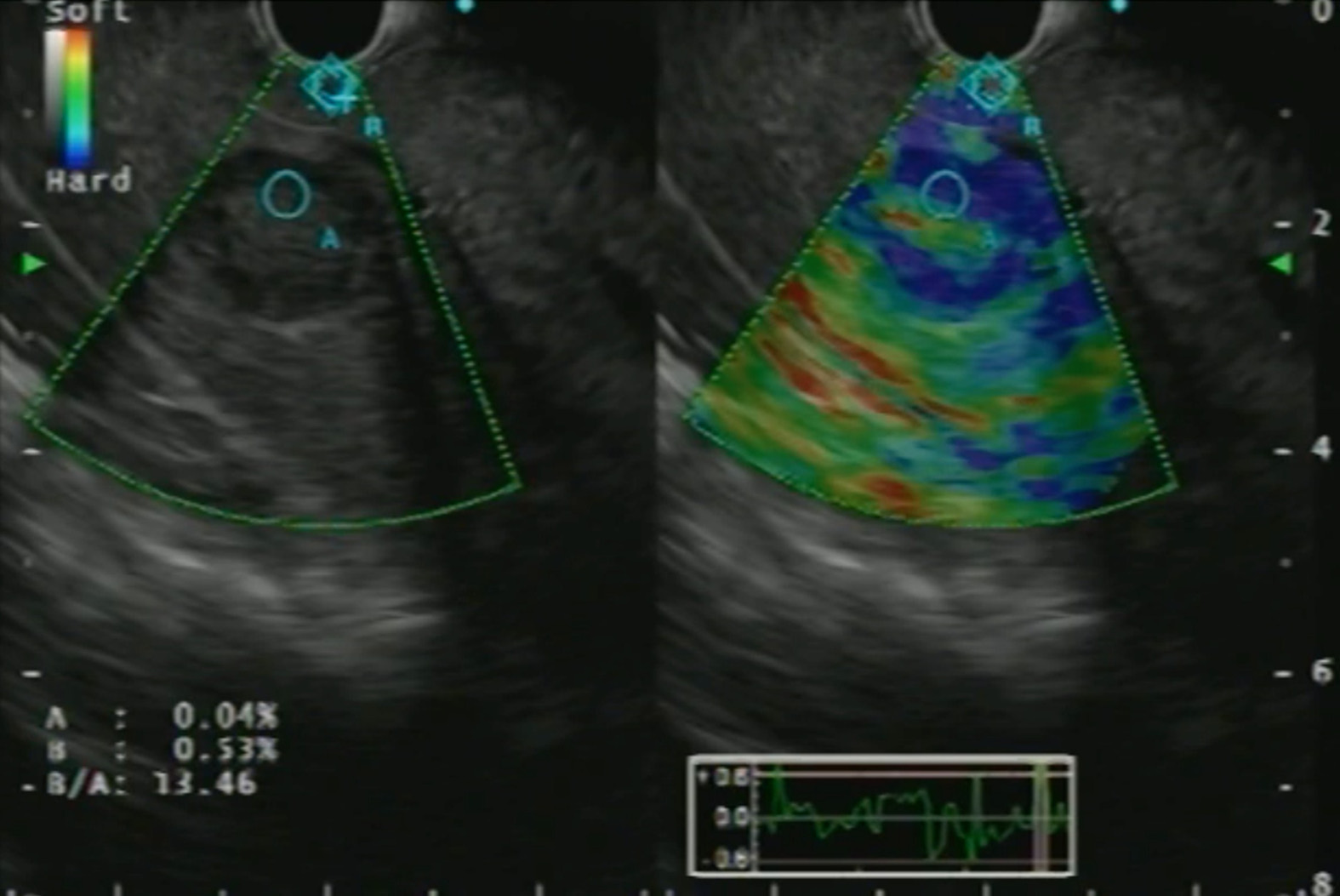
Figure 6 Endoscopic ultrasound elastography of a focal liver lesion with strain ratio calculation.
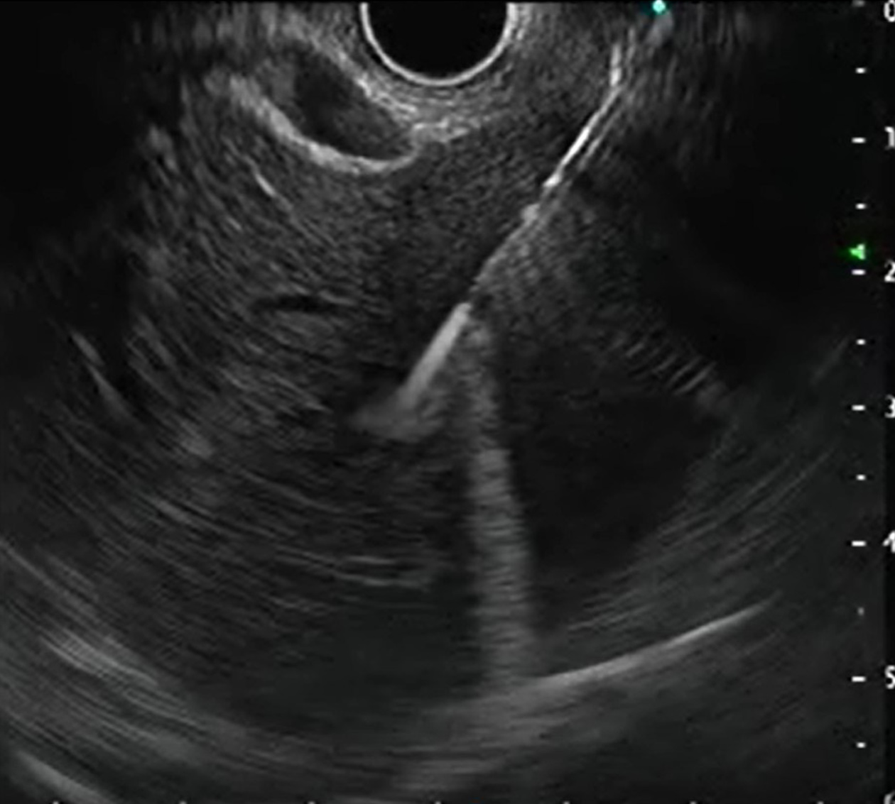
Figure 7 Endoscopic ultrasound-guided liver biopsy.
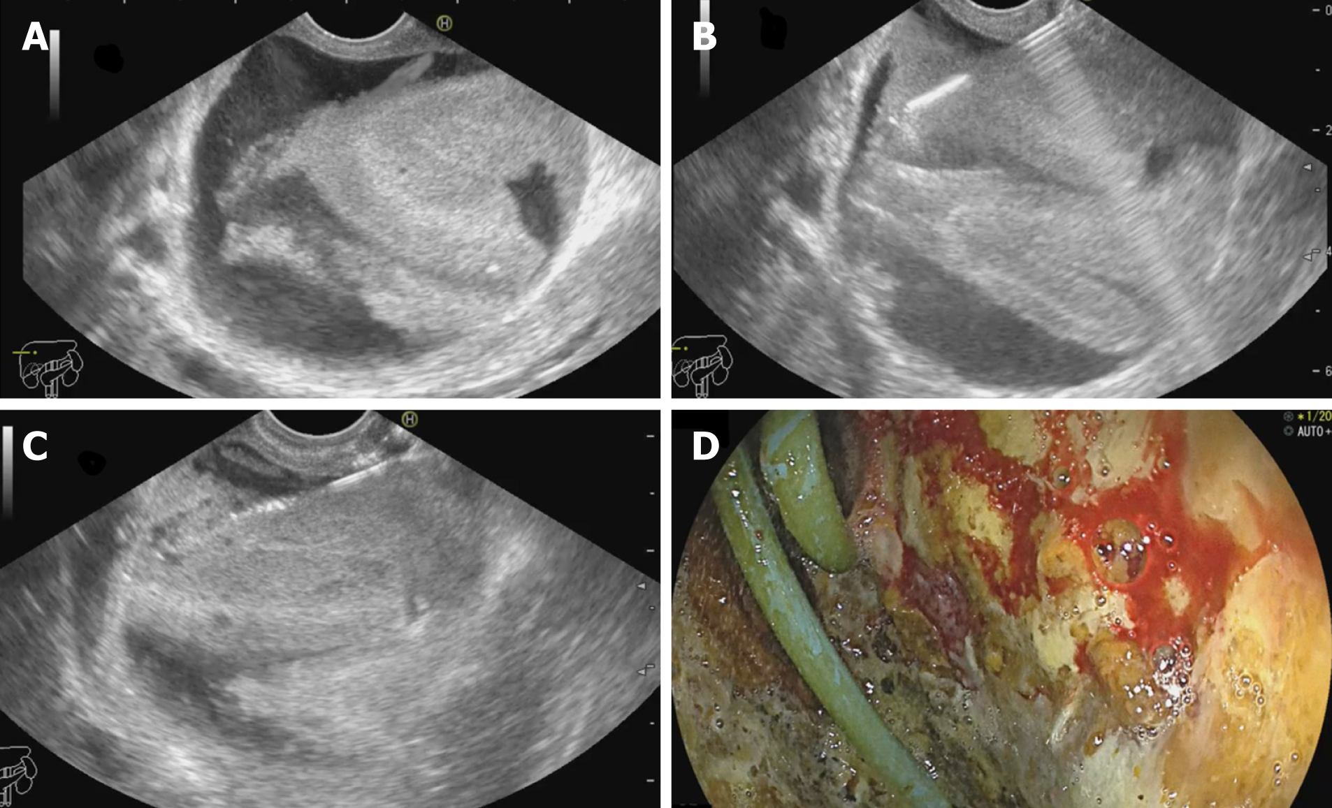
Figure 8 Endoscopic ultrasound-guided drainage of biloma.
A: Post-operative biloma noted on endoscopic ultrasound (EUS) with internal echoes; B: EUS-guided puncture of the biloma; C: Guidewire negotiated into the collection followed by placement of naso-cystic drain; D: Endoscopic view of the cavity entered with catheter noted in situ.
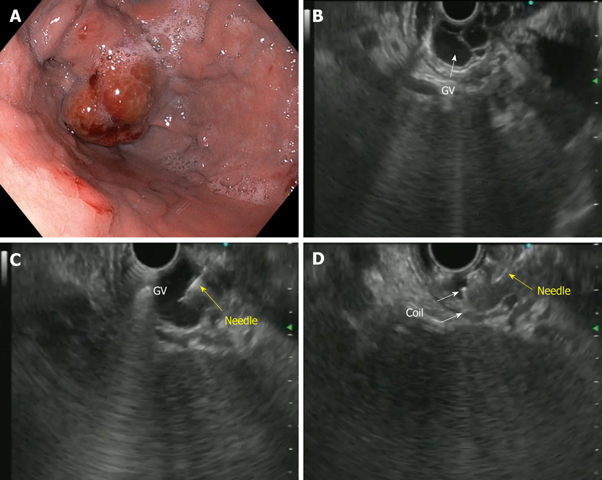
Figure 9 Endoscopic ultrasound-guided coil embolization of fundal varix.
A: Endoscopic view of the fundal varix; B: Endoscopic ultrasound (EUS) view of the fundal varix; C: EUS guided puncture of the varix with a 22-G needle; D: Coil deployment inside the varix. GV: Gastric varices.
- Citation: Dhar J, Samanta J. Role of endoscopic ultrasound in the field of hepatology: Recent advances and future trends. World J Hepatol 2021; 13(11): 1459-1483
- URL: https://www.wjgnet.com/1948-5182/full/v13/i11/1459.htm
- DOI: https://dx.doi.org/10.4254/wjh.v13.i11.1459


