Copyright
©The Author(s) 2021.
World J Hepatol. Oct 27, 2021; 13(10): 1269-1288
Published online Oct 27, 2021. doi: 10.4254/wjh.v13.i10.1269
Published online Oct 27, 2021. doi: 10.4254/wjh.v13.i10.1269
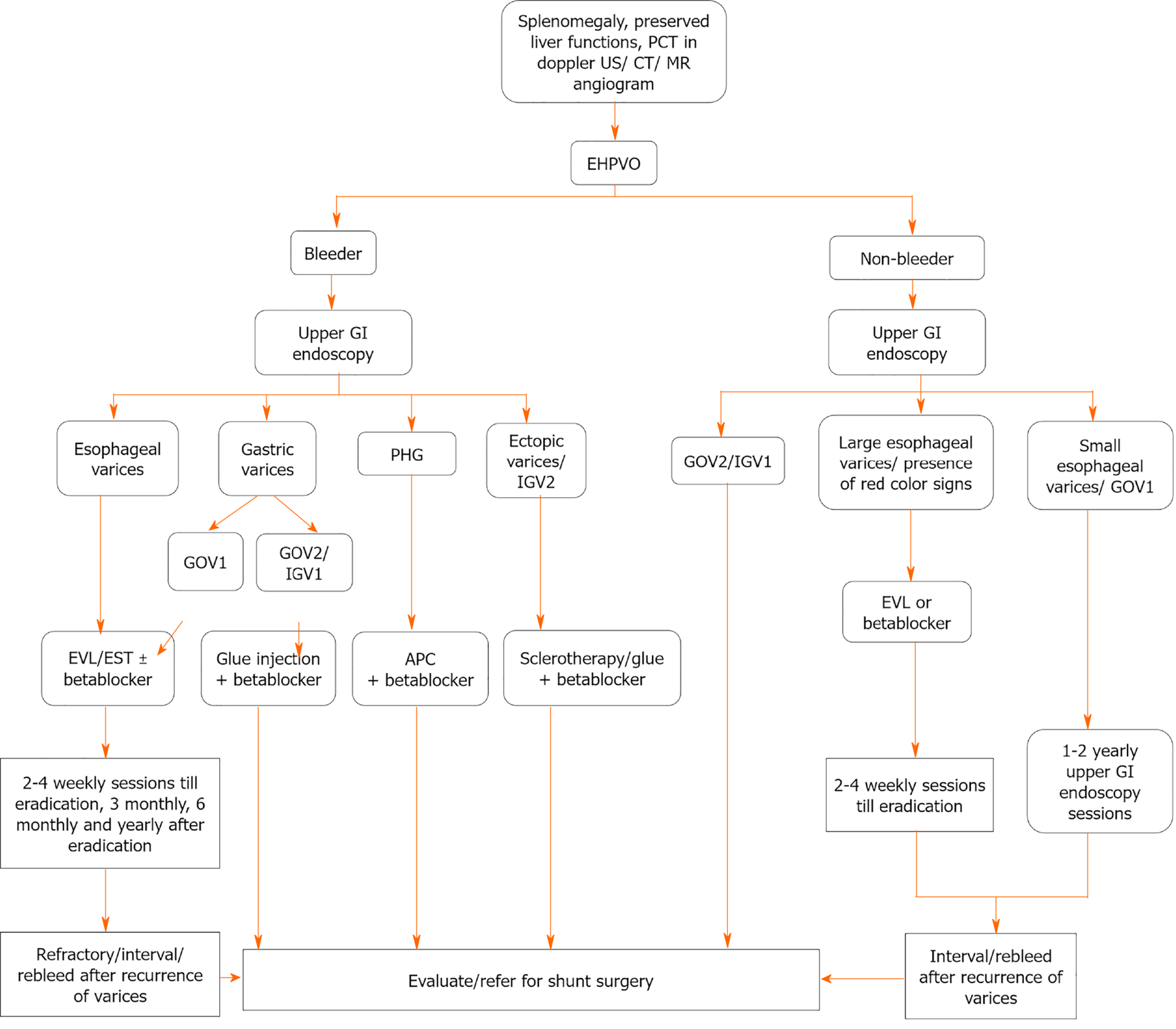
Figure 1 Algorithm for management of esophageal varices and gastric varices in extra-hepatic portal vein obstruction.
EHPVO: Extra-hepatic portal vein obstruction; PHG: Portal hypertensive gastropathy; GOV: Gastroesophageal varices; IGV: Isolated gastric varices; EVL: Endoscopic variceal ligation; EST: Endoscopic sclerotherapy; APC: argon plasma coagulation.
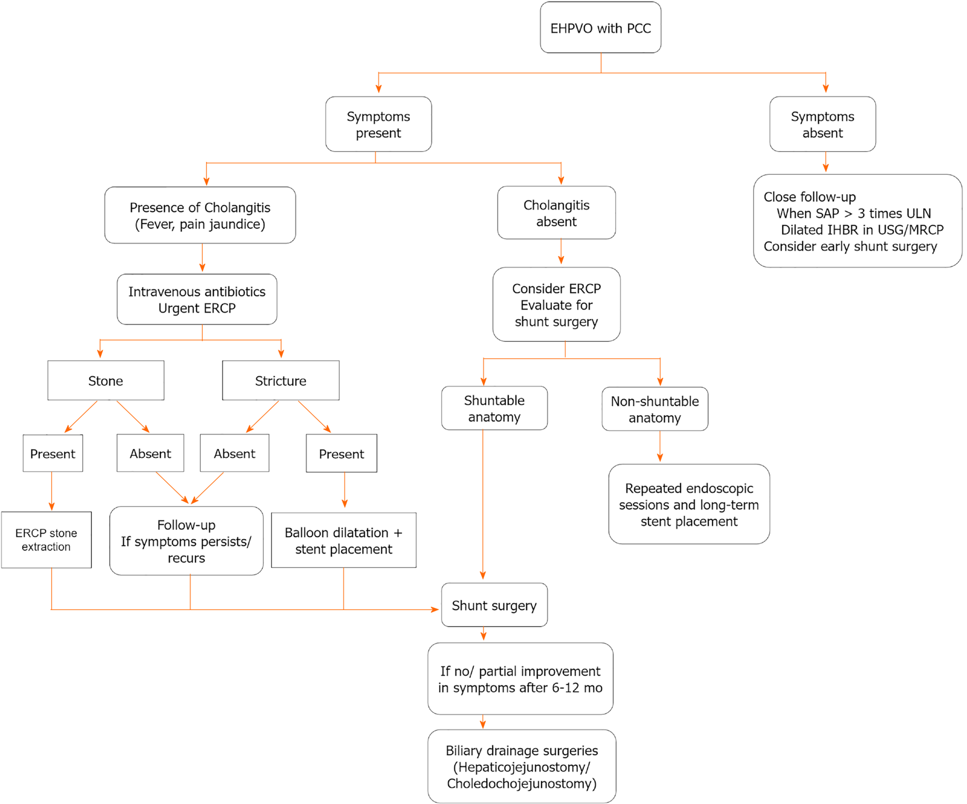
Figure 2 Algorithmic approach for management of portal cavernoma cholangiopathy in extra-hepatic portal vein obstruction.
EHPVO: Extra-hepatic portal vein obstruction; PCC: Portal cavernoma cholangiopathy; ERCP: Endoscopic retrograde pancreato-cholangiography; MRCP: Magnetic resonance cholangiopancreatography; SAP: serum alkaline phosphatase; IHBR: intrahepatic biliary radicle; USG: ultrasonography; ULN: upper limit of normal.
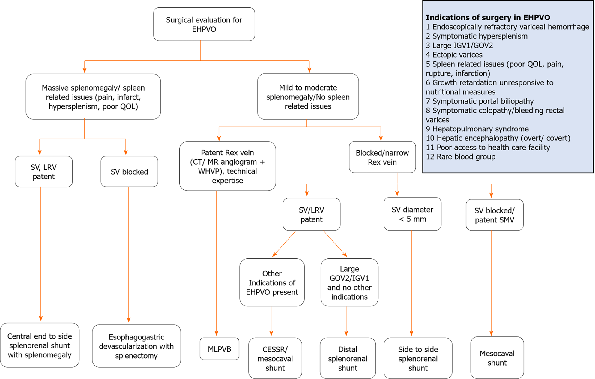
Figure 3 Indications of surgery in extra-hepatic portal vein obstruction and algorithmic approach for surgical management in extra-hepatic portal vein obstruction in developing countries.
EHPVO: Extra-hepatic portal vein obstruction; GOV: Gastroesophageal varices; IGV: Isolated gastric varices; EVL: Endoscopic variceal ligation; SV: splenic vein; SMV: superior mesenteric vein; EST: Endoscopic sclerotherapy; CT: Computed tomography, LRV: left renal vein; MR: magnetic resonance, CESSR: central end-to-side splenorenal shunt, QOL: quality of life; WVHP: wedge hepatic venous pressure; MLPVB: mesenterico left portal vein bypass (meso-Rex).
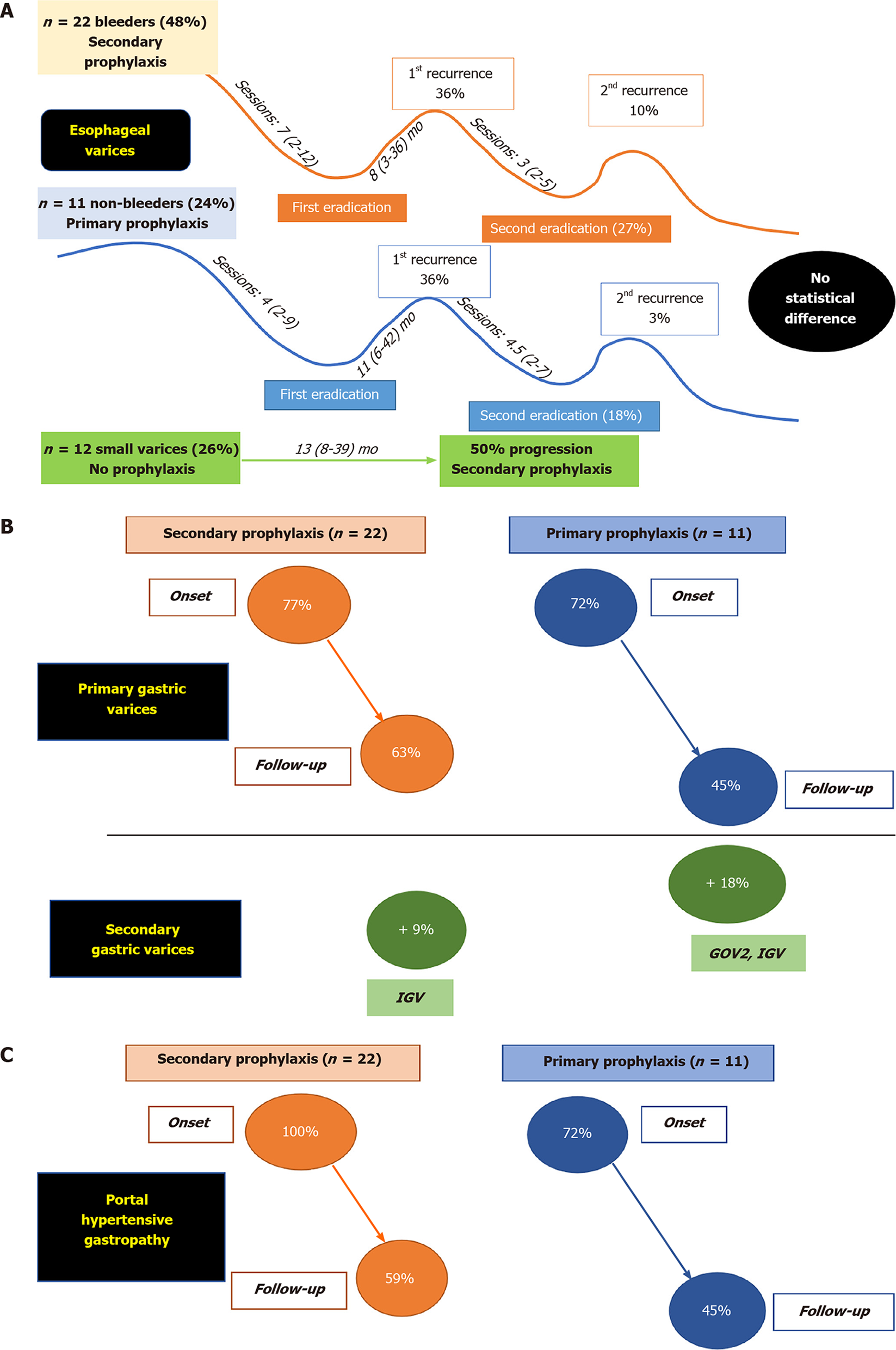
Figure 4 Natural history and follow-up outcome of esophageal varices, gastric varices and portal hypertensive gastropathy in pediatric non-cirrhotic portal fibrosis in author's experience.
A: 22 children presented with variceal bleeding (bleeders), required Endoscopic Variceal Ligation and/or sclerotherapy. 11 children presented with large varices without bleeding (non-bleeders). The incidence of recurrence of varices following eradication is not statistically different between bleeders and non-bleeders. 50% small varices progressed to bleed in the follow-up (median time 13 mo); B: 77% bleeders and 72% non-bleeders had gastric varices (primary) at initial endoscopy. 9% bleeders and 18% non-bleeders develop gastric varices (secondary) in follow-up; C: 100% bleeders and 72% non-bleeders had portal hypertensive gastropathy at initial endoscopy, reduced to 59% and 45% respectively, in follow-up.
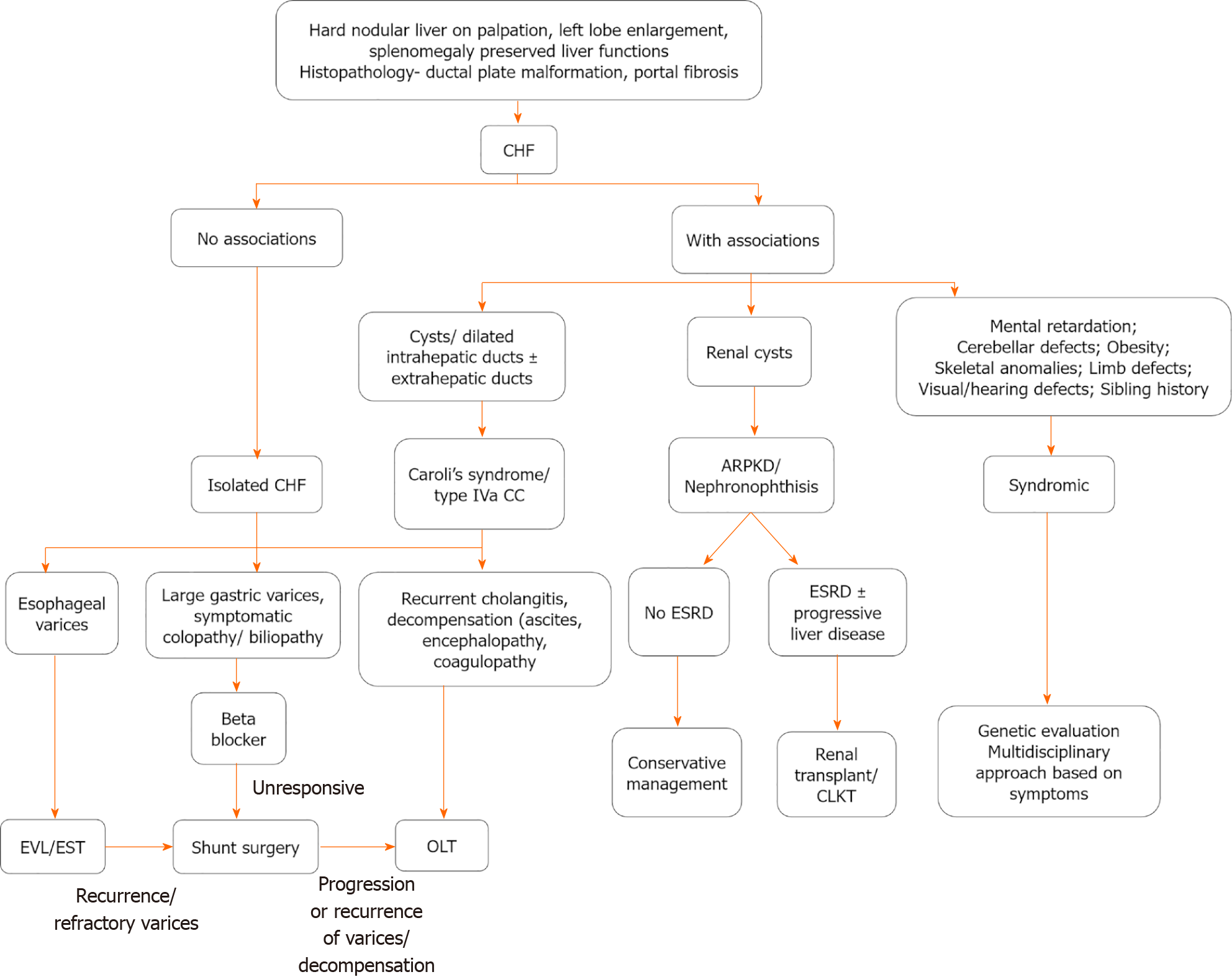
Figure 5 Algorithmic approach to diagnosis and management of congenital hepatic fibrosis.
CHF: Congenital hepatic fibrosis; CC: Choledochal cyst; ESRD: End-stage renal disease; EVL: Endoscopic variceal ligation; EST: Endoscopic sclerotherapy; CLKT: Combined liver-kidney transplantation; OLT: Orthotopic liver transplantation, ARPKD: autosomal recessive polycystic kidney disease.
- Citation: Sarma MS, Seetharaman J. Pediatric non-cirrhotic portal hypertension: Endoscopic outcome and perspectives from developing nations. World J Hepatol 2021; 13(10): 1269-1288
- URL: https://www.wjgnet.com/1948-5182/full/v13/i10/1269.htm
- DOI: https://dx.doi.org/10.4254/wjh.v13.i10.1269


