Copyright
©The Author(s) 2020.
World J Hepatol. Apr 27, 2020; 12(4): 160-169
Published online Apr 27, 2020. doi: 10.4254/wjh.v12.i4.160
Published online Apr 27, 2020. doi: 10.4254/wjh.v12.i4.160
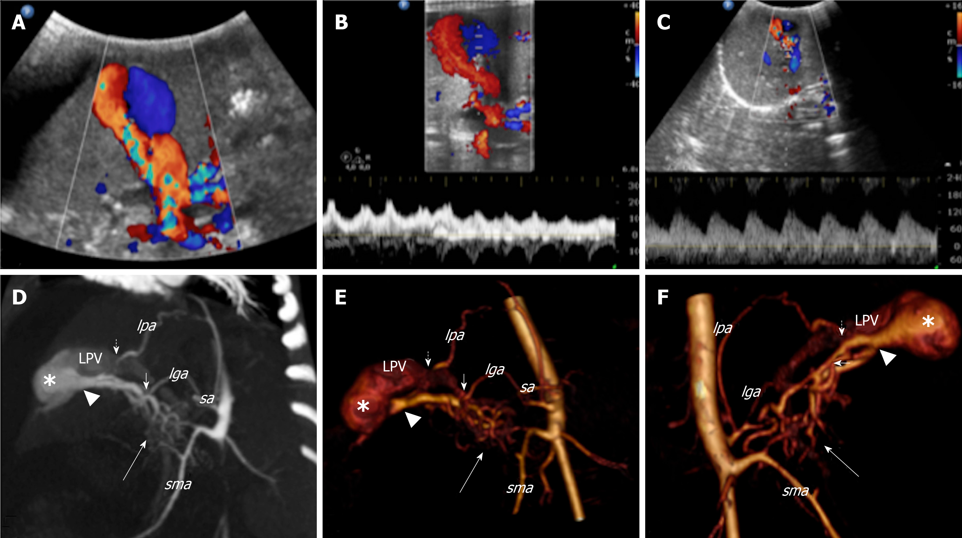
Figure 1 Colour Doppler ultrasound and computed tomography images that show the congenital intrahepatic arterioportal fistula.
A: Colour Doppler ultrasound images of the liver revealed aneurysmal dilatation of the left portal vein with turbulent bidirectional flow; B: Particularly, the venous flow appeared arterialised; C: Several dilated and tortuous branches of the hepatic artery with high flow rate supplied the dilated portal vein segment; D: Contrast enhanced multi-detector computer tomography images of the abdomen reformatted on both the left oblique: maximum intensity projection; E: 3D volume rendered; F: 3D-VR right oblique plane. Contrast enhanced multi-detector computer tomography shows the complex vascular malformation characterised by a dense dysplastic small arterial network that arose from the hepatic and superior mesenteric system (thick arrow) and directly converged to the left portal vein through one Y-shaped fistula within the Rex recess (arrow head). Two additional feeders to the vascular anomaly [from the phrenic (short dotted arrow) and left gastric (short arrow) artery, respectively] were also detected. lga: Left gastric artery; lpa: Left phrenic artery; sa: Splenic artery; sma: Superior mesenteric artery; LPV: Left portal vein.
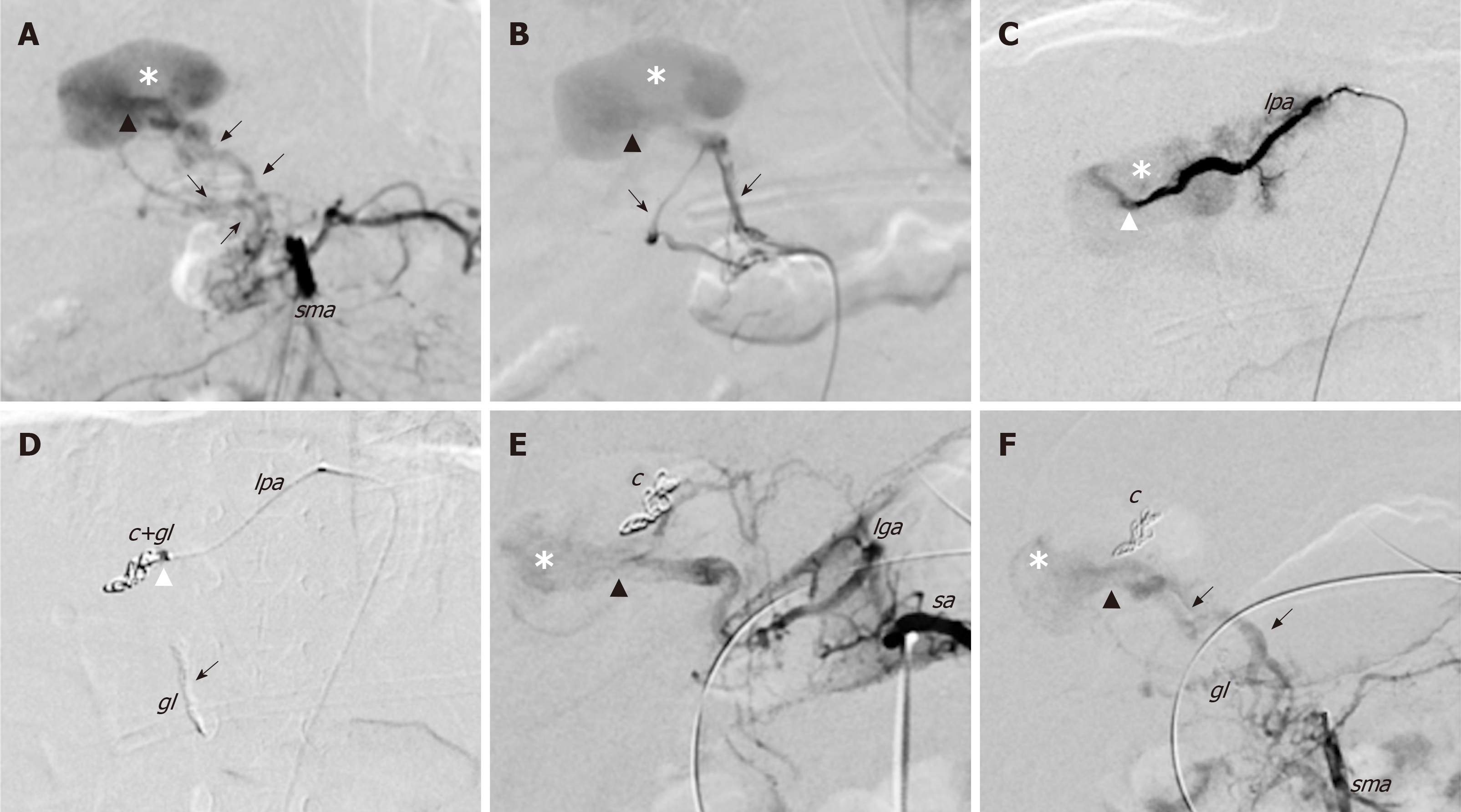
Figure 2 First angiography and endovascular embolization of the congenital intrahepatic arterioportal fistula.
Initial endovascular treatment of the malformation (digital subtraction angiograms). A and B: Angiograms from the superior mesenteric artery show dilated and tortuous dysplastic arteries (black arrows) that converged into the left aneurismal portal vein through one Y-shaped fistula within the Rex recess (black arrow head); C: Superselective catheterisation of a distal branch of the left phrenic artery that shows the additional shunt (white arrow head) into the venous aneurism; D: Embolisation of the shunts with glue and coils with glue cast; E and F: Angiographic control images from celiac trunk (E) and superior mesenteric artery (F) that show persistent patency of the fistula after the embolisation. c: Coils; gl: Glue; lga: Left gastric artery; lpa: Left phrenic artery; sma: Superior mesenteric artery; sa: Splenic artery.
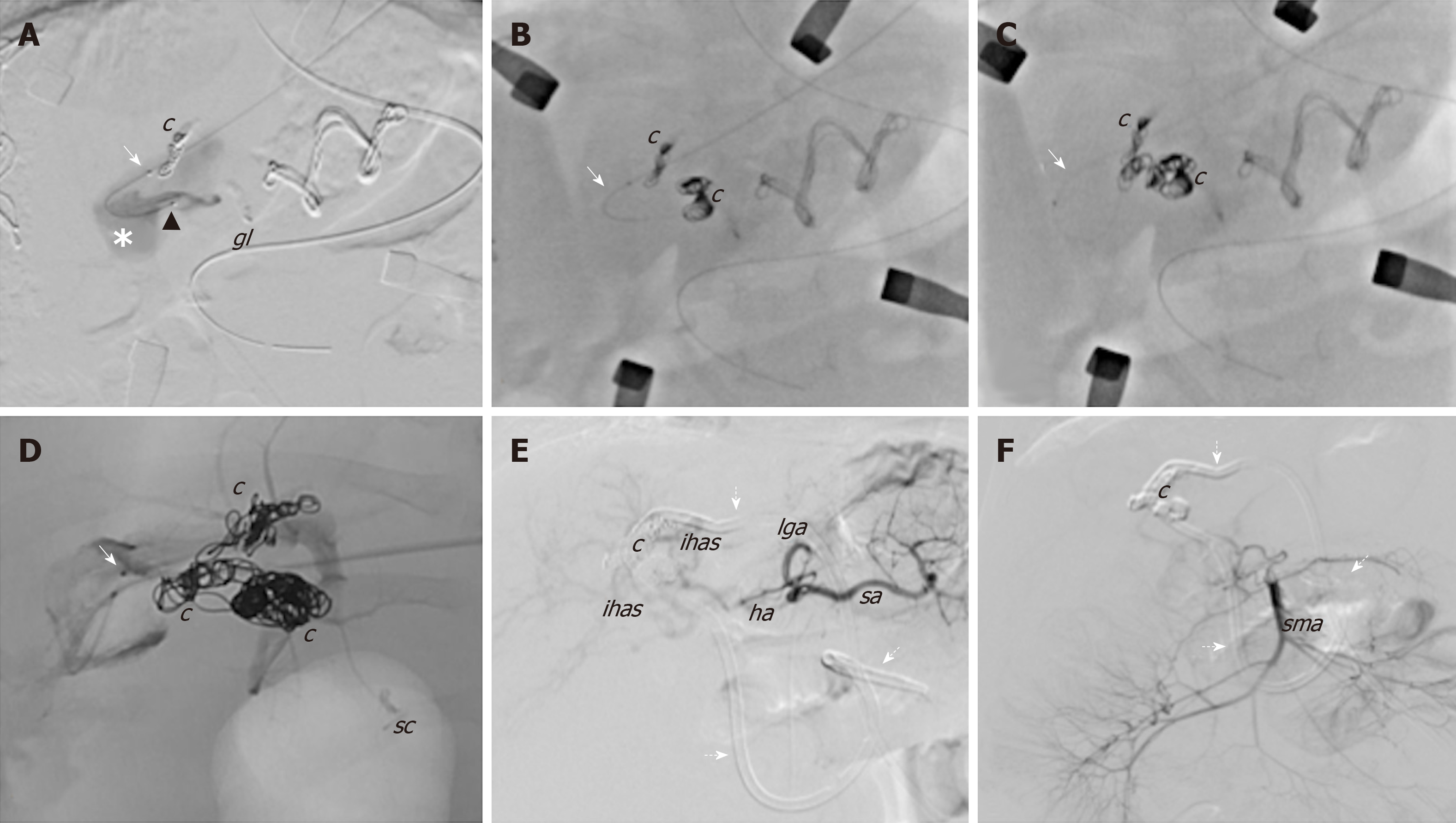
Figure 3 Angiographic images during the hybrid endovascular-surgical procedure of the congenital intrahepatic arterioportal fistula.
A-D: The initial attempts of selective anterograde catheterisation of the vascular malformation failed due to the small size of the arterial branches, and thus a hybrid procedure was performed. It consisted of (1) retrograde venous catheterisation of the fistula through direct left portal vein puncture (white arrow) and embolisation of the shunt with coils; and (2) surgical exposure of the Rex and selective surgical ligation of the small dysplastic arteries feeding the fistula; E and F: Final angiographic images from the celiac trunk and the superior mesenteric artery, which revealed complete closure of the shunt without signs of revascularisation. c: Coils; gl: Glue; ha: Hepatic artery; ihas: Intrahepatic arteries; lga: Left gastric artery; lpa: Left phrenic artery; sma: Superior mesenteric artery; sa: Splenic artery; sc: Surgical clips; Dotted arrows: External internal biliary drainage.
Figure 4 Cholangiogram.
A: Cholangiogram from the external biliary drainage shows marked dilatation of both intra- and extrahepatic biliary tree with tortuous and convoluted appearance of ducts and the stricture (arrow) of the distal tract of the choledocus without passage of contrast medium into the bowel system; B: Cholangiogram after positioning the external-internal biliary drainage showing slight reduction of the biliary dilatation, particularly within the right system; C: Cholangiogram after 3 mo of internal biliary stent placement that shows almost complete resolution of the biliary dilatation. c: Choledocus; cd: Cystic duct; chd: Common hepatic duct; djf: Duodenojejunal flexure; ebd: External biliary drainage; eibd: External-internal biliary drainage; g: Gallbladder; ibs: Internal biliary stent; lhd: Left hepatic duct; rhd: Right hepatic duct; asterisk: Coils; Arrow: Stricture within the distal choledocus.
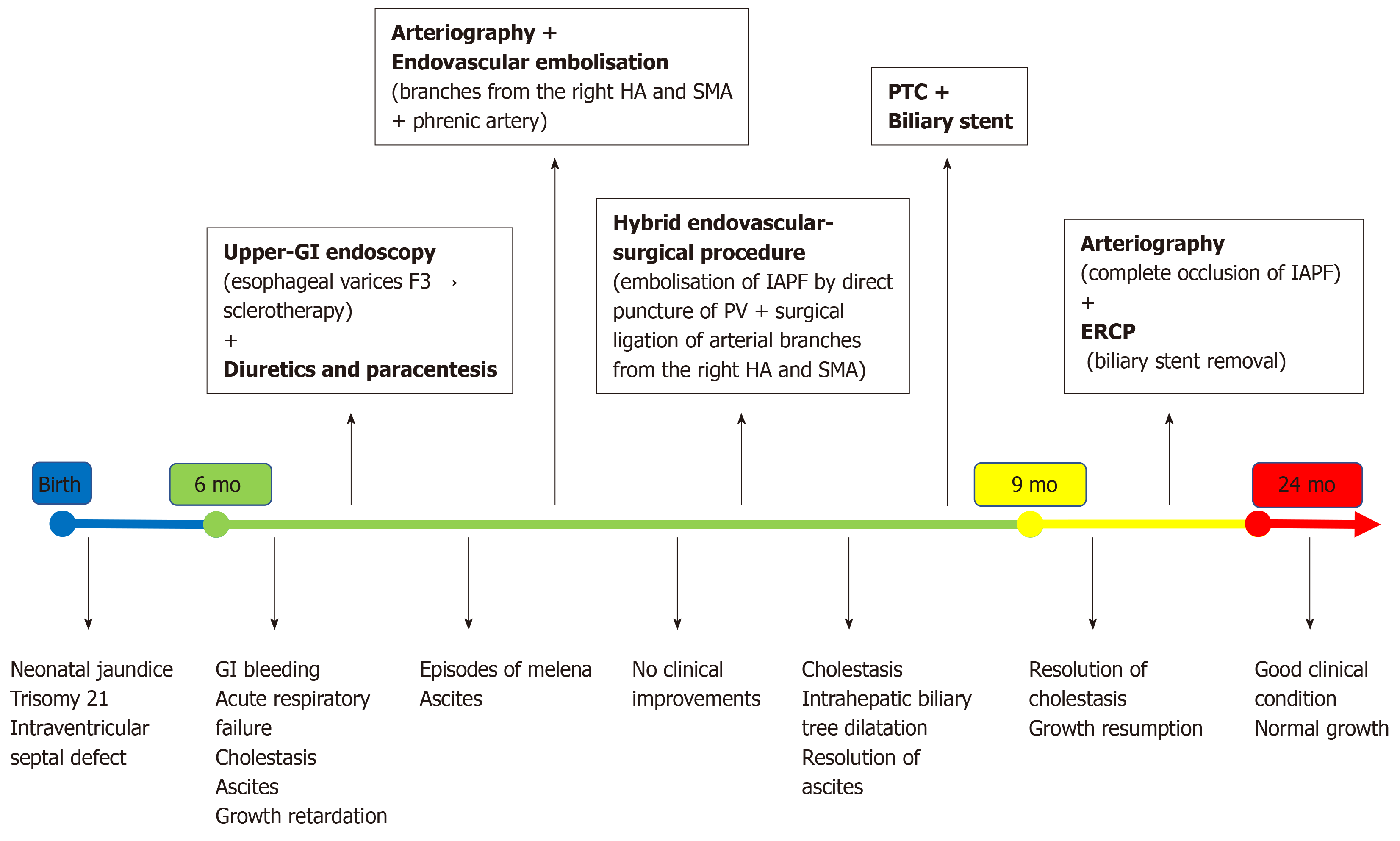
Figure 5 Summary of the patient’s clinical course and the therapeutic management.
Upper-GI: upper-gastrointestinal; ERCP: endoscopic retrograde cholangiopancreatography; HA: Hepatic artery; IAPF: Intrahepatic arterioportal fistula; PV: Portal vein; PTC: Percutaneous transhepatic cholangiography; SMA: Superior mesenteric artery.
- Citation: Angelico R, Paolantonio G, Paoletti M, Grimaldi C, Saffioti MC, Monti L, Candusso M, Rollo M, Spada M. Combined endovascular-surgical treatment for complex congenital intrahepatic arterioportal fistula: A case report and review of the literature. World J Hepatol 2020; 12(4): 160-169
- URL: https://www.wjgnet.com/1948-5182/full/v12/i4/160.htm
- DOI: https://dx.doi.org/10.4254/wjh.v12.i4.160


