Copyright
©The Author(s) 2019.
World J Hepatol. May 27, 2019; 11(5): 477-482
Published online May 27, 2019. doi: 10.4254/wjh.v11.i5.477
Published online May 27, 2019. doi: 10.4254/wjh.v11.i5.477
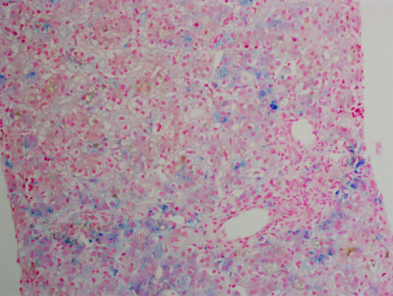
Figure 1 Approximately 10%-15% of the hepatocytes contained iron granules.
Iron deposition was identified in all three zones of the liver.
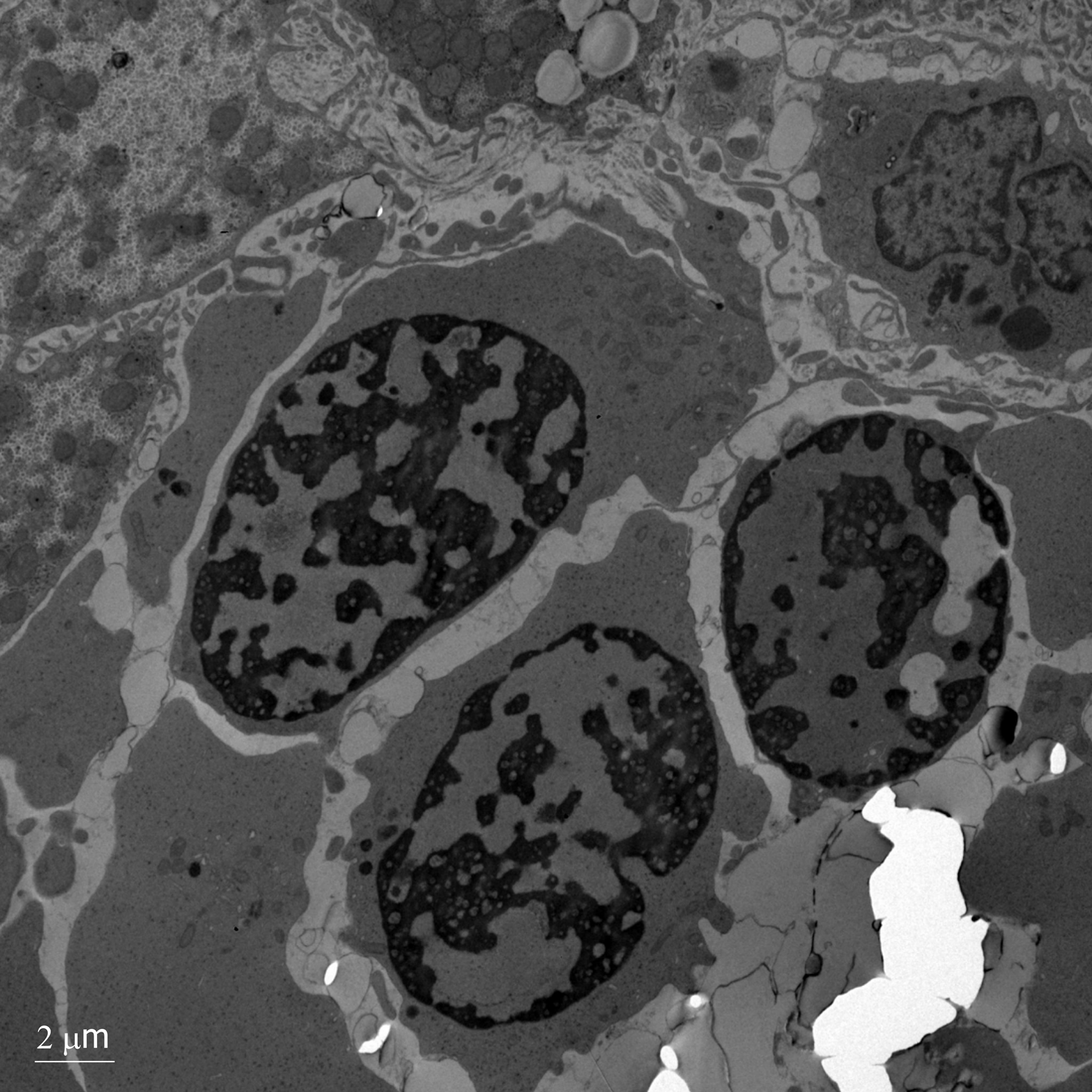
Figure 2 Electron microscopy showing erythroblasts with dense heterochromatin and translucent vacuoles.
There is widening of the nuclear pores with invagination of cytoplasm.
- Citation: Jaramillo C, Ermarth AK, Putnam AR, Deneau M. Neonatal cholestasis and hepatosplenomegaly caused by congenital dyserythropoietic anemia type 1: A case report. World J Hepatol 2019; 11(5): 477-482
- URL: https://www.wjgnet.com/1948-5182/full/v11/i5/477.htm
- DOI: https://dx.doi.org/10.4254/wjh.v11.i5.477


