Copyright
©The Author(s) 2019.
World J Hepatol. Mar 27, 2019; 11(3): 318-329
Published online Mar 27, 2019. doi: 10.4254/wjh.v11.i3.318
Published online Mar 27, 2019. doi: 10.4254/wjh.v11.i3.318
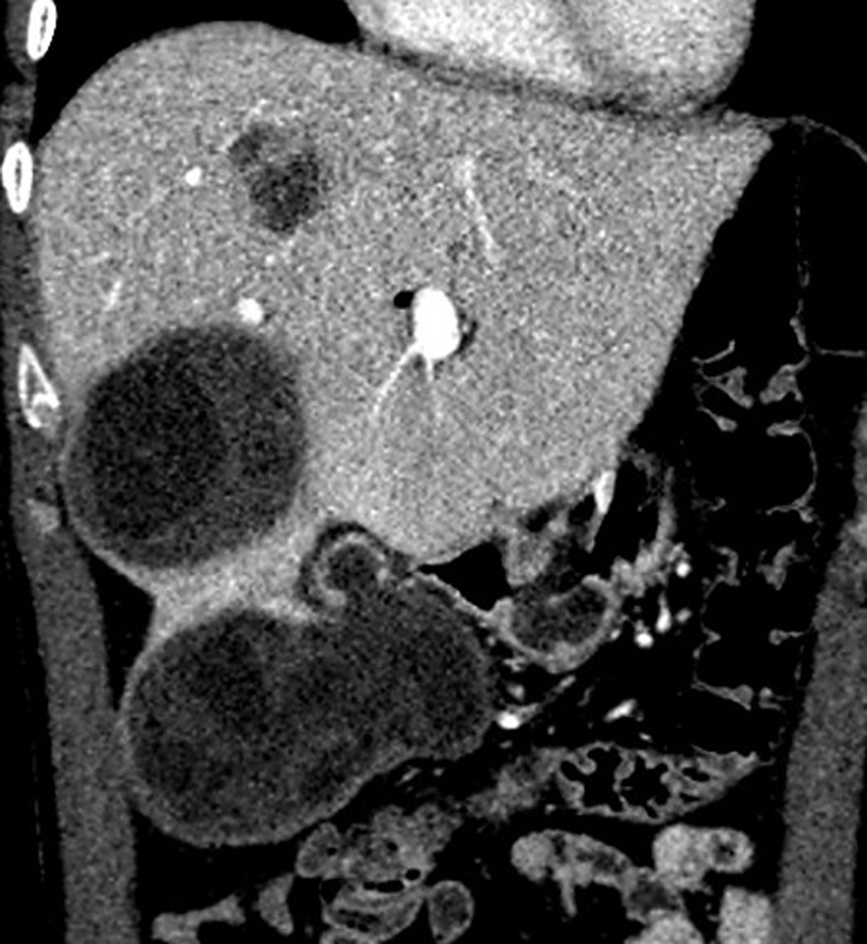
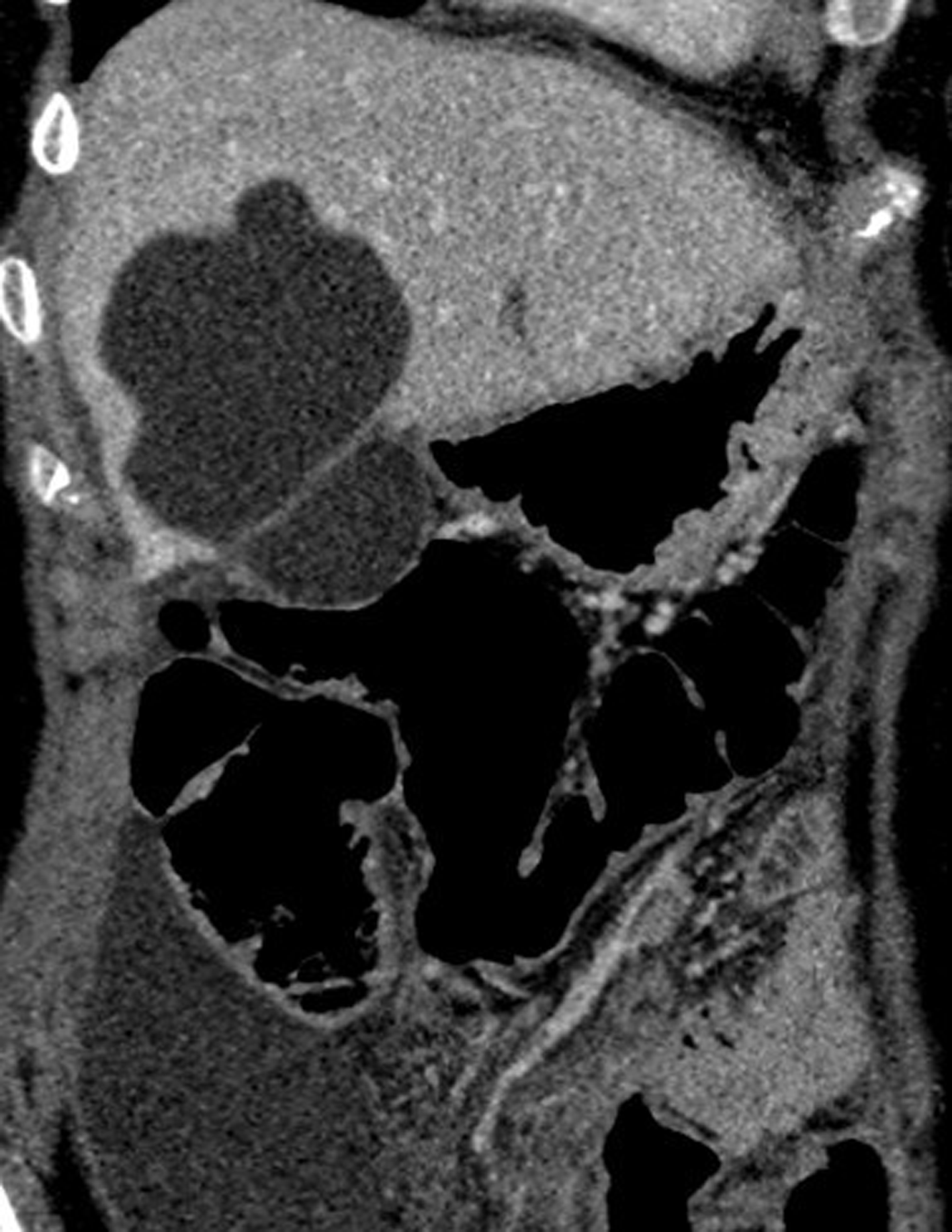
Figure 1 Coronal reformatted contrast-enhanced computed tomography shows that fluid collection and daughter vesicles adjacent to the hydatid cyst located in segment VI of the liver.
This finding is consistent with a perforated hydatid cyst.
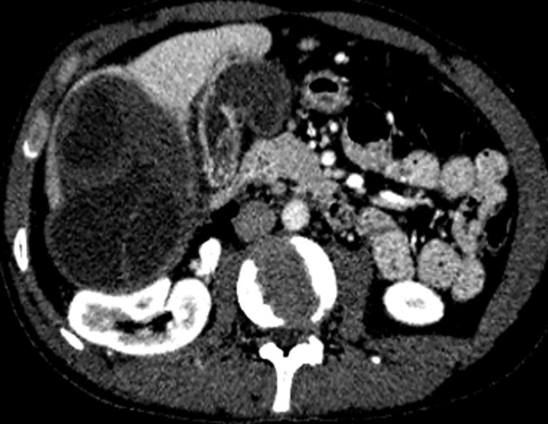
Figure 2 Axial plane computed tomography of the same patient shows exophytic extension of the giant hydatid cyst located in the right lobe of the liver.
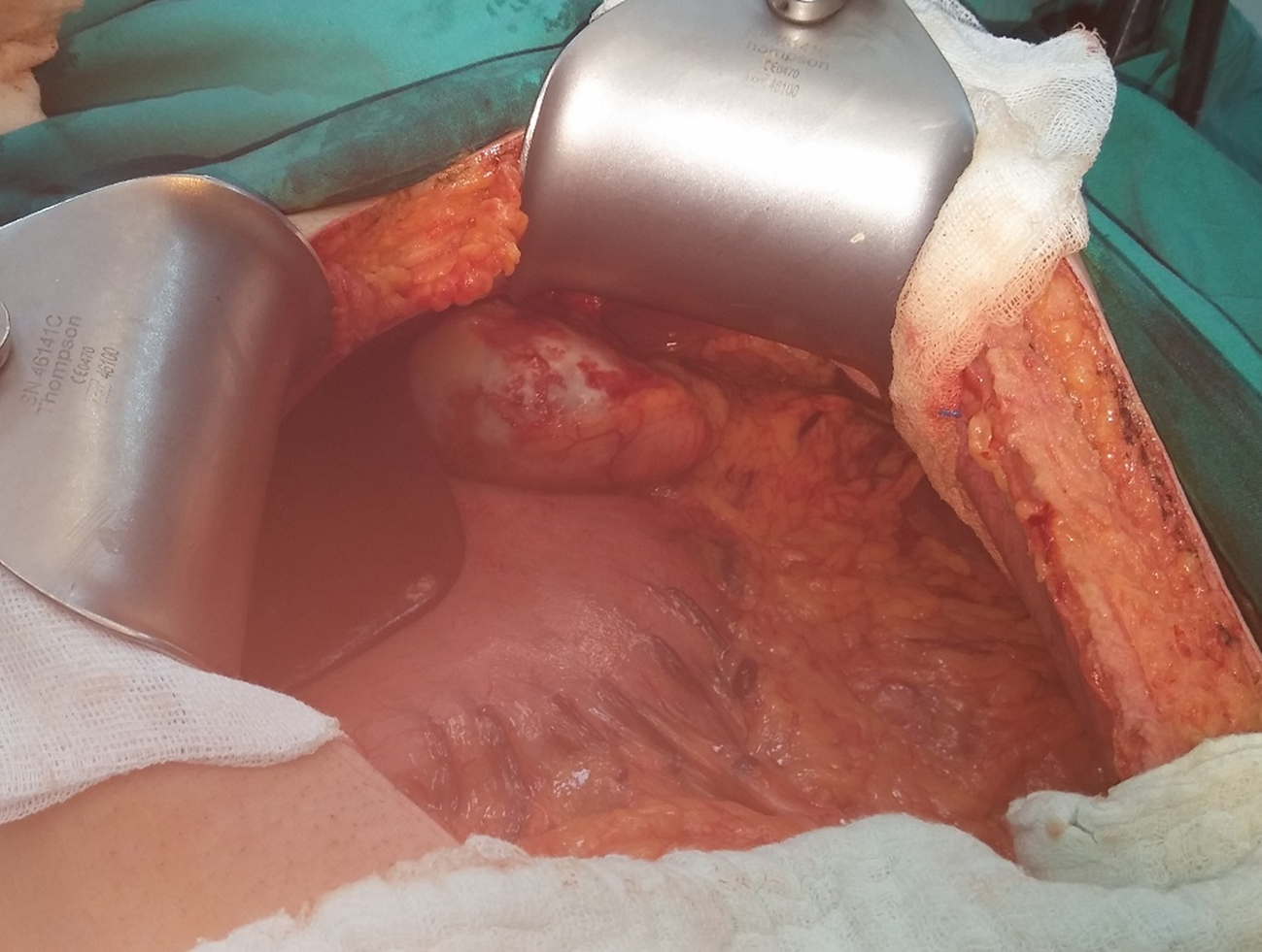
Figure 3 Intraoperative appearance of the hydatid cyst compatible lesion that originated and ruptured from the spleen.
This image was taken after aspiration of the hydatid cyst fluid in the abdominal cavity.
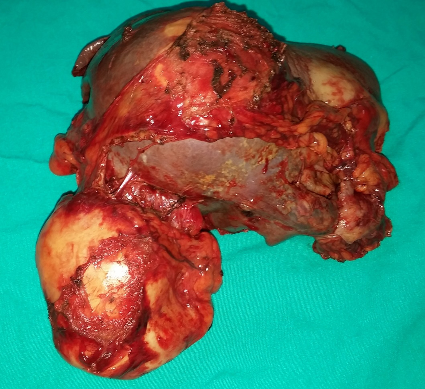
Figure 4 Apearance of the spleen and ruptured cyst specimens obtained from the same patient.
Figure 5 Coronal reformatted images of the venous phase of computed tomography shows perforated hydatid cyst located in segment V of the liver.
This image also shows fluid collection in the right paracolic area.
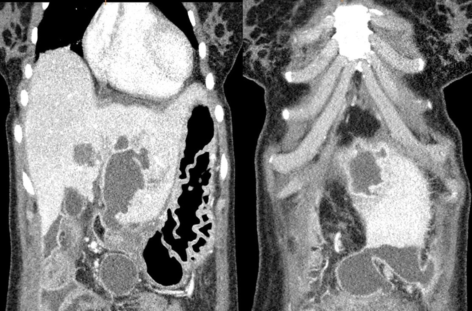
Figure 6 Two different coronal reformatted contrast-enhanced computed tomography images of the same patient show a perforated hydatid cyst located in segment III of the liver and fluid collection in the perihepatic/pelvic area.
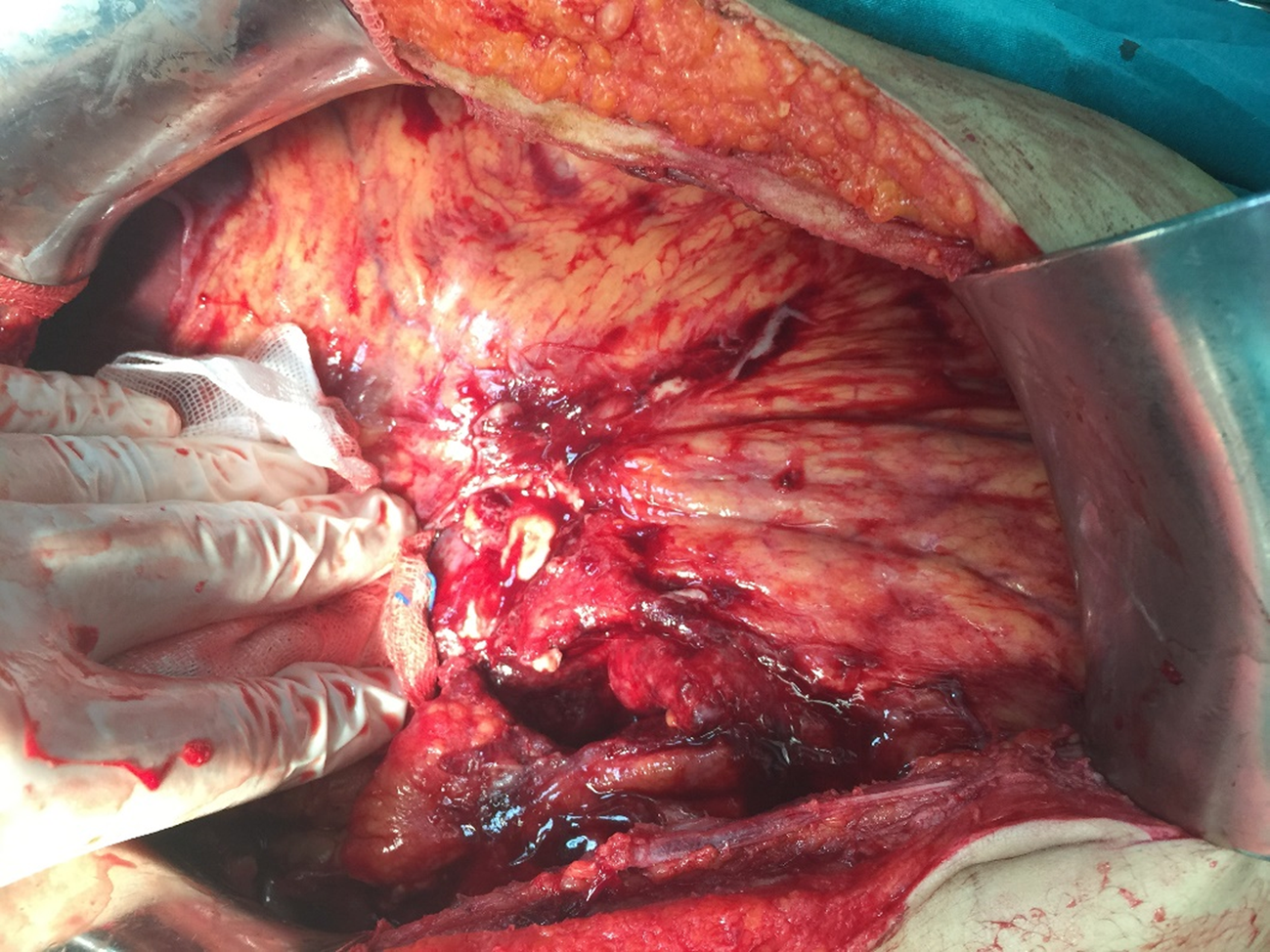
Figure 7 Intraoperative appearance of severe adhesions similar to sclerosing encapsulating peritonitis in the abdominal cavity secondary to hydatid cyst perforation.
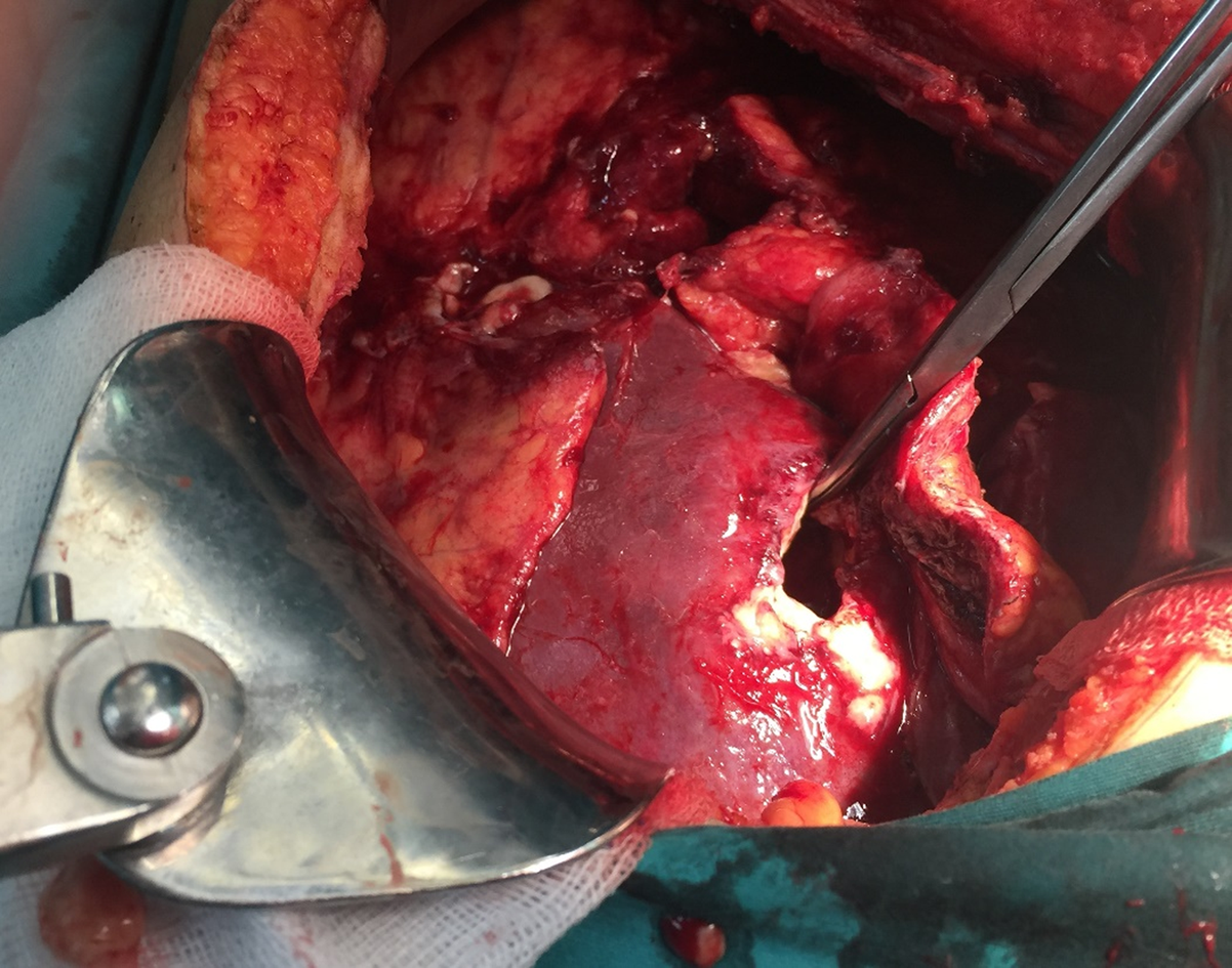
Figure 8 Intraoperative image obtained after evacuating the cystic contents.
- Citation: Akbulut S, Ozdemir F. Intraperitoneal rupture of the hydatid cyst: Four case reports and literature review. World J Hepatol 2019; 11(3): 318-329
- URL: https://www.wjgnet.com/1948-5182/full/v11/i3/318.htm
- DOI: https://dx.doi.org/10.4254/wjh.v11.i3.318


