Copyright
©The Author(s) 2019.
World J Hepatol. Feb 27, 2019; 11(2): 242-249
Published online Feb 27, 2019. doi: 10.4254/wjh.v11.i2.242
Published online Feb 27, 2019. doi: 10.4254/wjh.v11.i2.242
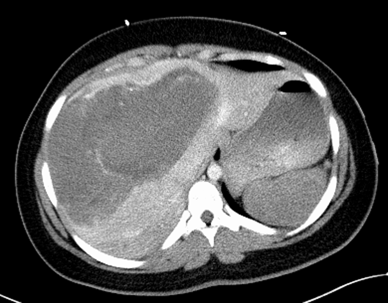
Figure 1 CT scan prior to interventions.
A CT of the abdomen and pelvis with contrast demonstrated a large 22 cm × 15 cm heterogenous, hypoattenuating mass encompassing nearly the entire liver. The mass demonstrated hypervascularity along the border and hyperattenuating areas, suggesting a large hemorrhagic liver mass with active hemorrhage.
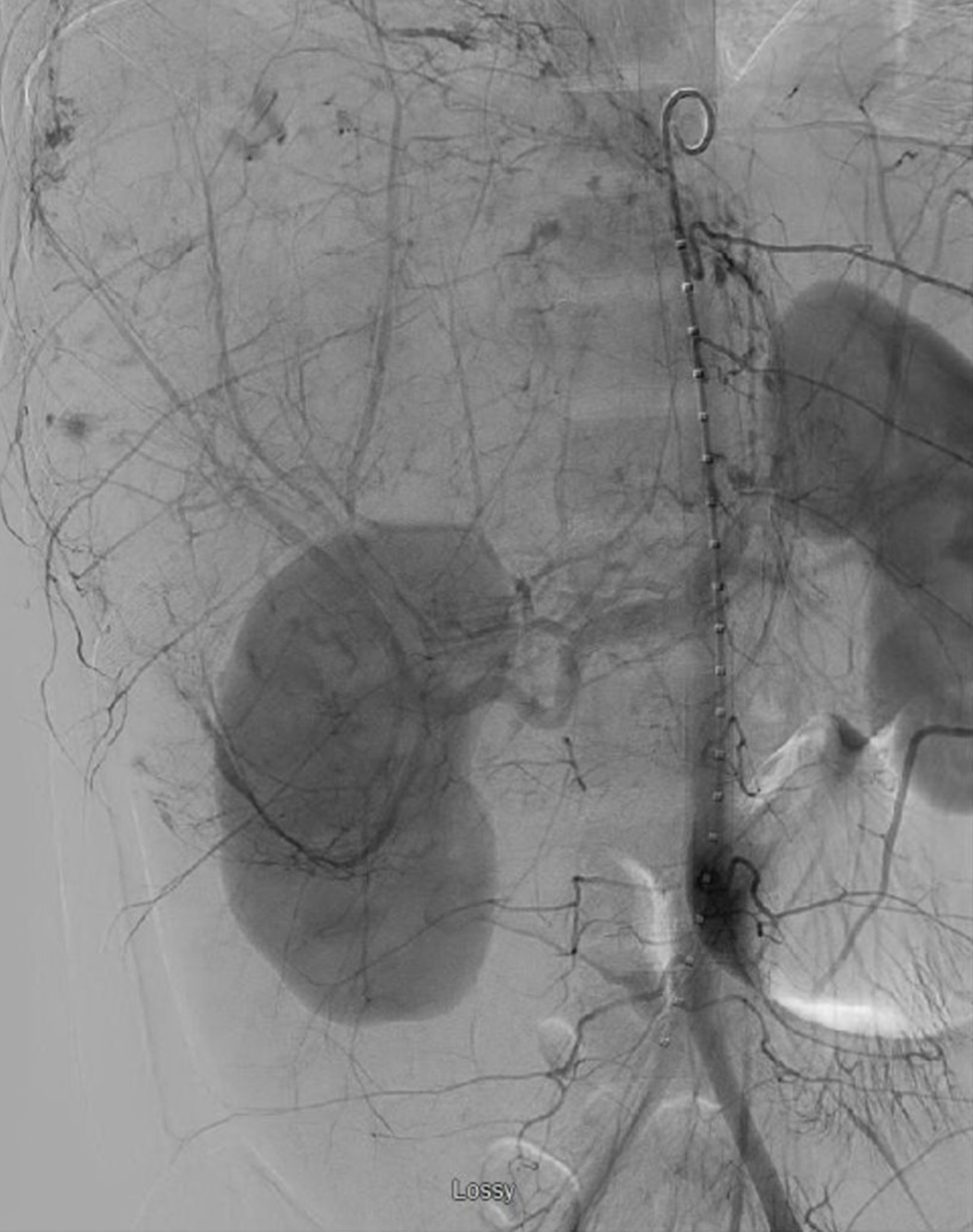
Figure 2 Mesenteric angiogram prior to transplant.
Mesenteric angiogram demonstrating a large right hepatic lobe with multiple areas of abnormal contrast accumulation indicative of ongoing hemorrhage. Gelfoam embolization of the right hepatic artery was performed.
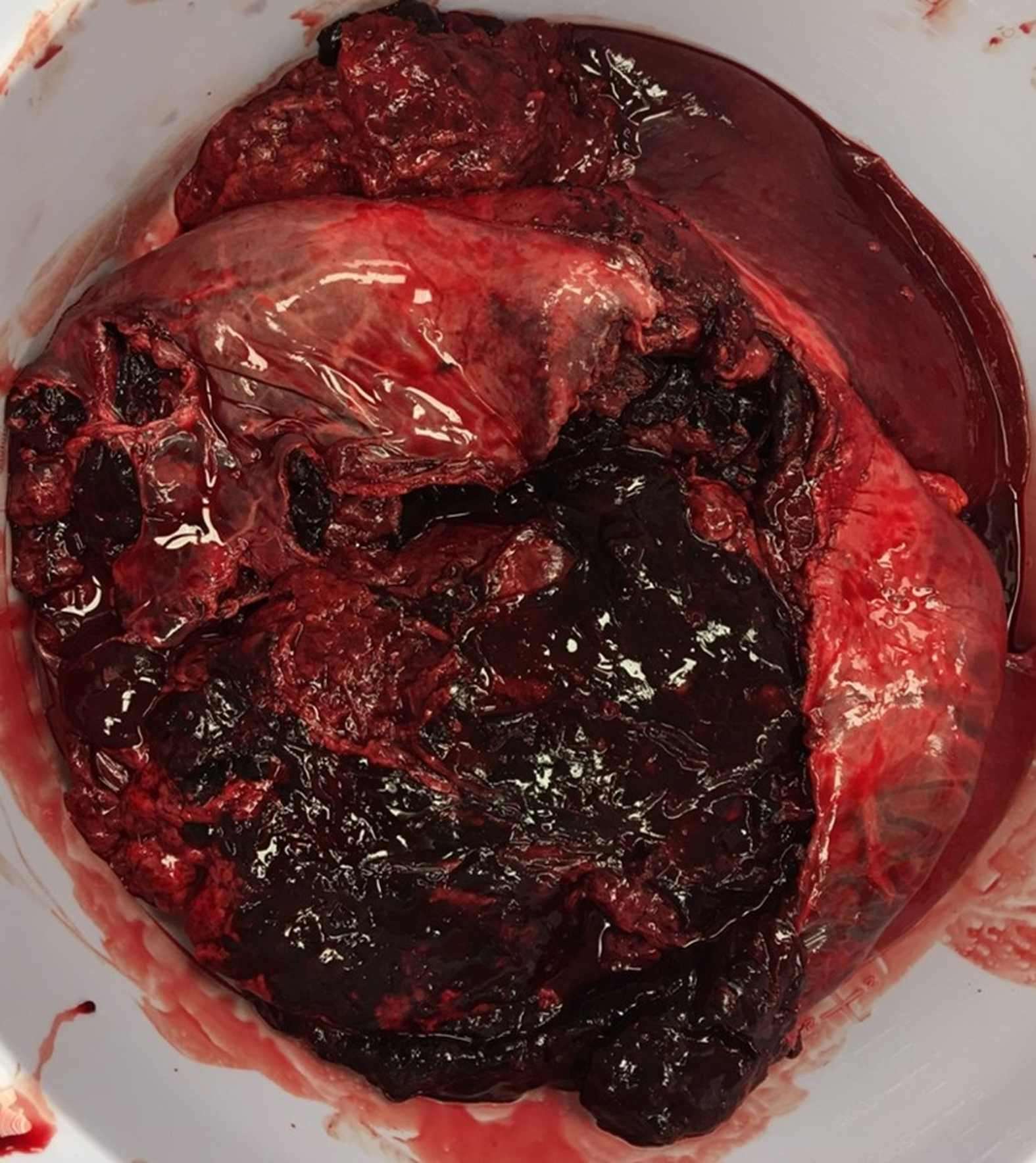
Figure 3 Explanted liver.
Explanted liver, measuring 34.5 cm × 22.5 cm × 8.5 cm, with a large surface disruption with adenomatous tissue and significant adherent clot.
- Citation: Salhanick M, MacConmara MP, Pedersen MR, Grant L, Hwang CS, Parekh JR. Two-stage liver transplant for ruptured hepatic adenoma: A case report. World J Hepatol 2019; 11(2): 242-249
- URL: https://www.wjgnet.com/1948-5182/full/v11/i2/242.htm
- DOI: https://dx.doi.org/10.4254/wjh.v11.i2.242


