Copyright
©The Author(s) 2018.
World J Hepatol. Jul 27, 2018; 10(7): 509-516
Published online Jul 27, 2018. doi: 10.4254/wjh.v10.i7.509
Published online Jul 27, 2018. doi: 10.4254/wjh.v10.i7.509
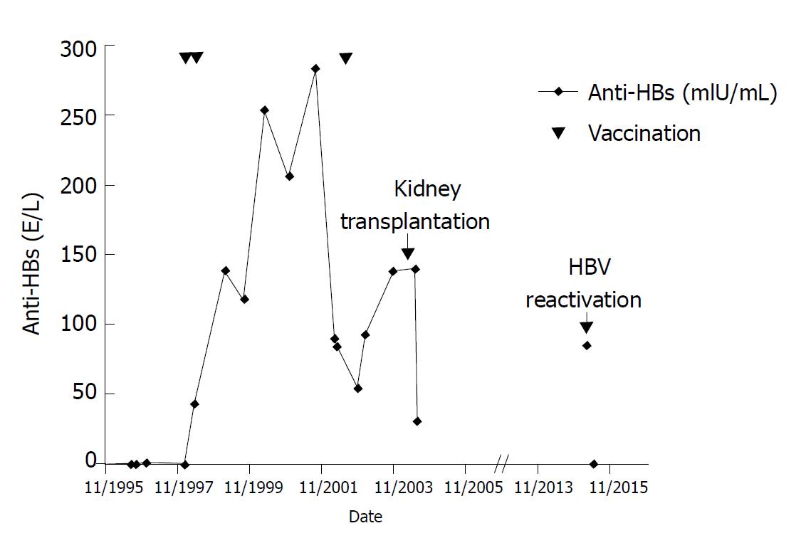
Figure 1 Course of anti-HBs levels since diagnosis of hepatitis B virus infection in 1995.
Vaccine was administered in January 1998, May 1998, and July 2002. Kidney transplantation and hepatitis B virus reactivation are indicated.
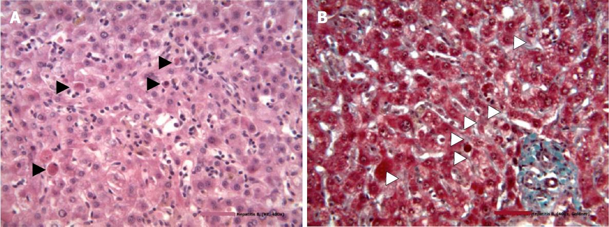
Figure 2 Hematoxylin and eosin stain of a liver specimen from the patient described in this study showing necrosis of hepatocytes (black triangles) without infiltrations and trichrome stain for fibrosis.
A: Necrosis of hepatocytes; B: Trichromestain for fibrosis. Periportal fibrosis is indicated by white triangles.
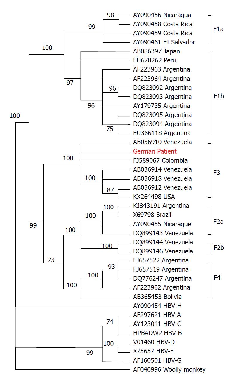
Figure 3 Phylogenetic tree of hepatitis B virus based on complete hepatitis B virus genomes.
The tree was constructed using the MEGAv6.0 software package[29], (http//:http://www.megasoftware.net), with the neighbor-joining method with p-distance and 1000 bootstrap replicates. Bootstrap values of at least 70 are shown at the nodes. Phylogenetic analysis was performed using reference sequences from GenBank, indicated in the tree by their GenBank accession numbers. Country of virus origin is included for all genotype F sequences. Woolly monkey HBV was used as the outgroup. The sequence from the patient described in this study is presented in red. HBV: Hepatitis B virus.
Figure 4 Vaccine escape mutations in the patient’s hepatitis B virus strain.
Alignment of the HBs antigen amino acid sequence (SHBs) of the patient´s strain with a HBV genotype F consensus sequence derived from the HIV-grade database (http://www.hiv-grade.de). Amino acids are named according to the one-letter code. Dots in the patient´s sequence represent amino acids identical to those in the consensus sequence. Positions relevant for drug resistance are indicated by blue boxes. Red asterisks denote positions in the patient´s strain (i.e., 109, 128, 129, 131, and 134) where both the amino acid of the consensus strain and the vaccine escape mutation were observed. HBV: Hepatitis B virus.
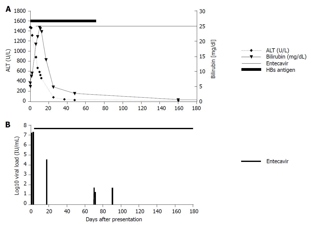
Figure 5 Course of amino transferases and bilirubin levels during acute hepatitis in March 2015 and HBV viremia levels after diagnosis of hepatitis B virus reactivation.
A: Course of amino transferases and bilirubin levels during acute hepatitis in March 2015; B: Course of HBV viremia levels after diagnosis of HBV reactivation. Start of antiviral treatment is indicated. ALT: Alanine aminotransferase.
- Citation: Schlabe S, Bremen KV, Aldabbagh S, Glebe D, Bremer CM, Marsen T, Mellin W, Cristanziano VD, Eis-Hübinger AM, Spengler U. Hepatitis B virus subgenotype F3 reactivation with vaccine escape mutations: A case report and review of the literature. World J Hepatol 2018; 10(7): 509-516
- URL: https://www.wjgnet.com/1948-5182/full/v10/i7/509.htm
- DOI: https://dx.doi.org/10.4254/wjh.v10.i7.509


