Copyright
©The Author(s) 2018.
World J Hepatol. Feb 27, 2018; 10(2): 297-307
Published online Feb 27, 2018. doi: 10.4254/wjh.v10.i2.297
Published online Feb 27, 2018. doi: 10.4254/wjh.v10.i2.297
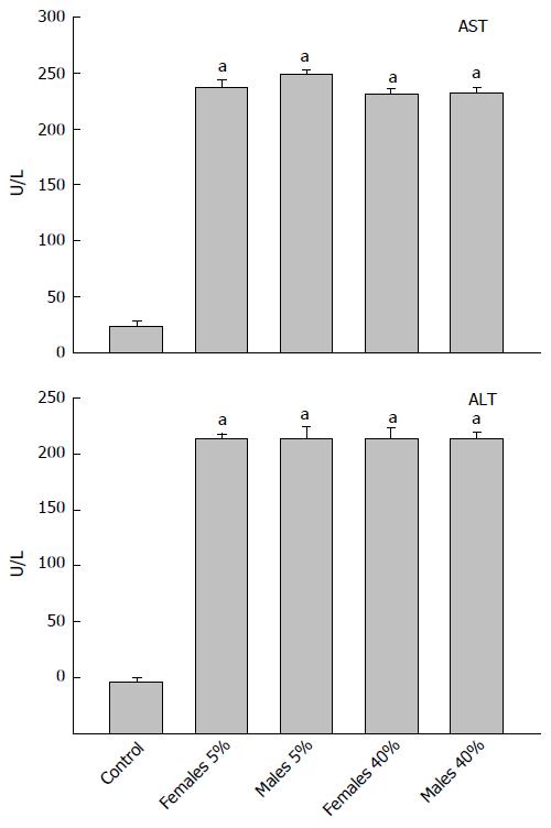
Figure 1 Activities of serum Aspartate and Alanine Aminotransferases after weekend alcohol consumption.
Values are expressed as the mean ± SEM in each experimental group (n = 3-6). aP < 0.05 vs the control group. AST: Aspartate Aminotransferase; ALT: Alanine Aminotransferase.
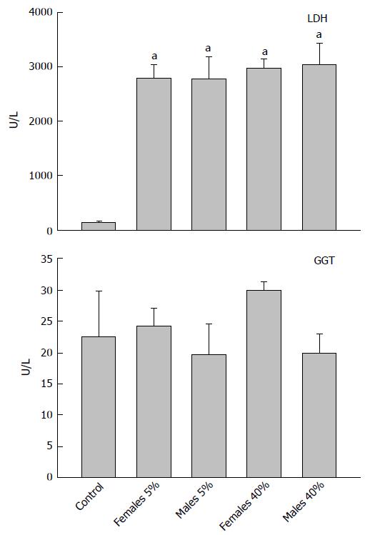
Figure 2 Activities of serum Lactate Dehydrogenase and Gamma-Glutamyltransferase after weekend alcohol consumption.
Values are expressed as the mean ± SEM in each experimental group (n = 3-6). aP < 0.05 vs the control group. LDH: Lactate Dehydrogenase; GGT: Gamma-Glutamyltransferase.
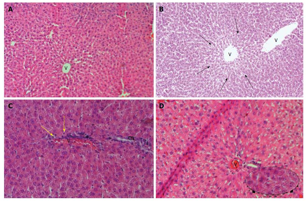
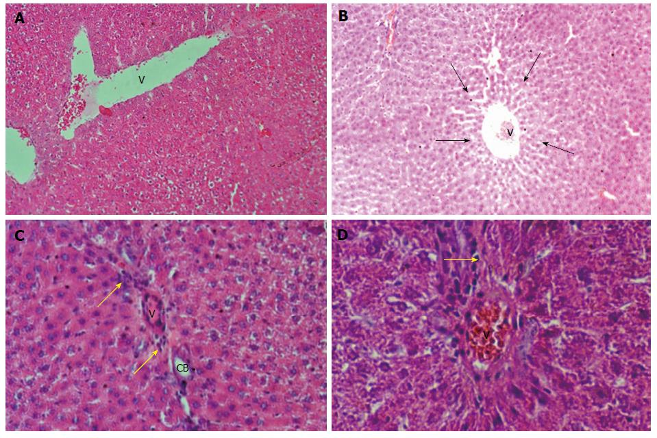
Figure 3 Hepatic histology of the group of females with weekend alcohol consumption at 5%.
A: Image of the control group; B-D: Images of the group of females with alcohol consumption at 5%. A: Hepatocytes were observed as formed in a line (40 ×); B: A zone of less pigmentation is observed, marked with black arrows, corresponding to periportal necrosis (40 ×); C: Slight inflammation with blue-colored cells (leukocytes) around the portal vein, which do not surpass the limiting plaque (yellow arrows) (60 ×); D: In the lower left part, steatosis is observed (60 ×). V: Portal vein; BC: Bile canaliculus. Hematoxylin and Eosin stain.
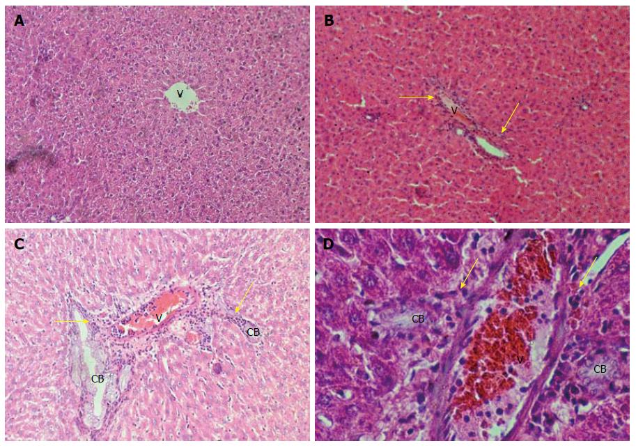
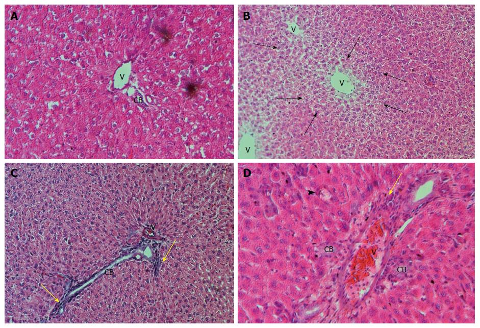
Figure 4 Hepatic histology of the group of males with weekend alcohol consumption at 5%.
A: Image of the control group; B-D: Images of the group of males with alcohol consumption at 5%. A: The uniformity of the structures is observed to be conserved (40 ×); B: Presence of fine and thick steatosis (pigmentation-diminution zone in the cytoplasm), inflammation present with leukocytes in single file around the portal vein (yellow arrows) (40 ×); C: Important inflammation is observed, represented by leukocytes around the portal vein and in the Bile Canaliculus (BC) (yellow arrows) (40 ×); D: Fine as well as thick steatosis is observed (pigmentation-diminution zone in the cytoplasm), as well as leukocyte infiltrate (yellow arrow) (100 ×). V: Portal Vein; BC: Bile canaliculus. Hematoxylin and Eosin stain.
Figure 5 Hepatic histology of the group of females with weekend alcohol consumption at 40%.
A: Image of the control group; B-D: Images of the group of females with alcohol consumption at 40%. A: The uniformity of the structures is observed to be conserved (40 ×); B: Periportal fibrosis, loss of cells around the portal vein (black arrows) (40 ×); C: Fine and thick steatosis and leukocytes in single file around the portal vein and some that emerge from the limiting plaque (yellow arrows) (60 ×); D: Both fine and thick steatosis and single-file leukocytes are observed (100 ×). V: Portal Vein; BC: Bile canaliculus. Hematoxylin and Eosin stain.
Figure 6 Hepatic histology of the group of males of 40%.
A: Image of the control group; B-D: Images of the group of males with alcohol consumption at 40%. A: The conserved, uniformized structures of the form and size of the hepatocytes are observed (40 ×); B: Periportal fibrosis is observed (black arrows) (40 ×); C: Leukocytes are observed in a cited single file (yellow arrows) (40 ×); D: Fine and thick steatosis is observed (zone with less pigment inside the cellular cytoplasm of the hepatocyte), leukocytes (yellow arrow), and the apoptotic cell (point of the black arrow) (60 ×). V: Portal Vein; BC: Bile canaliculus. Hematoxylin and Eosin stain.
- Citation: Morales-González JA, Sernas-Morales ML, Morales-González Á, González-López LL, Madrigal-Santillán EO, Vargas-Mendoza N, Fregoso-Aguilar TA, Anguiano-Robledo L, Madrigal-Bujaidar E, Álvarez-González I, Chamorro-Cevallos G. Morphological and biochemical effects of weekend alcohol consumption in rats: Role of concentration and gender. World J Hepatol 2018; 10(2): 297-307
- URL: https://www.wjgnet.com/1948-5182/full/v10/i2/297.htm
- DOI: https://dx.doi.org/10.4254/wjh.v10.i2.297


