Copyright
©2009 Baishideng.
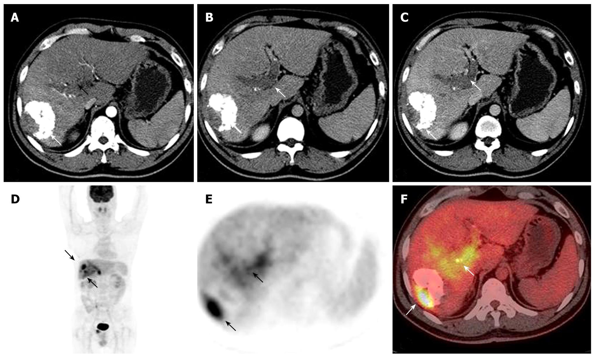
Figure 1 A 38-year man, who had TACE 1 mo before, underwent PET/CT to monitor response to the treatment.
Contrast-enhanced arterial-phase axial CT image showed the mass with partial accumulation of iodized oil in the right lobe of the liver (arrows, A, B). A filling defect was detected in the left branch during the portal phases (arrow, C). PET and PET/CT fused images (arrows, D, E and F) revealed residual viable tumor and a highly metabolically active tumor thrombus in the left branch of the portal vein.
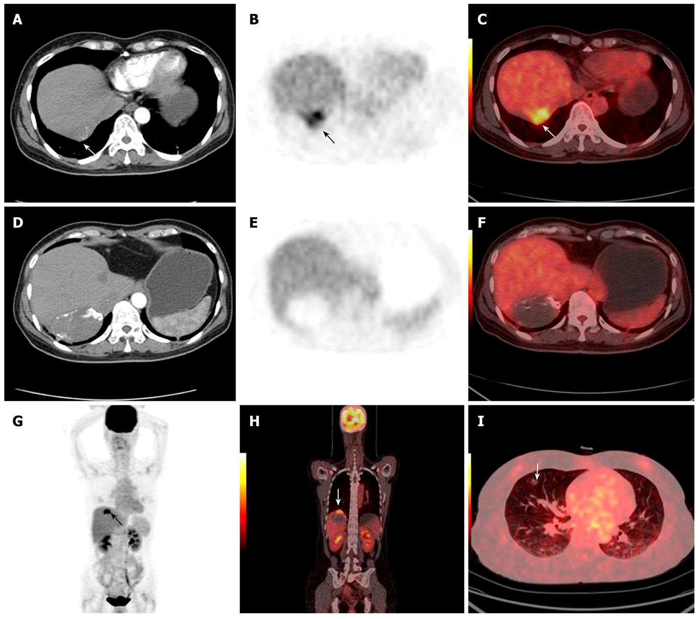
Figure 2 A 50-year woman who had HCC resection 2 years before had an intrahepatic HCC recurrence and received combined RFA and TACE treatment.
A highly metabolically active lesion was detected on the top of the lesion by PET and PET/CT fused images (white arrows, A-C and G). Contrast-enhanced CT image showed the non enhanced mass with faint accumulation of iodized oil in the right lobe of the liver (D-F). PET/CT fused images revealed a low FDG uptake benign lesion in the right lung (I). All findings were later verified by clinical follow-up.
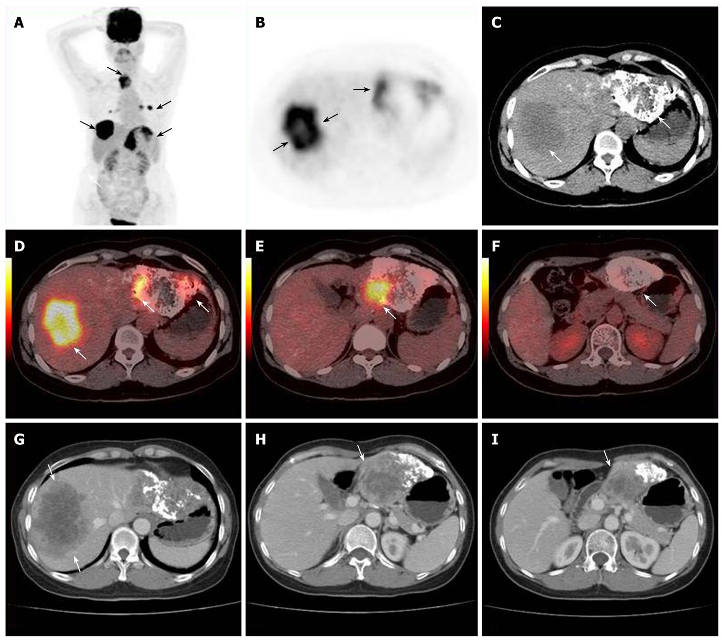
Figure 3 A 55-year woman suffering from HCC received TACE treatment.
Extrahepatic metastases (arrows, A) were detected by PET and PET/CT fused images. Highly metabolic new recurrent lesion of the right lobe (arrows, B-D) and a resident lesion of the left lobe (arrows, E, F) were detected by PET/CT fused imaging. Contrast-enhanced CT follow-up after 1 mo later confirmed the progression of lesions.
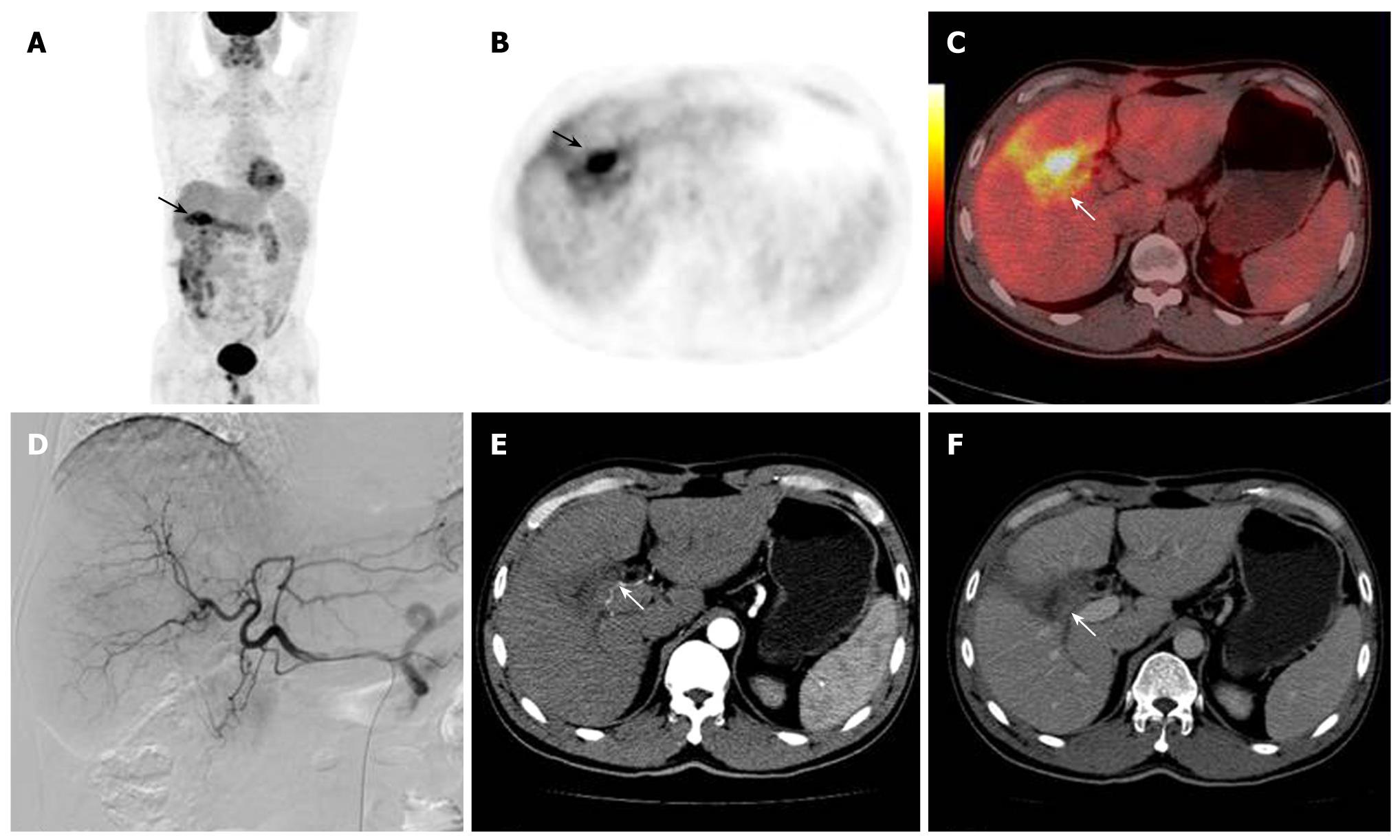
Figure 4 A 48-year man who had HCC resection 2 mo before the imaging, followed by TACE 40 d later.
PET and PET/CT fused images revealed highly metabolic lesions (arrows, A-C). Both contrast-enhanced axial CT imaging and arteriography failed to show lesions in the right lobe of the liver (arrows, D-F). These findings of PET/CT were later verified as inflammatory by post-operative tissue examination.
- Citation: Sun L, Guan YS, Pan WM, Luo ZM, Wei JH, Zhao L, Wu H. Metabolic restaging of hepatocellular carcinoma using whole-body 18F-FDG PET/CT. World J Hepatol 2009; 1(1): 90-97
- URL: https://www.wjgnet.com/1948-5182/full/v1/i1/90.htm
- DOI: https://dx.doi.org/10.4254/wjh.v1.i1.90


