Copyright
©2009 Baishideng.
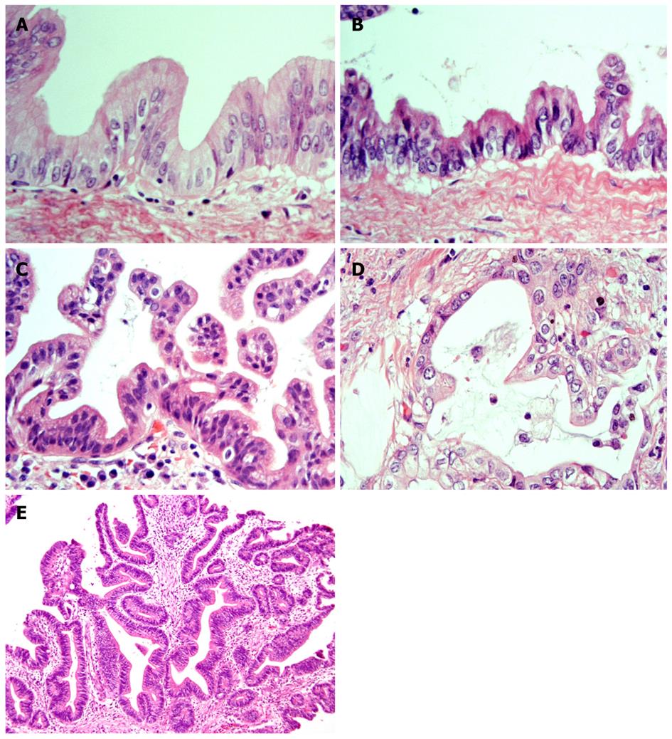
Figure 1 Representative histological features of neoplasms (HE staining).
A: BilIN-1; B: BilIN-2; BilIN-3; D: Invasive ICC; E: IPN-B. BilIN: biliary intraepithelial neoplasm; ICC: intrahepatic cholangiocarcinoma; IPN-B: intraductal papillary neoplasm of the bile duct[22].
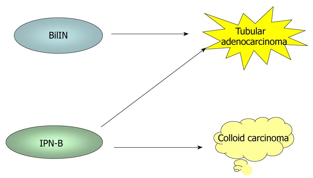
Figure 2 During biliary multistep carcinogenesis, BilIN is proposed to progresses to conventional ICC (tubular adenocarcinoma), whereas IPN-B is associated with colloid carcinoma (mucinous carcinoma) or conventional ICC.
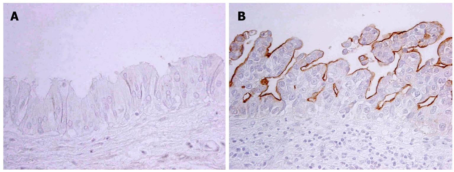
Figure 3 MUC1 is expressed in neoplastic cells of BilIN-3 (B) but not in those of BilIN1 (A) (Immunostaining).
A: BilIN-1; B: BilIN-3.
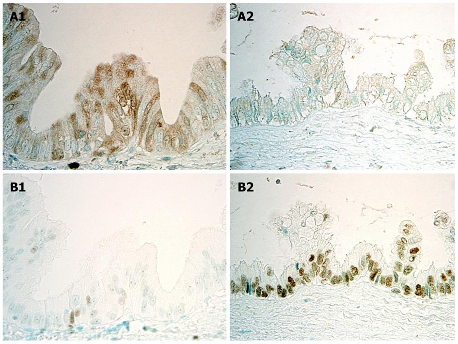
Figure 4 Immunohistochemical expression of p16INK4a and EZH2 in BilIN-1 and BilIN-2.
A: p16INK4a is intensely and frequently expressed in the cytoplasm and nuclei in BilIN-1 (left), but such expression is unclear in BilIN-2 (right); B: While EZH2 is occasionally expressed in the nuclei in BilIN-1 (left), this expression increases markedly in BilIN-2 (right)[22].
Figure 5 Semiquantitative evaluation of P16INK4a and EZH2 in BilIN-1, -2, and -3, and invasive cholangiocarcinoma (CC).
The labeling index (LI) reflects the percentage of cells immunohistochemically positive for P16INK4a or EZH2 in each lesion of BilIN-1, -2, and -3, and invasive CC. bP < 0.01 vs BilIN-1. A: The LI of P16INK4a expression (mean ± SD) decreases with the progressing of BilINs and is higher in invasive CC; B: Expression of EZH2 increases with the progression of BilINs and is greater in invasive CC[22].
- Citation: Nakanuma Y, Sasaki M, Sato Y, Ren X, Ikeda H, Harada K. Multistep carcinogenesis of perihilar cholangiocarcinoma arising in the intrahepatic large bile ducts. World J Hepatol 2009; 1(1): 35-42
- URL: https://www.wjgnet.com/1948-5182/full/v1/i1/35.htm
- DOI: https://dx.doi.org/10.4254/wjh.v1.i1.35


