修回日期: 2015-07-28
接受日期: 2015-08-04
在线出版日期: 2015-08-28
目的: 探讨Barrett食管与结直肠息肉的相关性.
方法: 收集Barrett食管组41例, 对照组176例, 比较两组中结直肠息肉的发生率, 息肉的病理类型及息肉发生部位.
结果: Barrett食管组中息肉发生率为41.5%, 高于对照组的25.6%, 二组之间差异有统计学意义(P = 0.042). 两组中炎性息肉及增生性息肉的发生率差异无统计学意义(P = 0.32, P = 0.18). 但Barrett食管组中腺瘤性息肉发生率显著高于对照组(P = 0.008). 两组中息肉发生部位无明显差异. 多因素Logistic回归分析显示Barrett食管是结直肠息肉的独立相关因素(OR = 2.397, 95%CI: 1.146-5.013, P = 0.020).
结论: Barrett食管与结直肠息肉发生相关, 对Barrett食管患者应重视结直肠息肉筛查和监视.
核心提示: 本研究采用病例对照研究的方法, 探讨Barrett食管与结直肠息肉的相关性. 结果表明Barrett食管患者息肉发生率高于对照组, 其中腺瘤性息肉发生率显著高于对照组. 多因素Logistic回归分析显示Barrett食管是结直肠息肉的独立相关因素.
引文著录: 易姗姗, 姜齐宏. Barrett食管与结直肠息肉的相关性. 世界华人消化杂志 2015; 23(24): 3899-3903
Revised: July 28, 2015
Accepted: August 4, 2015
Published online: August 28, 2015
AIM: To explore the correlation between Barrett's esophagus and colorectal polyps.
METHODS: A total of 41 patients with Barrett's esophagus and 176 controls were enrolled in the study. The incidence, pathological type and location of colorectal polyps were compared.
RESULTS: The incidence of polyps in patients with Barrett's esophagus was 41.5%, which was significantly higher than that of the control group (25.6%) (P = 0.042). The incidence of adenomatous polyps in patients with Barrett's esophagus was also significantly higher than that of the control group (P = 0.008), although there was no significant difference in the incidence of hyperplastic polyps and inflammatory polyps. The location of colorectal polyps showed no significant difference between the two groups. Logistic multivariate regression analysis revealed that Barrett's esophagus was an independent risk factor for colorectal polyps (OR = 2.397, 95%CI: 1.146-5.013, P = 0.020).
CONCLUSION: Patients with Barrett's esophagus have a higher incidence of colorectal polyps. Therefore, the screening and surveillance of colorectal polyps should be enhanced in patients with Barrett's esophagus.
- Citation: Yi SS, Jiang QH. Correlation between Barrett's esophagus and colorectal polyps. Shijie Huaren Xiaohua Zazhi 2015; 23(24): 3899-3903
- URL: https://www.wjgnet.com/1009-3079/full/v23/i24/3899.htm
- DOI: https://dx.doi.org/10.11569/wcjd.v23.i24.3899
Barrett食管是指食管下段的复层鳞状上皮被化生的单层柱状上皮所替代的一种病理现象, 可伴有或不伴有肠上皮化生. Barrett食管与食管腺癌密切相关. Sontag等[1]第一个报道Barrett食管与结肠癌相关, 引发了一些学者对Barrett食管和结直肠肿瘤关系的研究. Barrett食管可能会增加结直肠息肉和结直肠癌的发病风险[2,3]. 结直肠息肉中腺瘤性息肉是结直肠癌的癌前病变. 本研究旨在探讨Barrett食管与结直肠息肉发生的关系.
选取华中科技大学同济医学院附属普爱医院2009-09/2014-09住院患者, 行胃镜及肠镜检查的Barrett食管患者41例. 其中男性18例, 女性23例, 中位年龄54岁,平均年龄53.4岁±8.4岁. Barrett食管患者均经胃镜检查和组织病理学检查确诊. 根据我国Barrett食管诊治共识(2011年修订版), 其诊断标准为食管下段的复层鳞状上皮被化生的单层柱状上皮所替代, 可伴有或不伴有肠上皮化生[4]. 对照组176例: 同期住院行胃镜及肠镜检查排除Barrett食管患者, 其中男性74例, 女性102例, 中位年龄52岁,平均年龄52.2岁±9.1岁(两组均排除结直肠癌及结直肠息肉家族史、结直肠癌、炎症性肠病、肠结核、家族性腺瘤性息肉病及既往有结直肠息肉病史患者).
记录每个患者的性别、年龄. 胃镜下将Barrett食管分为: 全周型、岛型、舌型. 取活检时, 从胃食管结合处开始向上以2 cm间隔, 四象限活检, 并记录Barrett食管长度. 肠镜检查中, 记录肠道准备质量、息肉数量、大小、位置、息肉病理组织类型. 所有息肉均行病理组织检查. 息肉分为炎性息肉、增生性息肉、腺瘤性息肉, 腺瘤包括管状腺瘤、绒毛状腺瘤、管状绒毛状腺瘤. 左半结肠包括乙状结肠、降结肠和结肠脾曲, 右半结肠包括回盲部、升结肠、结肠肝曲和横结肠. 内镜检查由有丰富内镜操作经验的医师完成. 病理组织活检由高年资病理科医师阅片.
统计学处理 所有数据均采用SPSS16.0统计软件分析. 计量资料采用mean±SD表示, 计数资料采用百分比表示. 两组间频数比较采用χ2检验, 并用Logistic模型进行多因素分析, P<0.05为差异具有统计学意义.
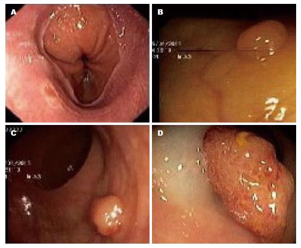
有217例入选者, 其中Barrett食管组41例, 对照组176例. Barrett食管组中息肉患者17例(41.5%), 而对照组中息肉患者例数45例(25.6%), 二组之间差异有统计学意义(P = 0.042)(图1).
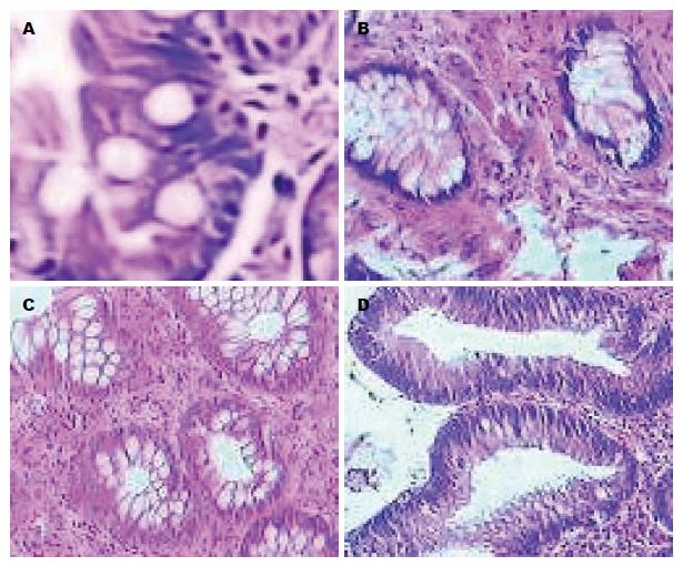
在Barrett食管组中腺瘤性息肉为13例(31.7%), 对照组腺瘤性息肉为25例(14.2%)(P = 0.008). Barrett食管组中炎性息肉为11例(26.8%), 增生性息肉为6例(14.6%), 对照组中炎性息肉为36例(20.5%), 增生性息肉为14例(8.0%), 但在两组中差异无统计学意义(P = 0.372, P = 0.183)(表1, 图2).
| 病理类型 | Barrett食管组 | 对照组 | P值 |
| 炎性息肉 | 11(26.8) | 36(20.5) | 0.372 |
| 增生性息肉 | 6(14.6) | 14(8.0) | 0.183 |
| 腺瘤性息肉 | 13(31.7) | 25(14.2) | 0.008 |
| 管状腺瘤 | 10(24.4) | 22(12.5) | 0.053 |
| 管状绒毛状腺瘤 | 3(7.3) | 4(2.3) | 0.100 |
| 绒毛状腺瘤 | 1(2.4) | 1(0.6) | 0.259 |
在Barrett食管组中左半结肠+直肠组息肉为13例(31.7%), 对照组中为35例(19.9%)(P = 0.101). 在Barrett食管组中右半结肠息肉为7例(17.1%), 对照组中为16例(9.6%), 两组中差异无统计学意义(P = 0.175).
控制年龄、性别两个变量进行多因素Logistic回归分析发现, Barrett食管与结直肠息肉的发病独立相关(OR = 2.397, 95%CI: 1.146-5.013, P = 0.020)(表2).
| 影响因素 | 赋值 | B | S.E. | Wald | P值 | OR | 95%CI |
| Barrett食管 | 1 = 有; 0 = 无 | 0.874 | 0.377 | 5.388 | 0.020 | 2.397 | 1.146-5.013 |
| 年龄 | 1 =≥60岁; 0 =<60岁 | 0.829 | 0.349 | 5.649 | 0.017 | 2.292 | 1.157-4.542 |
| 性别 | 1 = 男; 0 = 女 | 1.105 | 0.330 | 11.192 | 0.001 | 3.021 | 1.581-5.772 |
Barrett食管与食管腺癌相关, 被认为是食管腺癌的癌前病变. 但Barrett食管与结直肠肿瘤的关系并不确定. 有些学者研究显示Barrett食管患者中结直肠息肉和结直肠肿瘤患病率增高[2,3,5]. 也有一些研究[6-8]显示Barrett食管与结直肠肿瘤无相关性. 本研究发现Barrett食管患者发生结直肠息肉风险高于非Barrett食管患者(P = 0.042). 其中腺瘤性息肉的发生率为31.7%, 对照组腺瘤性息肉的发生率为14.2%, 两组之间差异有统计学意义(P = 0.008). 本研究进一步论证了Barrett食管与结直肠息肉存在相关性. 本研究还采用多因素Logistic回归分析方法, 在控制年龄、性别的影响因素下, 发现Barrett食管与结直肠息肉的相关仍有统计学意义. 结直肠息肉中腺瘤性息肉是结直肠癌公认的癌前病变. 本研究显示Barrett食管组中腺瘤性息肉发生率高, 因此应重视对Barrett食管患者行结直肠息肉的筛查.
对Barrett食管患者中结直肠息肉及结直肠肿瘤发病率高的潜在机制有一些可能的解释, 但仍不十分清楚. 肥胖是Barrett食管患者与结直肠肿瘤患者的危险因素[9,10]. 胆汁酸是导致结肠黏膜癌变的一种特殊致癌物质, 胆汁反流也会导致Barrett食管发生[11,12]. APC基因突变在结肠腺瘤的发生以及结肠腺瘤到结肠肿瘤的转化中起到一定作用, 这些改变在Barrett食管中也有所发现[13]. 在结肠癌及Barrett食管中也发现有Scr基因激活[14]. p53基因突变在结肠腺瘤到结肠癌的转化及Barrett食管中均有发现[15].
本研究显示Barrett食管与结直肠息肉相关, Barrett食管患者中结直肠息肉发病率高. 但本研究样本量不多, Barrett食管与结直肠息肉的关系还有待扩大样本量进一步验证. 对Barrett食管患者应重视结直肠息肉筛查和监视.
Barrett食管与食管腺癌密切相关. 但Barrett食管与结直肠息肉及结直肠肿瘤的关系并不确定. 结直肠息肉中腺瘤性息肉是结直肠癌的癌前病变, 因此研究Barrett食管与结直肠息肉特别是腺瘤性息肉的关系, 对Barrett食管患者的结直肠息肉筛查和监视有一定意义.
蔡全才, 副教授, 中国人民解放军第二军医大学附属长海医院临床流行病学与循证医学中心; 顾国利, 副主任医师, 中国人民解放军空军总医院普通外科
Barrett食管与食管腺癌之间的关系研究较多, 但Barrett食管与结直肠息肉及结直肠肿瘤关系的研究较少. Barrett食管与结直肠息肉及结直肠肿瘤的关系以及他们之间的可能机制还需要进一步研究.
有研究发现Barrett食管患者结直肠息肉及结直肠癌的患病率高. 但也有研究发现当与年龄、性别及其他危险因素对照后患病率是无差异的. Kumaravel等研究了519例患者, 其中有173例Barrett食管患者, 在Barrett食管患者中结直肠息肉发生率高于对照组(P = 0.003).
结直肠息肉中腺瘤性息肉是结直肠癌的癌前病变. 本研究发现Barrett食管患者腺瘤性息肉发生率显著高于对照组. 并采用多因素Logistic回归分析显示Barrett食管是结直肠息肉的独立相关因素.
本研究进一步论证了Barrett食管与结直肠息肉存在相关性. 因此对Barrett食管患者应重视结直肠息肉筛查和监视.
本研究国内报道较少, 具有一定的新颖性. 科学结论较明确, 实验证据比较充足.研究结果具有一定的临床预警意义.
编辑: 郭鹏 电编:闫晋利
| 1. | Sontag SJ, Schnell TG, Chejfec G, O'Connell S, Stanley MM, Best W, Chintam R, Nemchausky B, Wanner J, Moroni B. Barrett's oesophagus and colonic tumours. Lancet. 1985;1:946-949. [PubMed] [DOI] |
| 2. | Kumaravel A, Thota PN, Lee HJ, Gohel T, Kanadiya MK, Lopez R, Sanaka MR. Higher prevalence of colon polyps in patients with Barrett's esophagus: a case-control study. Gastroenterol Rep (Oxf). 2014;2:281-287. [PubMed] [DOI] |
| 3. | Sonnenberg A, Genta RM. Barrett's metaplasia and colonic neoplasms: a significant association in a 203,534-patient study. Dig Dis Sci. 2013;58:2046-2051. [PubMed] [DOI] |
| 5. | Andrici J, Tio M, Cox MR, Eslick GD. Meta-analysis: Barrett's oesophagus and the risk of colonic tumours. Aliment Pharmacol Ther. 2013;37:401-410. [PubMed] [DOI] |
| 6. | Laitakari R, Laippala P, Isolauri J. Barrett's oesophagus is not a risk factor for colonic neoplasia: a case-control study. Ann Med. 1995;27:499-502. [PubMed] |
| 7. | Murphy SJ, Anderson LA, Mainie I, Fitzpatrick DA, Johnston BT, Watson RG, Gavin AT, Murray LJ. Incidence of colorectal cancer in a population-based cohort of patients with Barrett's oesophagus. Scand J Gastroenterol. 2005;40:1449-1453. [PubMed] [DOI] |
| 8. | Cauvin JM, Goldfain D, Le Rhun M, Robaszkiewicz M, Cadiot G, Carpentier S, Rotenberg A, Mignon M, Boyer J, Galmiche JP. Multicentre prospective controlled study of Barrett's oesophagus and colorectal adenomas. Groupe d'Etude de l'Oesophage de Barrett. Lancet. 1995;346:1391-1394. [PubMed] [DOI] |
| 9. | Corley DA, Kubo A, Levin TR, Block G, Habel L, Zhao W, Leighton P, Quesenberry C, Rumore GJ, Buffler PA. Abdominal obesity and body mass index as risk factors for Barrett's esophagus. Gastroenterology. 2007;133:34-41; quiz 311. [PubMed] [DOI] |
| 10. | Lagergren J. Influence of obesity on the risk of esophageal disorders. Nat Rev Gastroenterol Hepatol. 2011;8:340-347. [PubMed] [DOI] |
| 11. | Bernstein H, Bernstein C, Payne CM, Dvorak K. Bile acids as endogenous etiologic agents in gastrointestinal cancer. World J Gastroenterol. 2009;15:3329-3340. [PubMed] [DOI] |
| 12. | McQuaid KR, Laine L, Fennerty MB, Souza R, Spechler SJ. Systematic review: the role of bile acids in the pathogenesis of gastro-oesophageal reflux disease and related neoplasia. Aliment Pharmacol Ther. 2011;34:146-165. [PubMed] [DOI] |
| 13. | Wang JS, Guo M, Montgomery EA, Thompson RE, Cosby H, Hicks L, Wang S, Herman JG, Canto MI. DNA promoter hypermethylation of p16 and APC predicts neoplastic progression in Barrett's esophagus. Am J Gastroenterol. 2009;104:2153-2160. [PubMed] [DOI] |
| 14. | Kumble S, Omary MB, Cartwright CA, Triadafilopoulos G. Src activation in malignant and premalignant epithelia of Barrett's esophagus. Gastroenterology. 1997;112:348-356. [PubMed] [DOI] |
| 15. | Wu TT, Watanabe T, Heitmiller R, Zahurak M, Forastiere AA, Hamilton SR. Genetic alterations in Barrett esophagus and adenocarcinomas of the esophagus and esophagogastric junction region. Am J Pathol. 1998;153:287-294. [PubMed] [DOI] |


