修回日期: 2012-03-02
接受日期: 2012-03-20
在线出版日期: 2012-04-28
腹内疝是一种少见的外科急腹症, 最常表现为小肠肠管进入正常或异常孔隙而导致的肠梗阻. 由于解剖结构的因素, 腹内疝有多种类型, 但嵌顿于膀胱子宫陷凹的腹内疝迄今未见报道. 本文报道嵌顿于膀胱子宫陷凹的腹内疝致小肠梗阻1例. 患者女性, 35岁, 以"下腹突发剧烈绞痛2 h"入院. 腹部彩超和CT显示小肠肠管扩张, 肠管内积液. 扩张肠管堆积位于子宫前方与膀胱后方. 急诊剖腹探查发现, 距回盲瓣40 cm处见一约50 cm小肠经由膀胱子宫陷凹处疝入, 因无肠管血运异常, 仅行肠粘连松解术治疗. 本病例提示, 超声和CT不仅有助于发现小肠梗阻的病因, 更有助于各型腹内疝的诊断.
引文著录: 黄颖秋. 嵌顿于膀胱子宫陷凹的腹内疝致小肠梗阻1例. 世界华人消化杂志 2012; 20(12): 1071-1073
Revised: March 2, 2012
Accepted: March 20, 2012
Published online: April 28, 2012
Intraperitoneal hernia is an uncommon acute abdominal disease that often manifests as an acute intestinal obstruction of small bowel loops that develops through normal or abnormal apertures. According to the anatomic structures, intraperitoneal hernia can be divided into many types. An incarcerated intraperitoneal hernia through the vesicouterine pouch is rarely seen. Here we report a case of incarcerated intraperitoneal hernia through the vesicouterine pouch in a 35-year-old female who presented to our hospital with severe lower abdominal cramps for 2 hours. Abdominal color ultrasound and CT scans showed dilated and fluid-filled small bowel loops. A cluster of dilated bowel loops was located in front of the uterine and behind the bladder. An emergency laparotomy was then performed. Approximately 50 cm of the small intestine, located 40 cm from the ileocecal valve, was found to be herniated through the vesicouterine pouch. Since no circulatory disorder was noted in the incarcerated intestine, only enterolysis was performed without enterectomy. Our case suggests that ultrasound and CT can help not only detect the cause of small bowel obstruction but also facilitate the diagnosis of a variety of internal hernias.
- Citation: Huang YQ. Small bowel obstruction caused by an incarcerated intraperitoneal hernia through the vesicouterine pouch: A case report. Shijie Huaren Xiaohua Zazhi 2012; 20(12): 1071-1073
- URL: https://www.wjgnet.com/1009-3079/full/v20/i12/1071.htm
- DOI: https://dx.doi.org/10.11569/wcjd.v20.i12.1071
腹内疝是临床少见或罕见的外科急腹症, 常引发绞窄性小肠梗阻, 病情凶险, 因腹内疝的临床症状差别很大, 术前诊断十分困难. 由于解剖结构的因素, 腹内疝有多种类型, 但嵌顿于膀胱子宫陷凹的腹内疝迄今未见国内外文献报道. 本文报道嵌顿于膀胱子宫陷凹的腹内疝致小肠梗阻1例, 以期对临床工作者有所借鉴.
病历号: 0644104. 患者, 女, 35岁, 以"下腹突发剧烈绞痛2 h"于2011-08-08入院. 患者于入院前2 h进食牛奶、面包后突然出现下腹剧烈疼痛, 呈持续性绞痛, 阵发性加剧, 伴恶心, 呕吐胃内容物1次, 无呕血, 无尿频、尿急、尿痛等膀胱刺激症状, 发病后未排便, 排气正常, 伴下腹坠胀感, 病来无发热. 月经正常. 无结核病史. 既往Ⅰ型糖尿病病史12年, 甲状腺炎术后1年, 腹膜外剖腹产术后9 mo.
入院查体: T 35.5 ℃, P 96次/分, R 18次/分, BP 140/80 mmHg, 神清, 急性痛苦病容, 辗转不安, 极度呻吟, 大声喊叫, 颜面苍白, 肢端发凉, 出冷汗, 皮肤黏膜无黄染及皮疹, 全身浅表淋巴结无肿大, 心肺检查无异常, 腹平坦, 无胃肠型, 下腹正中及左下腹压痛, 无反跳痛、肌紧张, 未触及腹部包块, 肝脾肋下未触及, 移动性浊音(-), 肠鸣音6次/分, 无气过水声, 双下肢无水肿, 双侧肌力5级, 双侧Babinski征(-).
实验室检查: 血白细胞(white blood cell, WBC) 10.86×10⁹/L, 中性粒细胞 76.11%, 红细胞(red blood cell, RBC) 4.91×10¹²/L, Hb 141 g/L, 血小板(Platelet, PLT) 298×109/L; 尿人绒毛膜促性腺激素(human chorionic gonadotropin, HCG)(-); 血糖13.6 mmol/L; 丙氨酸转氨酶16 U/L, 天门冬氨酸转氨酶24 U/L, 碱性磷酸酶113 U/L, 淀粉酶52 U/L, 总蛋白79.2 g/L, 白蛋白49.1 g/L, 总胆红素19.8 μmol/L; 尿素氮3.24 mmol/L, 肌酐42 μmol/L; HBsAb(-), HBsAg(-), HBeAg(-), HBeAb(-), HBcAb(-), S1(-), HAVAb(-), HCVAb(-); 凝血酶原时间12.0 s, 凝血酶原活动度78.5%; 结核抗体(-). 心电图示窦性心律, 心率96次/分.
腹部彩超: 肝胆脾胰双肾膀胱未见异常. 子宫大小约4.2×3.37×3.6 cm3, 肌层回声均匀, 内膜厚0.77 cm, 宫颈厚1.69 cm. 双卵巢显示清晰. 子宫前方与膀胱后方间可见范围约6.8×4.4 cm2不均质回声团, 其内可见无回声区及扩张肠管, 肠管内径3.2 cm, 可见蠕动.
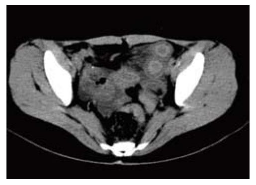
腹部CT: 肝表面光滑, 胆囊及胰腺大小、形态、密度均未见异常. 脾不大, 密度均匀. 双肾大小、形态、密度均未见异常. 膀胱充盈尚可, 壁不厚. 子宫大小形态正常, 壁不厚. 膀胱后区及子宫前区小肠扩张积液并堆积, 密度增高, 边缘模糊(图1).
根据上述资料, 考虑患者为膀胱子宫陷凹的腹内疝, 急性绞窄性肠梗阻, 转普外科行急诊手术治疗. 术中探查发现, 距回盲部40 cm处见一约50 cm小肠经由膀胱子宫陷凹处疝入, 呈紫红色, 经热敷及肠系膜封闭后颜色渐转红, 肠蠕动存在, 相应系膜终末动脉搏动良好, 判定无坏死, 冲洗下腹腔并于盆腔放置一引流管, 术毕关腹. 术中诊断: 腹内疝; 急性绞窄性肠梗阻.
腹内疝是指腹内脏器通过腹腔内孔隙偏离原来的位置, 形成隐匿于体内的异常突起, 其最常表现为小肠肠管进入孔隙而导致的肠梗阻, 而疝孔通常是已经存在的解剖结构, 如裂孔、隐窝和陷凹. 根据发生疝的解剖结构不同, 腹内疝的类型不一. 腹内疝是一种少见的外科急腹症, 常引发绞窄性肠梗阻, 病情凶险, 术前诊断十分困难, 除十二指肠旁疝[1]相对发病率较高外, 而其他的一些腹内疝类型在临床上罕见, 包括Winslow孔疝[2]、肠系膜疝[3]、网膜疝[4]、盲肠旁疝[5]、乙状结肠系膜疝[6]、膀胱上疝[7]、盆腔疝[8]、阔韧带疝[9]及腹膜后疝[10]等.
膀胱子宫陷凹是指女性膀胱子宫之间腹膜腔形成的生理性凹陷, 因陷凹较浅, 在此处形成腹内疝的可能性极低, 经文献检索, 迄今国内外尚无嵌顿于膀胱子宫陷凹的腹内疝的相关文献报道. 本病例是一种十分特殊的腹内疝类型. 该患以"下腹突发剧烈绞痛2 h"入院. 彩超提示子宫前方与膀胱后方间可见范围约6.8×4.4 cm2不均质回声团, 其内可见无回声区及扩张肠管, 肠管内径3.2 cm, 可见蠕动, 怀疑肠管疝入膀胱子宫之间形成腹内疝. 腹部CT进一步检查显示, 膀胱后区及子宫前区小肠扩张积液并堆积, 密度增高, 边缘模糊, 考虑为腹内疝致较窄性肠梗阻. 立即转普外科行急诊剖腹探查. 术中证实, 一段小肠嵌顿于膀胱子宫陷凹内, 形成较窄性小肠梗阻, 与术前诊断一致. 因诊断和治疗非常及时, 患者肠管未发生坏死, 仅行肠粘连松解术处理, 患者术后恢复良好. 该患缘何出现膀胱子宫陷凹的腹内疝, 原因尚不清楚, 可能与患者的膀胱子宫陷凹有先天畸形抑或是剖腹产导致的术后粘连有关.
本例患者为育龄女性, 以"下腹突发剧烈绞痛"入院, 病情十分凶险, 应与急性肠系膜动脉栓塞、卵巢囊肿扭转、黄体破裂、异位妊娠破裂、子宫扭转、膀胱结石绞痛、其他原因导致的肠套叠、腹内疝、肠梗阻等疾病鉴别. 本病例在第一时间准确诊断, 及时行外科手术治疗, 避免了肠坏死的发生, 得益于术前超声和CT的精确诊断.
总之, 本病例提示, 超声和CT不仅有助于发现小肠梗阻的病因, 更有助于腹内疝的诊断.
腹内疝是指腹腔内容物经腹膜或肠系膜凸入腹腔裂隙中, 常导致绞窄性小肠梗阻. 腹内疝在临床罕见, 类型不一, 但嵌顿于膀胱子宫陷凹的腹内疝迄今未见文献报道.
于则利, 教授, 首都医科大学附属北京同仁医院外科
本文首次报道了嵌顿于膀胱子宫陷凹的腹内疝致小肠梗阻的病例, 且术前既经超声和CT诊断, 后经手术证实.
该病例增加了腹内疝的新类型, 有助于拓宽临床诊断思路.
文章的科学性、创新性和可读性能较好地反映该作者的临床水平, 且该病例属于临床少见病例, 为临床提供了一种思路.
编辑: 张姗姗 电编: 闫晋利
| 1. | Lu CW, Liu LC. Right-side paraduodenal hernia: unexplained recurrent abdominal pain. Clin Imaging. 2012;36:68-71. [PubMed] [DOI] |
| 2. | Grisham A, Javan R. Image of the month. Foramen of Winslow hernia. Arch Surg. 2011;146:1329-1330. [PubMed] [DOI] |
| 3. | Kakimoto Y, Abiru H, Kotani H, Ozeki M, Tsuruyama T, Tamaki K. Transmesenteric hernia due to double-loop formation in the small intestine: a fatal case involving a toddler. Forensic Sci Int. 2012;214:e39-e42. [PubMed] [DOI] |
| 4. | Le Moigne F, Lamboley JL, de Charry C, Vitry T, Salamand P, Farthouat P, Michel P. An exceptional case of internal transomental hernia: correlation between CT and surgical findings. Gastroenterol Clin Biol. 2010;34:562-564. [PubMed] [DOI] |
| 5. | Nishi T, Tanaka Y, Kure T. A case of pericecal hernia with a hernial orifice located on the lateral side of the cecum. Tokai J Exp Clin Med. 2011;36:71-74. [PubMed] |
| 6. | Harrison OJ, Sharma RD, Niayesh MH. Early intervention in intersigmoid hernia may prevent bowel resection-A case report. Int J Surg Case Rep. 2011;2:282-284. [PubMed] [DOI] |
| 7. | Cissé M, Konaté I, Ka O, Dieng M, Dia A, Touré CT. Internal supravesical hernia as a rare cauase of intestinal obstruction: a case report. J Med Case Rep. 2009;3:9333. [PubMed] [DOI] |
| 8. | Forgues A, Junes F, Gateau T, Geissman A, Merignargues F, Ballanger P, Robert G. [Right obstructive pyelonephritis due to supra-piriform herniation of the pelvic ureter: a clinical case]. Prog Urol. 2011;21:887-890. [PubMed] [DOI] |
| 9. | Bangari R, Uchil D. Laparoscopic management of internal hernia of small intestine through a broad ligament defect. J Minim Invasive Gynecol. 2012;19:122-124. [PubMed] [DOI] |
| 10. | Leão P, Vilaça S, Oliveira M, Falcão J. Giant recurrent retroperitoneal liposarcoma initially presenting as inguinal hernia: Review of literature. Int J Surg Case Rep. 2012;3:103-106. [PubMed] [DOI] |


