修回日期: 2009-12-13
接受日期: 2009-12-14
在线出版日期: 2010-01-08
目的: 评价D-木糖吸收率在肝硬化门静脉高压进程中的变化规律, 欲探索一种非损伤性且简便实用的推测门静脉压力的方法.
方法: SD大鼠80只, ♂, 体质量180-220 g, 随机分为: 正常组5只, 正常饮水, 正常饮食. 肝硬化门脉高压诱导组75只, 共成模55只, 前5 wk使用0.03%硫代乙酰胺, 后5 wk使用0.04%硫代乙酰胺作为其饮水, 停药后6 wk仍继续观察肝组织及门静脉压力变化. 正常组及第2、4、6、8、10、11、12、13、14、15、16周共12组, 对每组大鼠检测尿D-木糖含量及门静脉压力, 并计算D-木糖吸收率与相应的门静脉压力的相关性.
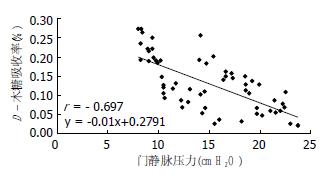
结果: 在肝硬化进程中自第8周纤维间隔明显增生, 肝小叶结构紊乱, 部分可见假小叶, 同时门静脉压力及D-木糖吸收率也是自第8周开始同步出现显著性差异. D-木糖吸收率与门静脉压力呈负相关, 具有统计学意义(r = -0.697, P<0.01), 同时得出两者的直线关系方程: y = -0.01x+0.2791.
结论: D-木糖吸收率可以反映肝硬化门静脉压力的变化趋势, 对于临床上判断门静脉压力的大小、评价治疗效果具有重要意义.
引文著录: 杨思贤, 赵金满, 徐珊珊. 肝硬化门静脉高压对D-木糖吸收率的影响及意义. 世界华人消化杂志 2010; 18(1): 9-13
Revised: December 13, 2009
Accepted: December 14, 2009
Published online: January 8, 2010
AIM: To evaluate the dynamic changes in D-xylose absorption rate during the progression of portal hypertension to find a noninvasive and practical method to evaluate portal pressure.
METHODS: Eighty male Sprague-Dawley rats, weighing 180-220 g, were randomly divided into two groups: normal control group (n = 5) and model group (n = 75). Fifty-five model rats were randomly divided into 11 subgroups (n = 5). The normal control group was given normal drinking water, while the model group was given 0.03% thioacetamide (TAA) for 5 weeks and 0.04% TAA for another 5 weeks. The pathological changes in the liver and portal pressure were monitored until 6 weeks after withdrawal of TAA. The D-xylose content of the urine and portal pressure were determined at weeks 2, 4, 6, 8, 10, 11, 12, 13, 14, 15, and 16, respectively. The correlation between D-xylose absorption rate and portal pressure was analyzed.
RESULTS: At week 8, apparent fibrous septum hyperplasia, disorganized hepatic lobule architecture, and even pseudolobules were observed. Meanwhile, portal vein pressure and D-xylose absorption rate began to show significant changes. The D-xylose absorption rate is inversely correlated with portal vein pressure (P < 0.01). The correlation coefficient is -0.697, and the linear equation is: y = -0.01x + 0.2791.
CONCLUSION: D-xylose absorption rate can reflect the changes in hepatic portal pressure and can be used to predict portal hypertension pressure and evaluate therapeutic effects in clinical practice.
- Citation: Yang SX, Zhao JM, Xu SS. Correlation of D-xylose absorption rate with portal pressure in rats with portal hypertension. Shijie Huaren Xiaohua Zazhi 2010; 18(1): 9-13
- URL: https://www.wjgnet.com/1009-3079/full/v18/i1/9.htm
- DOI: https://dx.doi.org/10.11569/wcjd.v18.i1.9
门静脉高压是各种肝病晚期的一种严重并发症[1,2], 死亡率高, 救治难度大, 而门脉压力测定是确定门脉高压的客观依据, 也是干预治疗效果评估的重要指标. 虽然门静脉压力的测定方法种类很多, 但是多为损伤性检测[3], 且有些方法存在一定危险性, 部分方法虽损伤较轻但操作较复杂[4-18], 严重影响临床推广应用. 本文通过研究门静脉压力与D-木糖吸收率间的关系, 评价D-木糖吸收与门静脉压力的相关性, 欲探索一种非损伤性且简单实用的监测门静脉压力的方法.
SD大鼠80只, ♂, 体质量180-220 g, 购于中国医科大学实验动物中心. 硫代乙酰胺(TAA)、D-木糖购于国药集团化学试剂有限公司. D-木糖试测定剂盒购于南京建成生物工程研究所, 1.33 mmol/L D-木糖标准品应用液配制方法: 取13.3 mmol/L的D-木糖标准品储备液以标准品稀释液作1:9稀释.
1.2.1 分组及造模: 随机分为: 正常组5只, 给予正常饮水, 正常饮食; 肝硬化门脉高压诱导组75只, 采用硫代乙酰胺法造模[19,20], 共成模55只, 1-5 wk使用0.03% TAA, 6-10 wk使用0.04% TAA作为其饮水, 停药后6 wk仍继续观察肝组织及门静脉压力变化.
1.2.2 门静脉压力的测定: 对于正常组及肝硬化门静脉高压诱导组(分别在第2、4、6、8、10、11、12、13、14、15、16周时分别取5只), 12组分别命名为0、1、2、3、4、5、6、7、8、9、10、11组. 应用标尺法测定门静脉压力. 5.5号头皮针穿刺门静脉成功后外接输液器, 输入少量液体至头皮针内充盈, 垂直提起头皮针管, 观察液面逐渐下移直至液柱稳定, 测量其高度, 以厘米(cmH2O)记录门静脉压力[21].
1.2.3 D-木糖吸收试验: 待测大鼠空腹14 h后每只灌服10%的D-木糖溶液1 mL, 放入代谢笼中收集5 h内的尿液, 并在开腹测压的同时膀胱穿刺将膀胱内残留的尿液取出, 计算尿液的总量. 根据D-木糖试剂盒要求, 采用间苯三酚法测定尿D-木糖总量[22], 以毫克(mg)记录尿D-木糖总量.
D-木糖吸收率的计算方法: 5 h尿中排出木糖(mg) = [(测定管A-测定空白管A)/(标准管A-试剂空白管A)]×标准管浓度(1.33 mmol/L)×样本测试前稀释倍数(10倍)×相对分子质量(150)×5 h尿总量(L); D-木糖吸收率 = (D-木糖口服量-5 h尿中排出木糖量)/D-木糖口服量×100%.
统计学处理 所有数据输入计算机建立数据库, 采用SPSS13.0软件包进行数据统计分析, 计量资料以mean±SD表示, 不同组D-木糖吸收率、门静脉压力组间比较用方差分析(ANOVA), 门静脉压力与D-木糖吸收率之间的密切程度采用Pearson相关分析, P<0.05为差异有统计学意义. 采用Excel软件将数据输入建立数据库, 以门静脉压力为X轴、D-木糖吸收率为Y轴绘制散点图.
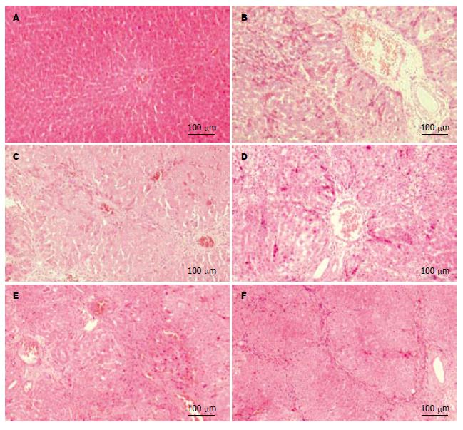
对肝组织石蜡切片进行HE染色, 光学显微镜下观察显示: 2 wk出现斑点状肝细胞坏死, 坏死区有炎细胞浸润, 自门静脉周围向外蔓延, 并有中央静脉周围炎性细胞的浸润, 肝细胞核增大, 肝板结构紊乱(图1B). 4 wk炎症反应进一步加重, 有肝细胞坏死区, 大量淋巴细胞中性白细胞聚集, 伴有肝细胞脂肪样变, 并可见部分大鼠肝脏有纤维化, 由图可见此视野内存在多个中央静脉, 且其间有纤维间隔形成(图1C). 8 wk纤维间隔明显增生, 肝小叶结构紊乱, 部分可见假小叶形成(图1D). 10 wk肝内假小叶形成, 光镜下未见恶变的肝组织(图1E). 16 wk肝内广泛假小叶形成, 未见恶变的肝组织(图1F).
以D-木糖吸收率为Y轴、门静脉压力为X轴制成散点图, 显示D-木糖吸收率随着压力的升高而下降(图2). 经统计学相关性分析D-木糖吸收率与门静脉压力的相关系数为-0.697, 并且具有统计学意义(P<0.01), 两者线性关系的直线方程为y = -0.01x+0.2791. 在大鼠肝硬化门静脉高压形成过程中各组尿D-木糖总量、D-木糖吸收率及门静脉压力的规律性动态变化(表1). 与正常组比较自第8周开始尿D-木糖含量、D-木糖吸收率开始出现显著性差异(P<0.01), 与正常组比较门静脉压力自第8周开始出现显著性差异(P<0.01).
| 分组 | D-木糖含量(mg) | P值 | D-木糖吸收率(%) | P值 | 门静脉压力(cmH2O) | P值 |
| 0 | 22.758±3.6 | 0.20±0.04 | 8.80±0.96 | |||
| 1 | 20.268±4.2 | 0.414 | 0.20±0.04 | 0.414 | 9.38±0.72 | 0.723 |
| 2 | 18.252±6.9 | 0.142 | 0.20±0.07 | 0.142 | 9.44±1.00 | 0.696 |
| 3 | 18.180±5.9 | 0.137 | 0.20±0.06 | 0.137 | 11.30±1.84 | 0.131 |
| 4 | 14.110±4.7 | 0.006 | 0.11±0.05 | 0.006 | 14.50±1.27 | 0.001 |
| 5 | 11.910±3.8 | 0.001 | 0.10±0.04 | 0.001 | 15.08±3.44 | 0.000 |
| 6 | 10.790±5.5 | 0.000 | 0.10±0.05 | 0.000 | 15.54±4.03 | 0.000 |
| 7 | 10.260±5.5 | 0.000 | 0.10±0.05 | 0.000 | 17.30±4.39 | 0.000 |
| 8 | 8.970±3.6 | 0.000 | 0.10±0.04 | 0.000 | 17.66±2.52 | 0.000 |
| 9 | 7.976±4.6 | 0.000 | 0.10±0.04 | 0.000 | 18.48±2.52 | 0.000 |
| 10 | 6.140±3.5 | 0.000 | 0.10±0.04 | 0.000 | 19.82±2.90 | 0.000 |
| 11 | 5.660±5.1 | 0.000 | 0.10±0.10 | 0.000 | 22.36±1.87 | 0.000 |
D-木糖吸收率及门静脉压力组间两两比较显示第0、1、2、3组的D-木糖吸收率及门静脉压力均无显著差异, 但是D-木糖吸收率仍呈下降趋势. 自第4组起各组与第0、1、2、3组D-木糖吸收率、门静脉压力组间两两比较存在显著性差异(P<0.05). D-木糖吸收率第4组与第10、11组存在显著性差异, 同时门静脉压力第4组与第9、10、11组也存在显著性差异. 即当D-木糖吸收率出现显著性差异时门静脉压力也同步出现显著性差异(P<0.05).
门静脉高压是肝硬化出现腹水、食管静脉曲张的直接因素. 肝硬化门静脉高压引起脾大, 脾功能亢进, 侧支循环形成时可出现出血、门体分流性脑病、门脉高压性胃肠血管病等严重并发症, 威胁到患者生命[23,24]. 1991年kozarek等[25]提出门静脉高压性肠病概念, 指继发于门静脉高压以肠道血管扩张为特征的一种病变. 在小肠内镜下, 几乎所有肝硬化门静脉高压患者小肠黏膜均有不同程度异常, 表现为小肠绒毛水肿、萎缩、血管异常甚至小肠静脉曲张[13]. 在胶囊内镜下, 门静脉高压患者的马赛克征和静脉曲张阳性率为68%[26]. 部分病理学研究也证实了在肝硬化门静脉高压时小肠绒毛会发生不同程度的改变[27-31], 且有研究提出门静脉高压性肠病的发生率与肝功能损害无关, 而与门静脉压力的增高有关[32,33]. 所以本文以门静脉高压时小肠血管曲张瘀血, 黏膜水肿影响小肠吸收且随着门静脉压力的升高以上变化更为严重为基础, 通过小肠黏膜的吸收变化情况间接监测门静脉压力的变化. D-木糖是一种五碳糖, 与其他单糖不同, 他在小肠通过易化扩散被不完全吸收, D-木糖吸收试验是检测小肠黏膜吸收功能的最好的非损伤性检测方法, 口服一定量D-木糖后, 50%的D-木糖被小肠吸收, 大约一半在体内代谢, 即大约25%摄入的D-木糖由尿排出. 本研究在制作大鼠肝硬化门静脉高压模型的过程中, 参考相关文献报道的门脉压力变化规律[34], 确定测量门静脉压力的时点, 测压前测定尿D-木糖含量及吸收率通过分析观察两者的相关性, 评价通过D-木糖吸收率检测门静脉压力变化趋势的可行性.
本实验结果显示, 随着门静脉压力的升高D-木糖吸收率逐渐下降, 两者呈负相关, 相关系数为-0.697. 与正常组相比自第8周开始D-木糖吸收率及门静脉压力均出现显著性差异, 第8周开始尿D-木糖总量小于口服量的18%, 说明小肠黏膜已经发生吸收不良, 并且随着用药时间的延长尿D-木糖总量逐渐下降. D-木糖吸收率及门静脉压力组间两两比较示在D-木糖吸收率发生显著变化时门静脉压力也出现了显著性的变化, 自第11周起D-木糖吸收率各组间变化无显著性差异, 这与门静脉压力变化的幅度有关, 当门静脉压力升高幅度较小时, D-木糖吸收率变化不显著, 但是仍然是呈下降趋势. 由此得出在一定程度上D-木糖吸收率可以反映肝硬化门静脉压力的变化趋势. 研究得出的D-木糖吸收率与门静脉压力关系的直线方程y = -0.01x+0.2791, 有助于根据木糖的吸收率估算门静脉压力的值, 对于临床上判断门静脉压力的大小, 评价治疗效果具有重要意义. 本文通过肝硬化引起门静脉高压性肠病进而影响肠吸收功能, 后者可影响D-木糖吸收率的设想进行试验, 发现D-木糖吸收率和门静脉压力呈负相关, 目前国内外尚未见有关D-木糖吸收率与门静脉压力关系的基础研究报道. 笔者曾进行了心肝核素显影比值与门静脉高压相关性的研究, 发现心肝核素显影感兴趣区的确定受主观因素影响较大. 而D-木糖吸收率检测不受主观因素影响, 且操作简便, 用于门静脉压力检测值得深入研究.
门静脉高压是各种肝病晚期的一种严重并发症, 死亡率高, 救治困难, 而门脉压力测定是确定门脉高压的客观依据, 也是干预治疗效果评估的重要指标. 虽然门静脉压力的测定方法种类很多, 但是多为损伤性检测, 且有些方法存在一定的危险性, 部分方法虽损伤较轻但操作较复杂, 因此门脉高压症的研究存在很大的困难.
李瑜元, 教授, 广州市第一人民医院内科.
本课题组曾进行心肝核素显影比值与门静脉高压相关性的研究, 发现心肝核素显影感兴趣区的确定受主观因素影响较大. 认为D-木糖吸收率不受主观因素影响, 且操作简便, 用于门静脉压力检测值得深入研究.
本文通过门静脉高压引起门静脉高压性肠病(PHE)进而影响肠吸收功能, 后者可影响D-木糖吸收率的设想进行试验. 结果表明D-木糖吸收率和门静脉压力呈负相关.
根据木糖的吸收率估算门静脉压力的值, 对于临床上判断门静脉压力的大小, 评价治疗效果具有重要意义.
本研究立题有一定创新性, 尚无同类报道, 学术价值较好, 但实验干扰因素多, 需进一步研究.
编辑: 李军亮 电编:吴鹏朕
| 1. | Bosch J, Garcia-Pagán JC, Berzigotti A, Abraldes JG. Measurement of portal pressure and its role in the management of chronic liver disease. Semin Liver Dis. 2006;26:348-362. [PubMed] [DOI] |
| 2. | Skill NJ, Theodorakis NG, Wang YN, Wu JM, Redmond EM, Sitzmann JV. Role of cyclooxygenase isoforms in prostacyclin biosynthesis and murine prehepatic portal hypertension. Am J Physiol Gastrointest Liver Physiol. 2008;295:G953-G964. [PubMed] [DOI] |
| 3. | Liao WC, Hou MC, Lin HC, Tan PC, Liao TM, Li HS, Lee SD. Feasibility of needle puncture measurement of variceal pressure in patients undergoing endoscopic variceal ligation. Hepatogastroenterology. 2009;56:392-402. [PubMed] |
| 4. | Nanashima A, Sumida Y, Abo T, Sakamoto I, Ogawa Y, Sawai T, Takeshita H, Hidaka S, Nagayasu T. Usefulness of measuring hepatic functional volume using Technetium-99m galactosyl serum albumin scintigraphy in bile duct carcinoma: report of two cases. J Hepatobiliary Pancreat Surg. 2009;16:386-393. [PubMed] [DOI] |
| 6. | 苏 成海, 毕 佩英, 张 玮, 桑 士标, 陈 卫昌. 无创伤性测定门静脉压力与肝血流二法联合应用研究肝硬化的价值. 苏州大学学报(医学版). 2002;22:706-708. |
| 8. | Zhang L, Duan YY, Li JM, Yin JK. Hemodynamic features of Doppler ultrasonography in patients with portal hypertension: intraoperative direct measurement of portal pressure in the portal venous system. J Ultrasound Med. 2007;26:1689-1696. [PubMed] |
| 9. | Kumar A, Sharma P, Sarin SK. Hepatic venous pressure gradient measurement: time to learn! Indian J Gastroenterol. 2008;27:74-80. [PubMed] |
| 10. | Bosch J, Abraldes JG, Berzigotti A, García-Pagan JC. The clinical use of HVPG measurements in chronic liver disease. Nat Rev Gastroenterol Hepatol. 2009;6:573-582. [PubMed] [DOI] |
| 11. | Vorobioff JD. Hepatic venous pressure in practice: how, when, and why. J Clin Gastroenterol. 2007;41 Suppl 3:336-343. [PubMed] |
| 12. | Buscaglia JM, Shin EJ, Clarke JO, Giday SA, Ko CW, Thuluvath PJ, Magno P, Dray X, Kantsevoy SV. Endoscopic retrograde cholangiopancreatography, but not esophagogastroduodenoscopy or colonoscopy, significantly increases portal venous pressure: direct portal pressure measurements through endoscopic ultrasound-guided cannulation. Endoscopy. 2008;40:670-674. [PubMed] [DOI] |
| 13. | Kodama M, Uto H, Numata M, Hori T, Murayama T, Sasaki F, Tsubouchi N, Ido A, Shimoda K, Tsubouchi H. Endoscopic characterization of the small bowel in patients with portal hypertension evaluated by double balloon endoscopy. J Gastroenterol. 2008;43:589-596. [PubMed] [DOI] |
| 14. | Parikh S. Hepatic Venous Pressure Gradient: Worth Another Look? Dig Dis Sci. 2009;54:1178-1183. [PubMed] [DOI] |
| 15. | Giday SA, Clarke JO, Buscaglia JM, Shin EJ, Ko CW, Magno P, Kantsevoy SV. EUS-guided portal vein catheterization: a promising novel approach for portal angiography and portal vein pressure measurements. Gastrointest. Endosc. 2008;67:338-342. [PubMed] [DOI] |
| 16. | Vizzutti F, Arena U, Rega L, Romanelli RG, Colagrande S, Cuofano S, Moscarella S, Belli G, Marra F, Laffi G. Performance of Doppler ultrasound in the prediction of severe portal hypertension in hepatitis C virus-related chronic liver disease. Liver Int. 2007;27:1379-1388. [PubMed] |
| 17. | Singal AK, Ahmad M, Soloway RD. Duplex Doppler Ultrasound Examination of the Portal Venous System: An Emerging Novel Technique for the Estimation of Portal Vein Pressure. Dig Dis Sci. 2009; Jul 23. [Epub ahead of print]. [PubMed] |
| 18. | Chang HK, Park YJ, Koh H, Kim SM, Chung KS, Oh JT, Han SJ. Hepatic fibrosis scan for liver stiffness score measurement: a useful preendoscopic screening test for the detection of varices in postoperative patients with biliary atresia. J Pediatr Gastroenterol Nutr. 2009;49:323-328. [PubMed] [DOI] |
| 22. | Eberts TJ, Sample RH, Glick MR, Ellis GH. A simplified, colorimetric micromethod for xylose in serum or urine, with phloroglucinol. Clin Chem. 1979;25:1440-1443. [PubMed] |
| 23. | El Atti EA, Nevens F, Bogaerts K, Verbeke G, Fevery J. Variceal pressure is a strong predictor of variceal haemorrhage in patients with cirrhosis as well as in patients with non-cirrhotic portal hypertension. Gut. 1999;45:618-621. [PubMed] |
| 24. | Sass DA, Chopra KB. Portal hypertension and variceal hemorrhage. Med Clin North Am. 2009;93:837-853 vii-viii. [PubMed] |
| 25. | Kozarek RA, Botoman VA, Bredfeldt JE, Roach JM, Patterson DJ, Ball TJ. portal colopathy:prospective study of colonoscopy in patients with portal hypertension. Gastroenterology. 1991;101:1192-1197. [PubMed] |
| 26. | Figueiredo P, Almeida N, Lérias C, Lopes S, Gouveia H, Leitão MC, Freitas D. Effect of portal hypertension in small bowel: an endoscopic approach. Dig Dis Sci. 2008;53:2144-2150. [PubMed] [DOI] |
| 27. | Misra V, Misra SP, Dwivedi M, Gupta SC. Histomorphometric study of portal hypertensive enteropathy. Am J Clin Pathol. 1997;108:652-657. [PubMed] |
| 28. | Such J, Guardiola JV, de Juan J, Casellas JA, Pascual S, Aparicio JR, Solá-Vera J, Pérez-Mateo M. Ultrastructural characteristics of distal duodenum mucosa in patients with cirrhosis. Eur J Gasroenterol Hepatol. 2002;14:371-376. [PubMed] [DOI] |
| 29. | Barakat M, Mostafa M, Mahran Z, Soliman AG. Portal hypertensive duodenopathy: clinical, endoscopic, and histopathologic profiles. Am J Gastroenterol. 2007;102:2793-2802. [PubMed] [DOI] |
| 30. | Higaki N, Matsui H, Imaoka H, Ikeda Y, Murakami H, Hiasa Y, Matsuura B, Onji M. Characteristic endoscopic features of portal hypertensive enteropathy. J Gastroenterol. 2008;43:327-331. [PubMed] [DOI] |
| 31. | Goulas S, Triantafyllidou K, Karagiannis S, Nicolaou P, Galanis P, Vafiadis I, Tzivras M, Mavrogiannis C. Capsule endoscopy in the investigation of patientswith portal hypertension and anemia. Can J Gastroenterol. 2008;22:469-474. [PubMed] [DOI] |
| 32. | Yamakado S, Kanazawa H, Kobayashi M. Portal hypertensive colopathy: endoscopic findings and the relation to portal pressure. Intern Med. 1995;34:153-157. [PubMed] [DOI] |


