修回日期: 2009-03-04
接受日期: 2009-03-09
在线出版日期: 2009-03-28
目的: 观察家兔生后发育期间十二指肠胰岛淀粉样多肽(islet amyloid polypeptide, IAPP)的表达及分布特点.
方法: 家兔60只, 分为生后5、15、25、35、60、90 d共6组, 每组10只, 应用免疫组织化学SABC法及图像分析方法检测IAPP在十二指肠的表达及分布特点.
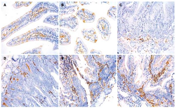
结果: 不同发育阶段家兔十二指肠中均有IAPP的表达, 阳性产物呈棕黄色, 存在于胞质. 生后5、15 d, IAPP阳性细胞散在分布于黏膜上皮细胞间, 细胞数量少且染色浅淡; 25-90 d, IAPP阳性细胞数量增多, 且细胞染色深, 主要分布于固有层结缔组织中, 少量分散存在于黏膜上皮细胞间. 图像分析结果表明, IAPP阳性细胞的数量从第35天开始逐渐增多(F = 24.19, P = 0.0001), 平均灰度值从第15天开始逐渐下降(F = 72.42, P = 0.004).
结论: IAPP作为一种营养因子, 可能对生后家兔十二指肠黏膜的生长发育起重要作用.
引文著录: 胡赟, 梁文妹. 胰岛淀粉样多肽在生后发育期间家兔十二指肠中的表达及意义. 世界华人消化杂志 2009; 17(9): 910-913
Revised: March 4, 2009
Accepted: March 9, 2009
Published online: March 28, 2009
AIM: To investigate the expression and distribution features of islet amyloid polypeptide (IAPP) in duodenum of rabbits during the postnatal development period.
METHODS: Sixty rabbits were equally divided into the 5th, 15th, 25th, 35th, 60th, and 90th d groups. Immunohistochemical SABC staining and image analysis were performed to detect the expression of IAPP.
RESULTS: The expression of IAPP was found in the duodenum of rabbits at different postnatal development stages. Positive production was brownish-yellow and resided in the cytoplasm. In the 5th and 15th d groups, a few IAPP positive cells scattered over epithelium mucosa cells, and the staining was weaker. During 25th to 90th d, the number of IAPP positive cells increased and staining became stronger gradually. The positive cells were mainly distributed in the connective tissue of tunica propria, and some cells scattered over epithelium mucosa cells. The result of image analysis indicated the number of IAPP positive cells increased from 35th to 90th d (F = 24.19, P = 0.0001), and the mean grey values decreased gradually from 15th d (F = 72.42, P = 0.004).
CONCLUSION: IAPP maybe involve in the development of tunica mucosa in rabbit duodenum as a nutrition factor.
- Citation: Hu Y, Liang WM. Expression of islet amyloid polypeptide in duodenum of rabbits during the postnatal development period. Shijie Huaren Xiaohua Zazhi 2009; 17(9): 910-913
- URL: https://www.wjgnet.com/1009-3079/full/v17/i9/910.htm
- DOI: https://dx.doi.org/10.11569/wcjd.v17.i9.910
IAPP又称为糖尿病相关肽(diabetes-associated peptide, DAP)或淀粉不溶素(amylin), 自从1986年被提纯以来, 已引起众多学者的关注. 除由胰岛细胞分泌外, 也可由胃肠内分泌细胞分泌[1]. 已有研究证明, 人胎胃、小肠、结肠、直肠和阑尾均有IAPP的表达. 但现有研究中对生后不同发育阶段家兔十二指肠中是否有IAPP表达尚无定论. 本研究应用免疫组织化学及图像分析方法, 对家兔十二指肠生后发育期间IAPP的定位及分布特点进行了较详细的定性和定量分析, 为深入研究IAPP的生理作用及其对生后发育的影响提供更多的形态学依据.
生后5、15、25、35、60、90 d家兔各10只, 体质量分别为0.072±0.006、0.151±0.003、0.209±0.021、0.345±0.091、1.21±0.152、2.001±0.093 kg, 由贵阳医学院动物实验动物中心提供. 经股动脉放血处死后, 迅速取其十二指肠组织入Bouin液固定, 梯度酒精脱水, 常规石蜡包埋, 制成4 µm厚连续切片.
1.2.1 免疫组织化学SABC法: 显示IAPP阳性细胞. 其主要步骤为: 切片常规脱蜡至水, 甲醇-过氧化氢封闭10 min, 正常羊血清(1:50)室温30 min, 兔抗IAPP(1:2000, Peninsula Lab INC产品)4℃孵育过夜, 羊抗兔IgG(1:100)37℃孵育20 min, SABC复合物37℃孵育20 min, DAB-H2O2液显色, 苏木精复染, 中性树胶封片. 方法对照: 以PBS缓冲液代替IAPP抗血清, 余步骤同上.
1.2.2 图像分析: 随机选取各发育阶段家兔十二指肠切片3张, 应用BioMias图像分析系统进行检测. 在40倍物镜下, 每张切片随机选取5个视野, 计数IAPP阳性细胞数量, 并测得其平均灰度值.
统计学处理 应用SPSS11.0统计学软件对所得数据进行显著性分析. 组间比较用单因素方差分析(One-way analysis of variance, ANOVA)(mean±SD), P<0.05具有统计学意义.
生后发育不同阶段的家兔十二指肠中均有IAPP阳性细胞, 阳性产物呈棕黄色, 存在于胞质(图1). 生后5 d及15 d, IAPP阳性细胞散在于黏膜上皮细胞间, 多呈柱状, 细胞数量少且染色浅淡(图1A-B); 25 d到35 d, IAPP阳性细胞则位于固有层结缔组织中, 圆形或卵圆形, 且数量逐渐增多, 免疫染色变深(图1C-D); 60 d及90 d时IAPP阳性细胞不仅分布于固有层结缔组织中, 也分散存在于黏膜上皮细胞间, 且细胞数量多, 染色深(图1E-F). 方法对照切片上未见阳性细胞.
IAPP阳性细胞数量在5 d、15 d、25 d时变化不明显(P>0.05), 从35 d开始, 细胞数量开始增多(F = 24.19, P<0.05); IAPP阳性细胞的平均灰度值在15 d、25 d时呈逐渐降低趋势(F = 72.42, P<0.05), 但35 d以后变化不明显(P>0.05, 表1).
| 生后(d) | 测量指标 | |||
| 细胞数量 | P | 平均灰度值 | P | |
| 5 | 11.56±3.54 | 186.51±11.03 | ||
| 15 | 10.01±4.13 | 0.3120 | 171.18±11.29c | 0.004 |
| 25 | 10.67±2.51 | 0.5470 | 152.38±26.58c | 0.015 |
| 35 | 28.37±4.27a | 0.0001 | 151.19±29.92 | 0.348 |
| 60 | 45.36±2.09a | 0.0001 | 150.41±24.93 | 0.111 |
| 90 | 52.48±6.22a | 0.0001 | 150.84±25.66 | 0.173 |
IAPP是由胰岛B细胞分泌的一种37肽, 具有抑制餐后胰高血糖分泌、抑制胃排空、抑制食欲等多种生理功能[2]. 生理状态下, IAPP与胰岛素共存于胰岛B细胞内, 与胰岛素及其他葡萄糖调节因子一起调节体内糖的代谢, 对维持体内血糖水平稳定起重要作用[3-4]. IAPP是2型糖尿病发生的重要病理因素之一, 可通过启动细胞内凋亡信号蛋白[5]、氧化应激[6]、诱导细胞膜离子样通道形成[7]等机制引起B细胞凋亡及功能衰竭. 另有研究证实, IAPP具有与降钙素基因相关肽及降钙素相似的结构, 是降钙素家族成员之一, 能促进成骨细胞增殖及骨形成, 同时还能抑制破骨细胞的活性及骨吸收, 对骨的生长和改建具有重要的调节作用[8].
此外, IAPP还与胃肠道的功能密切相关. 能抑制胃酸分泌, 减少肥大细胞的脱颗粒现象, 增加肠系膜淋巴管收缩的频率和幅度, 促进胃溃疡的愈合等, 对胃肠黏膜的生长和修复有重要的调节作用[9]. IAPP能以旁分泌、自分泌方式调节胃黏膜的内分泌活动[1], 或作为一种新的脑肠肽参与维持消化过程中血糖稳定的内分泌反应[10]. 已有的研究证明, 人胎小肠中仅十二指肠上皮及固有层有IAPP的表达, 且与胎龄有关. 本研究结果表明, 生后不同发育阶段家兔十二指肠均有IAPP表达, 其定位从上皮逐渐向固有层过渡, 与胎儿十二指肠IAPP表达的结果相似. 综合以上研究结果, 我们认为在生长发育的过程中, 随着IAPP的表达由上皮向固有层迁移, IAPP逐渐参与了十二指肠消化和内分泌功能的调节过程, 与十二指肠的生长发育密切相关.
目前有关IAPP在胃肠道表达的文献报道均只涉及其分布特征方面的研究, 本研究除定性外, 还尝试应用图像分析方法测量了IAPP阳性表达强度的改变. 我们发现随生后发育时间的延长, 兔十二指肠IAPP阳性细胞数量逐渐增多, 平均灰度值逐渐下降, 表明IAPP阳性表达的强度增强, IAPP含量增多. 我们推测这可能是对胃肠道所接受的食物刺激越来越复杂的一种适应性反应.
German et al[9]的研究表明IAPP能促进胃肠黏膜的生长和修复, 在减少有害因素对胃黏膜造成损伤的同时刺激保护因素发挥作用, 从而促进胃溃疡的愈合. 综合现有文献报道及本组研究结果, 我们推测随家兔生后发育时间延长, 十二指肠分泌增加的IAPP可能作为一种营养因子, 促进小肠黏膜的生长发育, 并且通过肠-胰岛轴调节机制影响胰岛激素对糖代谢的调节, 同时对生后发育期间骨骼的构建起调节作用.
胰岛淀粉样多肽由胰岛细胞分泌, 也见于胃肠道. 目前尚未见其在生后发育家兔十二指肠表达的报道. 本研究较详细的观察了其在生后不同时段家兔十二指肠的定位及分布特点, 充实了有关IAPP的研究资料.
洪天配, 教授, 北京大学第三医院内分泌科
胰岛淀粉样多肽与糖尿病发生发展的相关机制一直是研究热点, 近年来逐渐深入并涉及到肥胖、骨质疏松等领域, 但这些作用机制仍不是很明确.
已有研究证明, 人胎胃、小肠、结肠、直肠和阑尾及大鼠胃肠道均有胰岛淀粉样多肽的表达, 而人胎小肠中仅十二指肠上皮及固有层有表达, 空肠及回肠均未见表达.
本文的相关工作为胰岛淀粉样多肽生物学功能研究提供了实验依据. 在家兔十二指肠的生后发育过程中, 胰岛淀粉样多肽的表达、形态及定量结果改变与消化和内分泌功能的调节密切相关, 为研究其在十二指肠生后发育中的作用提供了形态学资料.
本文选题较好, 设计合理, 结果可靠, 具有较好的学术价值.
编辑: 李军亮 电编:吴鹏朕
| 1. | Mulder H, Lindh AC, Ekblad E, Westermark P, Sundler F. Islet amyloid polypeptide is expressed in endocrine cells of the gastric mucosa in the rat and mouse. Gastroenterology. 1994;107:712-719. [PubMed] [DOI] |
| 2. | Gebre-Medhin S, Olofsson C, Mulder H. Islet amyloid polypeptide in the islets of Langerhans: friend or foe? Diabetologia. 2000;43:687-695. [PubMed] [DOI] |
| 3. | Yan LM, Tatarek-Nossol M, Velkova A, Kazantzis A, Kapurniotu A. Design of a mimic of nonamyloidogenic and bioactive human islet amyloid polypeptide (IAPP) as nanomolar affinity inhibitor of IAPP cytotoxic fibrillogenesis. Proc Natl Acad Sci U S A. 2006;103:2046-2051. [PubMed] [DOI] |
| 4. | Knight JD, Williamson JA, Miranker AD. Interaction of membrane-bound islet amyloid polypeptide with soluble and crystalline insulin. Protein Sci. 2008;17:1850-1856. [PubMed] [DOI] |
| 5. | Zhang S, Liu J, Dragunow M, Cooper GJ. Fibrillogenic amylin evokes islet beta-cell apoptosis through linked activation of a caspase cascade and JNK1. J Biol Chem. 2003;278:52810-52819. [PubMed] [DOI] |
| 6. | Konarkowska B, Aitken JF, Kistler J, Zhang S, Cooper GJ. Thiol reducing compounds prevent human amylin-evoked cytotoxicity. FEBS J. 2005;272:4949-4959. [PubMed] [DOI] |
| 7. | Quist A, Doudevski I, Lin H, Azimova R, Ng D, Frangione B, Kagan B, Ghiso J, Lal R. Amyloid ion channels: a common structural link for protein-misfolding disease. Proc Natl Acad Sci U S A. 2005;102:10427-10432. [PubMed] [DOI] |
| 8. | Naot D, Cornish J. The role of peptides and receptors of the calcitonin family in the regulation of bone metabolism. Bone. 2008;43:813-818. [PubMed] [DOI] |
| 9. | German SV, Zhuĭkova SE, Komarov FI, Kopylova GN, Kuper GJ, Luk'iantseva GV, Samonina GE, Smirnova EA, Umarova BA. [Pancreatic hormone amylin and integrity of the gastric mucosa]. Vestn Ross Akad Med Nauk. 2001;34-38. [PubMed] |
| 10. | Guidobono F. Amylin and gastrointestinal activity. Gen Pharmacol. 1998;31:173-177. [PubMed] [DOI] |


