修回日期: 2007-08-25
接受日期: 2007-09-01
在线出版日期: 2007-09-08
目的: 分析胰腺神经鞘瘤的特点并探讨其诊治方法.
方法: 对文献上报告的49例及我院收治的1例胰腺神经鞘瘤患者的临床特点进行回顾性分析.
结果: 胰头病变25例, 胰体9例, 胰体/尾部6例, 胰尾部5例, 胰头/体部2例, 定位不明的2例, 胰颈部1例. 实性肿物18例, 囊性25例, 7例患者肿瘤的性质不能确定. 49例患者手术切除, 胰十二指肠切除术13例, 远端胰腺切除术12例, 肿物局部切除术12例, 单纯肿物核除术8例, 术式不明2例, 肿物无法切除1例, 活检1例, 拒绝手术1例. 随访时间最长的达到9 a, 无肿瘤复发和转移.
结论: 胰腺神经鞘瘤术前诊断困难, 绝大部分为良性, 恶性少见, 确诊靠病理, 良性者可行肿瘤核除术, 恶性者可根治性切除, 术中快速病理切片有助于决定手术方式.
引文著录: 李新星, 郑继慧, 戴朝六, 闫兆鹏, 张逸婧. 胰腺神经鞘瘤50例. 世界华人消化杂志 2007; 15(25): 2741-2746
Revised: August 25, 2007
Accepted: September 1, 2007
Published online: September 8, 2007
AIM: To analyze the incidence of pancreatic schwannoma and investigate its diagnosis and treatment.
METHODS: A retrospective study of clinical data from 50 patients with pancreatic schwannoma.
RESULTS: The lesion was located in the head of pancreas in 25 patients, the body in 9 (18%), the body and tail in 6, the tail in 5, the head and body in 2, the neck in 1, and the location was not specified in 2 patients. Solid tumors were found in 18 patients, cystic tumors in 25, and the nature of the tumor was not specified in 7 patients. Forty-nine patients received operative therapy. Thirteen patients underwent a Whipple operation, 12 distal pancreatectomy, 12 local resection, 8 simple enucleation, and in 2 patients the surgical procedure was not specified. The tumor in 1 patient was unresectable. One patient underwent biopsy. One patient refused surgery. The longest follow-up was 9 years, and there have been no reports of local recurrence or metastasis.
CONCLUSION: Preoperative diagnosis of pancreatic schwannoma is difficult. Definite diagnosis depends on pathological examination. Benign tumors can be effectively treated by local excision but those with malignancy should be treated by simple enucleation. Intraoperative frozen sections are useful for determining which kind of operation should be undertaken.
- Citation: Li XX, Zheng JH, Dai CL, Yan ZP, Zhang YJ. Analysis of 50 cases of pancreatic schwannoma. Shijie Huaren Xiaohua Zazhi 2007; 15(25): 2741-2746
- URL: https://www.wjgnet.com/1009-3079/full/v15/i25/2741.htm
- DOI: https://dx.doi.org/10.11569/wcjd.v15.i25.2741
胰腺神经鞘瘤(雪旺细胞瘤Schwannoma)临床少见, 自1948年Cornil首次报道本病以来, 至2007-03国外共发表有关文献36篇, 总计42例. 国内文献报道共7例, 最近我院收治1例, 现结合文献就国内外报告的50例加以综合分析.
收集国外1948/2007-03发表的有关胰腺神经鞘瘤文献36篇(42例)(表1), 国内文献7篇(7例)及我院收治的1例患者, 共计8例(表1).
| 病例 | 年份 | 性别 | 年龄 | 主要症状 | 部位 | 性状 | 大小 | 术式 | 恶变 | 随访(mo) |
| 国外 | ||||||||||
| 1[3] | 2007 | M | 36 | 上腹不适 | 钩突 | 囊性 | 4.0 | 核除 | - | 无 |
| 2[12] | 2006 | M | 42 | 无症状 | 体 | 实性 | 2.5 | 远端胰腺,脾切除 | - | 无 |
| 3[7] | 2005 | F | 46 | 衰弱,体质量减轻 | 头 | 实性伴中心坏死 | 3.0 | 胰十二指肠切除 | - | 24 |
| 4[11] | 2005 | M | 71 | 上腹痛,食欲减退 | 头 | 囊性 | 1.0 | 核除 | - | 10 |
| 5[9] | 2005 | F | 64 | 无症状 | 头 | 实性 | 2.5 | 核除 | - | 24 |
| 6[4] | 2004 | F | 69 | 左上腹痛伴恶心呕吐 | 头 | 囊性 | 5.0 | 肿物局部切除1 | - | 无 |
| 7[2] | 2004 | F | 77 | 上腹痛,体质量减轻 | 体 | 实性 | 3.5 | 核除 | - | 3 |
| 8[6] | 2004 | F | 55 | 无症状 | 头 | 囊性 | 8.0 | 胰十二指肠切除 | - | 10 |
| 9[8] | 2004 | F | 67 | 无症状 | 头 | 囊性 | 5.0 | 胰十二指肠切除 | - | 43 |
| 10[10] | 2003 | M | 46 | 右上腹痛 | 头 | 囊性 | 2.2 | 胰十二指肠切除 | - | 无 |
| 11[1] | 2001 | F | 47 | 后背痛 | 头 | 实性 | 5.5 | 胰十二指肠切除 | - | 15 |
| 12[1] | 2001 | F | 73 | 急性腹痛 | 头 | 囊性 | 3.0 | 胰十二指肠切除 | - | 17 |
| 13[13] | 2001 | F | 63 | 上腹痛 | 尾 | 囊性 | 10 | 远端胰腺切除 | - | 6 |
| 14[14] | 1999 | F | 50 | 上腹痛 | 体/尾 | 囊性 | 9.5 | 远端胰腺,脾切除 | - | 7 |
| 15[15] | 1998 | M | 52 | 无症状 | 体 | 囊性 | 5.5 | 肿物局部切除1 | - | 无 |
| 16[15] | 1998 | M | 69 | 无症状 | 头 | 囊性 | 6 | 胰十二指肠切除 | - | 无 |
| 17[16] | 1998 | F | 70 | 上腹痛 | 体/尾 | 囊性 | 18.0 | 肿物局部切除1 | - | 24 |
| 18[17] | 1997 | M | 63 | 无症状 | 体 | 实性 | 2 | 核除 | - | 24 |
| 19[17] | 1997 | F | 54 | 腹痛并向背部放散 | 头 | 实性 | 2 | 核除 | - | 24 |
| 20[18] | 1997 | F | 46 | 上腹痛 | 头 | 囊性 | 12.0 | 肿物局部切除1 | - | 无 |
| 21[19] | 1995 | M | 47 | 右腹痛 | 体 | NS | 3.5 | 远端胰腺切除 | - | 48 |
| 22[19] | 1995 | M | 63 | 腹痛 | 体 | 囊性 | NS | 远端胰腺切除 | - | 无 |
| 23[19] | 1995 | F | 68 | 上腹痛 | 头/体 | 囊性 | NS | NS | - | 6 |
| 24[20] | 1995 | M | 41 | 腹痛 | 钩突 | 囊性 | 1.5 | 胰十二指肠切除 | - | 无 |
| 25[21] | 1994 | M | 59 | 无症状 | 钩突 | 实性 | 4 | 胰十二指肠切除 | - | 10 |
| 26[22] | 1993 | M | 89 | 上腹痛,发热,呕吐 | 体/尾 | 囊性 | 20 | 远端胰腺切除 | - | 无 |
| 27[23] | 1993 | M | 46 | 右腹痛 | 钩突 | 囊性 | 6 | 肿物局部切除1 | - | 无 |
| 28[24] | 1992 | F | 56 | 右下腹痛 | 体 | 囊性 | 4 | 远端胰腺切除 | - | 无 |
| 29[25] | 1992 | M | 73 | 右上腹痛 | 体/尾 | 实性 | 2 | 肿物局部切除1 | - | 无 |
| 30[26] | 1990 | F | 72 | 贫血,黑便 | 头 | 实性伴中心坏死 | 7 | 胰十二指肠切除 | + | 无 |
| 31[27] | 1990 | F | 75 | 急性腹痛,呕吐,腹泻 | 头 | 实性 | 7 | 拒绝手术 | - | 7 |
| 32[28] | 1989 | F | 35 | 腹痛,贫血,黑便 | 头 | NS | NS | 胰十二指肠切除 | + | 24 |
| 33[29] | 1988 | F | 46 | 胆绞痛 | 头 | 实性 | 4 | 核除 | - | 108 |
| 34[30] | 1987 | F | 40 | 上腹痛,黄疸,体质量减轻 | 头 | 实性伴中心坏死 | 10 | 胰十二指肠切除 | + | 9 |
| 35[31] | 1984 | M | 70 | 腹痛,腹胀,便秘 | 体 | 囊性 | 4 | 肿物局部切除1 | - | 无 |
| 36[32] | 1982 | M | 60 | 腰背痛,体质量减轻 | 体/尾 | 囊性 | 17.5 | 无法切除 | + | 4 |
| 37[33] | 1982 | F | 48 | 上腹不适,嗳气 | 体/尾 | 囊性 | 26 | 远端胰腺切除 | - | 无 |
| 38[34] | 1978 | F | 50 | 腹部无痛性肿物 | 尾 | 实性 | 19 | 远端胰腺切除 | - | 13 |
| 39[35] | 1969 | F | 66 | 无症状 | NS | 囊性 | NS | 肿物局部切除1 | - | 无 |
| 40[36] | 1962 | F | 65 | 腹痛 | 尾 | NS | NS | 远端胰腺切除 | - | 12 |
| 41[36] | 1962 | F | 32 | 腹痛 | 尾 | NS | NS | 远端胰腺切除 | - | 12 |
| 42[2] | 1948 | M | 36 | 黑便 | 头 | NS | NS | 活检 | - | 6 |
| 国内 | ||||||||||
| 43本院 | 2007 | M | 42 | 无症状 | 头 | 实性 | 6.0 | 核除 | - | 无 |
| 44[5] | 2006 | M | 72 | 上腹痛 | 头/体 | 实性 | 1.0 | 肿物局部切除1 | - | 无 |
| 45[37] | 2005 | M | 17 | 上腹痛,呕吐 | NS | 实性伴中心坏死 | 4.0 | 肿物切除,十二指肠侧侧吻合术 | - | 无 |
| 46[38] | 2005 | M | 72 | 上腹胀痛 | 颈 | NS | 1.0 | NS | - | 无 |
| 47[39] | 2004 | M | 65 | 左上腹不适 | 尾 | 囊性 | 4.0 | 远端胰腺,脾切除 | - | 无 |
| 48[40] | 1999 | M | 44 | 无症状 | 头 | 实性 | 6.0 | 肿物局部切除1 | - | 无 |
| 49[41] | 1996 | M | 50 | 上腹不适 | 体 | 囊性 | 4.2 | 肿物局部切除1 | - | 无 |
| 50[42] | 1990 | F | 38 | 右上腹隐痛 | 头 | NS | 12.0 | 胰十二指肠切除 | - | 无 |
总结国内外报告的50例胰腺神经鞘瘤患者的临床资料, 分析其临床病理特点, 探讨胰腺神经鞘瘤的临床诊治原则.
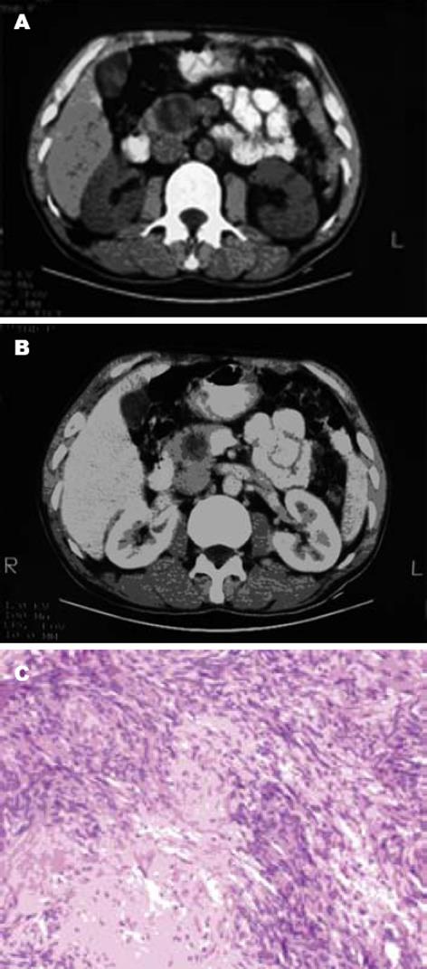
我院1例患者, 男, 42岁, 1 mo前体检超声发现胰头肿块. 无腹痛、体质量下降、黄疸, 既往无酗酒、创伤及胰腺炎病史. 查体: 皮肤巩膜无黄染, 腹部未扪及包块. 实验室检查(包括血糖、血清胰岛素、血清淀粉酶及脂肪酶等)均正常. 血清肿瘤标记物(CEA、CA199、CA125等)亦在正常范围. CT平扫示胰头内圆形低密度灶, 大小约7.3 cm×4.1 cm×4.0 cm(图1A), 增强CT肿物略增强, 密度不均, CT值10-36 HU(图1B). 剖腹手术见肿瘤位于胰腺头部, 周围无炎症征象. 术中冰冻切片提示良性神经鞘瘤, 遂行单纯肿物核除术. 大体标本见肿物呈灰红色, 大小6 cm×4 cm×3 cm, 包膜完整, 切面呈灰黄色, 实性, 质韧. 镜下, 瘤细胞呈梭形, 排列成漩涡状及栅栏状(图1C), 病理诊断: 胰头神经鞘瘤.
50例胰腺神经鞘瘤患者的详细情况见表1. 患者平均年龄55.94(范围17-89)岁, 表明胰腺神经鞘瘤易发于中老年人群. 男女比例24∶26. 最常见的主诉为腹痛, 见于29例患者(58%). 平均大小是6.60(范围1.0-26) cm. 腹痛组肿瘤的平均大小小于非腹痛组(6.09 cm vs 7.25 cm), 表明腹痛不依赖于肿瘤的大小, 且为胰腺神经鞘瘤的早期症状之一. 胰头部病变25例, 占50%, 胰体部9例, 占18%, 胰体/尾部6例, 占12%, 胰尾部5例, 占10%, 胰头/体部2例, 占4%, 定位不明2例, 占4%, 胰颈部1例, 占2%, 实性肿物18例, 占36%, 囊性肿物25例, 占50%, 7例患者肿瘤的性质不确定.
25例胰头病变的患者中, 行胰十二指肠切除术13例, 6例单纯肿物核除术, 4例肿物局部切除术, 1例活检, 1例拒绝手术. 9例胰体病变的患者中, 行远端胰腺切除术4例, 3例肿物局部切除术, 2例单纯肿物核除术. 6例胰体/尾病变的患者中, 行远端胰腺切除术3例, 2例肿物局部切除术, 1例肿物无法切除. 5例胰尾病变的患者, 均实施了远端胰腺切除术. 2例胰头/体部病变患者中, 1例行肿物局部切除术, 1例术式不明. 2例占位不明患者均行肿物局部切除术. 1例胰颈病变的患者术式不明.
英文文献42例中25例随访了平均19.6±21.65(3-108) mo, 另外17例未随访. 国内文献病例随访不明. 随访病例中无复发或死亡.
4例恶性患者, 均来自英文文献, 有3例肿瘤在胰头部, 行胰十二指肠切除术, 1例在胰体/尾部, 已无法切除, 但均无远处转移. 3例患者随访期间(4-24) mo无复发或死亡.
神经鞘瘤又称雪旺细胞瘤(schwannoma), 是由神经鞘膜增生形成, 来源于神经外胚层的Schwann细胞和中胚层的神经内膜, 可发生于任何部位有Schwann细胞膜的神经, 尤好发于脊髓和脑, 四肢、颈部等部位大神经也较多见, 发生于消化系统少见, 发生于胰腺甚为少见[1]. 胰腺神经鞘瘤源自迷走神经分支的自主性交感神经或副交感神经纤维[2]. 国外Cornil et al于1948年首先报道, 至今共计42例. 国内至今共报道7例. 患者的年龄从17岁至89岁不等, 无性别差异, 多为单发病灶, 且大多数肿瘤位于胰腺头部.
胰腺神经鞘瘤可发生于胰腺的任何部位, 以胰腺头部多见, 其次是胰体、胰体/尾部[3]. 患者一般无特异临床表现, 常表现为不同程度的上腹胀满、不适、隐痛, 也可伴有体质量降低、恶心、黑便、黄疸等, 少数患者无临床症状. 胰腺神经鞘瘤多为单发, 生长缓慢, 表面一般有完整的包膜, 略半透明, 如有囊性变、出血时可呈粉红色、黄色、灰褐色. 肿瘤由Schwann细胞和不同含量的网状纤维组成, Schwann细胞是支持周围神经的主要元件, 除了产生和维持髓鞘外, 他还包绕并机械性地保护无髓鞘神经元轴突, 引导神经纤维的再生. Schwann细胞能产生两种类型的肿瘤: 神经鞘瘤或Schwann细胞瘤, 神经纤维瘤. 神经鞘瘤是一种典型的包膜完整的神经鞘膜瘤, 不含神经细胞和胶原, 在显微镜下, 观察切片免疫过氧化物酶染色, S-100蛋白、弹性蛋白、CD56为强阳性, 而包括索蛋白、细胞角蛋白AE1/AE3、中间蛋白、平滑肌肌球蛋白、CD34和CD117在内的其他肿瘤标记物为阴性[4]. 组织学上分析肿瘤组织有2种结构: (1) Antoni A区: 高度细胞化, 有致密的细胞结构和网状纤维, 细胞呈栅栏样编织状或旋涡状排列, 无有丝分裂的迹象; (2)Antoni B区: 典型的细胞减少样, 肿瘤细胞多形性, 基质缺少纤维, 细胞核稀少或仅在边缘有平行排列的网状结构, 通常表现为退化性变, 例如淀粉样变、囊形成、间质出血、钙化, 多数胰腺神经鞘瘤可见到A、B两区共存或两区相互移行状态[4].
胰腺神经鞘瘤的术前诊断很困难, CT扫描通常是确定胰腺肿瘤的最佳选择. 在CT扫描中, 肿瘤常为边界清晰, 包膜完整, CT值在0-30 HU的低密度病灶, 通常呈均一密度或花斑状[5]. 这些低密度区是由多种因素混合造成的, 包括低细胞质以及囊性退变. 病理分型与CT表现密切相关, 以Antoni A区为主的胰腺神经鞘瘤, 由于肿瘤脂质成分高, 而网状血管成分使其对比增强, 病变看起来质地不均, 呈复合体样, 有时可见多处分隔及不同程度的中心坏死或出血的低密度灶, 而以Antoni B区为主的胰腺神经鞘瘤, 由于疏松细胞不成形的结构、无明显纤维成分的基质, CT呈囊性外观, 无对比增强[5-6].
胰腺神经鞘瘤在磁共振成像上具有特征性的表现: 典型的包膜, 在T1加权上为低信号, T2加权上为高信号[7]. 除了信号强度的变化, 磁共振可以提示涉及血管的情况, 这可以更进一步确定病灶的恶变潜能. 胰腺神经鞘瘤在T2加权上为高信号, 且相对于其他胰腺组织的特征性对比增强, 有助于与腺癌的鉴别.
由于临床上本病常无特异症状, 与其他胰腺肿瘤不易鉴别. 囊性胰腺神经鞘瘤与胰腺假性囊肿非常相似, 因此胰腺炎的有无非常重要; 一个残留囊肿通常继发于慢性胰腺炎. 棘球蚴疾病, 可有囊壁钙化, 及其他部位受累, 容易根据临床和实验室检查诊断. 小细胞质液性腺瘤呈蜂窝状, 中央星形瘢痕, 中央或周围有钙化是其可靠指标. 囊性淋巴管瘤密度同水, 多分叶状, 偶有板层钙化, 与胰腺神经鞘瘤不易区别. 无功能胰岛细胞肿瘤最常见于胰体部, CT示其质地为均匀的低密度影, 因此很难与胰腺神经鞘瘤鉴别.
此外, 孤立性神经纤维瘤等具有梭形细胞特征的胰腺非上皮性肿瘤, 也与胰腺神经鞘瘤类似. 然而, 神经纤维瘤没有Antoni A和Antoni B特点, 且对S-100蛋白的染色不均匀[8]. 胰腺神经鞘瘤多有包膜, 而神经纤维瘤则没有. 在磁共振上见到沿肿瘤边缘的低信号影, 可以作为胰腺神经鞘瘤与神经纤维瘤鉴别诊断的标准. 胰腺神经鞘瘤和神经纤维瘤的鉴别非常重要, 因为神经纤维瘤与Recklinghausen病(多发性神经纤维瘤病)明显相关, 极易恶变[8]. 胰腺神经鞘瘤可以是良性或恶性, 但良性恶变极为少见[9]. 继发于神经纤维瘤的恶变类似梭形细胞肉瘤, 而胰腺神经鞘瘤的恶变通常呈上皮样表现, 这种上皮样表现提示可能为转移癌[10]. 恶性胰腺神经鞘瘤现在被称为恶性周围神经鞘瘤, 以区别于良性胰腺神经鞘瘤恶变. 肿瘤生长迅速、周围组织侵袭、实性不均匀, 边界不清、CT呈特征性的对比增强, 以及血栓形成可能均为恶性的常见特征. 细胞角蛋白阳性而S-100蛋白阴性, 缺少经典的Antoni A或Antoni B特征可以帮助区别良恶性胰腺神经鞘瘤.
尽管胰腺神经鞘瘤的最终确诊有赖于肿瘤的病理学检查, 但影像学检查, 可以帮助确定病灶的良性本质, 明确解剖定位, 缩小鉴别诊断范围. 细针穿刺细胞学检查或术中冰冻切片可以提高术前诊断的准确性[3].
胰腺神经鞘瘤最好的治疗就是外科切除. 因为胰腺神经鞘瘤的恶变发生率极低, 所以该肿瘤提倡创伤小的单纯肿瘤核除术; 肿瘤的早期发现与早期治疗可以提高核除术的比例以及避免创伤性很大的胰十二指肠切除术[11]. 50例中, 只有8例进行了单纯肿瘤核除术(大小: 1-4 cm之间). 绝大多数胰腺神经鞘瘤可根据他们的大小和部位行核除术. 术中冰冻切片活检有助于确立良性胰腺神经鞘瘤的诊断, 这对于术式的选择很有意义, 可以将其限于核除术而非进一步的根治切除. 胰腺神经鞘瘤切除术后再复发极为罕见, 因此, 术中为避免损伤神经或周围组织, 即使肿瘤包膜不能完整切除, 甚至行肿瘤次全切除术也是可以的[9]. 如肿瘤大位于胰头部、侵袭壶腹部或脾门部均可根据具体情况行根治性手术. 最近有报告: 胰腺神经鞘瘤对放疗有较好反应, 但具体疗效仍不清楚[12].
文献中, 随访时间最长者达到9 a, 无肿瘤复发和转移. 4例活检为恶性的患者, 其确诊方法并不一致, 有3例未采用或无法采用免疫荧光染色. 获得随访的3例(1例没有报道)在术后4-24 mo均无瘤或带瘤生存. Paranjape et al[2]认为可能是误诊的神经纤维瘤恶变.
胰腺神经鞘瘤虽属罕见, 但应注意与胰腺其他疾病鉴别. 术前影像检查有助于诊断; 确诊需要组织学检查. 对于良性肿瘤, 建议行单纯肿瘤核除术, 但恶性肿瘤需行根治性切除术. 无法切除者, 可行放疗, 但疗效还有待观察.
胰腺神经鞘瘤(雪旺细胞瘤Schwan-noma)临床少见, 可发生于任何部位有Schwann细胞膜的神经, 尤好发于脊髓和脑, 四肢、颈部等部位大神经也较多见, 发生于消化系统少见, 发生于胰腺甚为少见, 国外Cornil et al于1948年首先报道, 至今共计42例. 国内至今共报道7例. 患者的年龄从17岁至89岁不等, 无性别差异, 多为单发病灶, 且大多数肿瘤位于胰腺头部.
对于良性肿瘤, 建议行单纯肿瘤核除术, 但恶性肿瘤需行根治性切除术, 术中快速病理切片有助于决定手术方式, 无法切除者, 可行放疗, 但疗效还有待观察.
神经鞘瘤又称雪旺细胞瘤(schw-annoma), 是由神经鞘膜增生形成, 来源于神经外胚层的Schwann细胞和中胚层的神经内膜, 可发生于任何部位有Schwann细胞膜的神经, 尤好发于脊髓和脑, 四肢、颈部等部位大神经也较多见, 发生于消化系统少见, 发生于胰腺甚为少见. 胰腺神经鞘瘤源自迷走神经分支的自主性交感神经或副交感神经纤维.
本文总结和归纳了胰腺神经鞘瘤的临床病理特点, 治疗, 预后. 全文层次清楚, 逻辑性强, 分析全面深入, 有较强的临床实用性.
编辑: 程剑侠 电编:郭海丽
| 1. | Almo KM, Traverso LW. Pancreatic schwannoma: an uncommon but important entity. J Gastrointest Surg. 2001;5:359-363. [PubMed] [DOI] |
| 2. | Paranjape C, Johnson SR, Khwaja K, Goldman H, Kruskal JB, Hanto DW. Clinical characteristics, treatment, and outcome of pancreatic Schwannomas. J Gastrointest Surg. 2004;8:706-712. [PubMed] [DOI] |
| 3. | Fasanella KE, Lee KK, Kaushik N. Clinical challenges and images in GI. Benign schwannoma of the pancreatic head. Gastroenterology. 2007;132:489, 830. [PubMed] |
| 4. | Bui TD, Nguyen T, Huerta S, Gu M, Hsiang D. Pancreatic schwannoma. A case report and review of the literature. JOP. 2004;5:520-526. [PubMed] |
| 5. | Yu RS, Sun JZ. Pancreatic schwannoma: CT findings. Abdom Imaging. 2006;31:103-105. [PubMed] [DOI] |
| 6. | von Dobschuetz E, Walch A, Werner M, Hopt UT, Adam U. Giant ancient schwannoma of pancreatic head treated by extended pancreatoduodenectomy. Pancreatology. 2004;4:505-508. [PubMed] [DOI] |
| 7. | Novellas S, Chevallier P, Saint Paul MC, Gugenheim J, Bruneton JN. MRI features of a pancreatic schwannoma. Clin Imaging. 2005;29:434-436. [PubMed] [DOI] |
| 8. | Akiyoshi T, Ueda Y, Yanai K, Yamaguchi H, Kawamoto M, Toyoda K, Hayashi T, Ohuchida J. Melanotic schwannoma of the pancreas: report of a case. Surg Today. 2004;34:550-553. [PubMed] [DOI] |
| 9. | Soumaoro LT, Teramoto K, Kawamura T, Nakamura N, Sanada T, Sugihara K, Arii S. Benign schwannoma of the pancreas. J Gastrointest Surg. 2005;9:288-290. [PubMed] [DOI] |
| 10. | Tan G, Vitellas K, Morrison C, Frankel WL. Cystic schwannoma of the pancreas. Ann Diagn Pathol. 2003;7:285-291. [PubMed] [DOI] |
| 11. | Wu YL, Yan HC, Chen LR, Chen J, Gao SL, Li JT. Pancreatic benign schwannoma treated by simple enucleation: case report and review of literature. Pancreas. 2005;31:286-288. [PubMed] [DOI] |
| 12. | Di Benedetto F, Spaggiari M, De Ruvo N, Masetti M, Montalti R, Quntini C, Ballarin R, Di Sandro S, Costantini M, Gerunda GE. Pancreatic schwannoma of the body involving the splenic vein: Case report and review of the literature. Eur J Surg Oncol. 2007;33:926-928. [PubMed] [DOI] |
| 13. | Lee JS, Kim HS, Jung JJ, Han SW, Kim YB. Ancient schwannoma of the pancreas mimicking a cystic tumor. Virchows Arch. 2001;439:697-699. [PubMed] [DOI] |
| 14. | Morita S, Okuda J, Sumiyoshi K, Taketani M, Moriguchi A, Katsu K, Tanigawa N. Pancreatic Schwannoma: report of a case. Surg Today. 1999;29:1093-1097. [PubMed] [DOI] |
| 15. | Brown SZ, Owen DA, O'Connell JX, Scudamore CH. Schwannoma of the pancreas: a report of two cases and a review of the literature. Mod Pathol. 1998;11:1178-1182. [PubMed] |
| 16. | Hsiao WC, Lin PW, Chang KC. Benign retro-peritoneal schwannoma mimicking a pancreatic cystic tumor: case report and literature review. Hepatogastroenterology. 1998;45:2418-2420. [PubMed] |
| 17. | Feldman L, Philpotts LE, Reinhold C, Duguid WP, Rosenberg L. Pancreatic schwannoma: report of two cases and review of the literature. Pancreas. 1997;15:99-105. [PubMed] [DOI] |
| 18. | Todd KE, Lewis MP, Gloor B, Kusske AM, Ashley SW, Reber HA. Management decisions for unusual periampullary tumors. Am Surg. 1997;63:927-932. [PubMed] |
| 19. | Ferrozzi F, Bova D, Garlaschi G. Pancreatic schwannoma: report of three cases. Clin Radiol. 1995;50:492-495. [PubMed] [DOI] |
| 20. | Sugiyama M, Kimura W, Kuroda A, Muto T. Schwannoma arising from peripancreatic nerve plexus. AJR Am J Roentgenol. 1995;165:232. [PubMed] [DOI] |
| 21. | Steven K, Burcharth F, Holm N, Pedersen IK. Single stage pancreaticoduodenectomy (Whipple's procedure), radical cystectomy and bladder substitution with the urethral Kock reservoir. Case report. Scand J Urol Nephrol. 1994;28:199-200. [PubMed] [DOI] |
| 22. | Melato M, Bucconi S, Marus W, Spivach A, Perulli A, Mucelli RP. The schwannoma: an uncommon type of cystic lesion of the pancreas. Ital J Gastroenterol. 1993;25:385-387. [PubMed] |
| 24. | Urban BA, Fishman EK, Hruban RH, Cameron JL. CT findings in cystic schwannoma of the pancreas. J Comput Assist Tomogr. 1992;16:492-493. [PubMed] [DOI] |
| 25. | Burd DA, Tyagi G, Bader DA. Benign schwannoma of the pancreas. AJR Am J Roentgenol. 1992;159:675. [PubMed] [DOI] |
| 26. | Coombs RJ. Case of the season. Malignant neurogenic tumor of duodenum and pancreas. Semin Roentgenol. 1990;25:127-129. [PubMed] [DOI] |
| 27. | Liessi G, Barbazza R, Sartori F, Sabbadin P, Scapinello A. CT and MR imaging of melanocytic schwannomas; report of three cases. Eur J Radiol. 1990;11:138-142. [PubMed] [DOI] |
| 28. | Walsh MM, Brandspigel K. Gastrointestinal bleeding due to pancreatic schwannoma complicating von Recklinghausen's disease. Gastroenterology. 1989;97:1550-1551. [PubMed] [DOI] |
| 29. | Barcena Barros JM, Manuel Palazuelos JC, Concha Garcia A, Val Bernal JF, Naranjo Gomez A, Casado Martin F. Benign schwannoma of the pancreas. Rev Quir Esp. 1988;15:158-159. [PubMed] |
| 30. | Eggermont A, Vuzevski V, Huisman M, De Jong K, Jeekel J. Solitary malignant schwannoma of the pancreas: report of a case and ultrastructural examination. J Surg Oncol. 1987;36:21-25. [PubMed] [DOI] |
| 31. | Veroux G, Cannizzaro MA, Amodeo C, Nicotra P, Imme A. Neurinoma of the pancreas. Minerva Chir. 1984;39:1219-1228. [PubMed] |
| 32. | Moller Pedersen V, Hede A, Graem N. A solitary malignant schwannoma mimicking a pancreatic pseudocyst. A case report. Acta Chir Scand. 1982;148:697-698. [PubMed] |
| 33. | Wimmer B, Gospos C, Ruf G, Wagner R. Neurilemmoma of the pancreas. Rofo. 1982;137:731-734. [PubMed] [DOI] |
| 34. | Ladouce P, Ravisse P, Triquet J, Wermynck J. An isolated neurinoma in the pancreas (author's transl). J Chir (Paris). 1978;115:285-288. [PubMed] |
| 35. | Doutre LP, Paccalin J, Traissac D, Traissac FJ. Exogastric schwannoma considered and treated as a posttraumatic pseudocyst of the pancreas. Arch Fr Mal App Dig. 1969;58:563-567. [PubMed] |
| 36. | Frileux C, Terquem J, Wiel R, Henry JG. On schwannomas of the pancreas. (Apropos of 2 cases). Arch Mal Appar Dig Mal Nutr. 1962;51:721-725. [PubMed] |


