修回日期: 2006-07-25
接受日期: 2006-07-31
在线出版日期: 2006-10-08
目的: 研究结肠癌患者结肠黏膜中白介素-8(IL-8)、白介素-15(IL-15)的表达并探讨其临床应用价值.
方法: 采用免疫组化法检测结肠癌患者结肠黏膜中IL-8, IL-15的表达情况.
结果: IL-8染色阳性44例, 占66.7%. IL-15染色阳性40例, 占60.6%. IL-8和IL-15表达水平与结肠癌临床分期(IL-8: r = 0.437, P = 0.006; IL-15: r = 0.317, P = 0.014)、浸润深度(IL-8: r = 0.332, P = 0.003; IL-15: r = 0.312, P = 0.015)、淋巴结转移(IL-8: r = 0.316, P = 0.042; IL-15: r = 0.236, P = 0.017)、病理分级(IL-8: r = 0.826, P = 0.0001; IL-15: r = 0.368, P = 0.001)均呈显著正相关.
结论: 检测IL-8, IL-15的表达可做为判断结肠癌恶性程度有价值的指标.
引文著录: 高伟, 吴瑜, 司雁菱. 白介素-8、白介素-15在结肠癌患者结肠黏膜的表达及意义. 世界华人消化杂志 2006; 14(28): 2806-2809
Revised: July 25, 2006
Accepted: July 31, 2006
Published online: October 8, 2006
AIM: To investigate the expression of interleukin-8 (IL-8) and IL-15 in colonic mucosa from colon cancer patients and study their relationships with colon cancer.
METHODS: Immunohistochemical technique was used to detect the expression of IL-8 and IL-15 in 66 patients with colon cancer.
RESULTS: The positive rates of IL-8 and IL-15 were 66.7% (44/66) and 60.6% (40/66), respectively. Significant correlations existed between expression of IL-8, IL-15 and the following factors: clinical stages (IL-8: r = 0.437, P = 0.006; IL-15: r = 0.317, P = 0.014), invasive depth (IL-8: r = 0.332, P = 0.003; IL-15: r = 0.312, P = 0.015), regional lymph node metastasis (IL-8: r = 0.316, P = 0.042; IL-15: r = 0.236, P = 0.017), histologic grades (IL-8: r = 0.826, P = 0.0001; IL-15: r = 0.368, P = 0.001).
CONCLUSION: Detection of IL-8 and IL-15 expression is helpful in assessing the malignant degrees of colon cancer.
- Citation: Gao W, Wu Y, Si YL. Expression and significances of interleukin-8 and interleukin-15 in colonic mucosa of colon cancer patients. Shijie Huaren Xiaohua Zazhi 2006; 14(28): 2806-2809
- URL: https://www.wjgnet.com/1009-3079/full/v14/i28/2806.htm
- DOI: https://dx.doi.org/10.11569/wcjd.v14.i28.2806
结肠癌的发病原因尚未完全明确, 其发病主要与环境等因素有关, 为多种因素共同作用的结果. 随着研究的深入, 细胞因子被认为在结肠癌的发病中具有极为重要的作用. IL-8和IL-15是2种重要的细胞因子, 他们参与多种疾病过程. 本研究旨在观察结肠癌患者结肠黏膜中IL-8和IL-15的表达情况并探讨其临床应用价值.
唐山工人医院2004-07/2005-103/4结肠镜活检常规病理学检查确诊为结肠癌并行手术治疗的病例共66例, 男32例, 女34例. 年龄23-79岁. 按WHO标准, 采用TNM分期: Ⅰ-Ⅱ期34例, Ⅲ-Ⅳ期32例; 组织学分级Ⅰ级21例, Ⅱ级22例, Ⅲ级23例. 所有病例2 mo内均未接受治疗. 各组间在年龄及性别等方面无统计学差异. 鼠抗人IL-8, IL-15 mAb购自北京中山公司, 免疫组化过氧化物酶标记的链霉卵白素(SP)试剂盒、二氨基联苯胺(DAB)显色试剂盒均购自福州迈新公司.
全部蜡块均行4 μm厚连续切片, 常规脱蜡至水, 室温下30 g/L甲醇过氧化氢去除内源性过氧化物酶, 微波修复抗原; 滴加一抗后4℃过夜, 滴加二抗后于37℃温箱孵育30 min; DAB-H2O2显色, 苏木素复染, 中性树胶封片. IL-8阳性表达多数位于结肠癌细胞质中, 部分位于核周, 呈棕黄色颗粒. IL-15阳性表达位于结肠癌细胞的胞质或细胞核内, 呈棕黄色颗粒. 随机选择10个高倍视野, 以阳性细胞占总数的百分率进行分级, <10%为-, 10%-50%为+, >50%为++.
统计学处理 采用SPSS 11.5统计软件, IL-8, IL-15与临床、病理特征的关系使用Spearman相关分析, P<0.05为显著性标准.
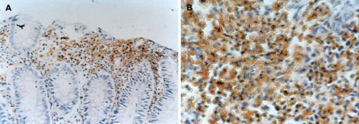
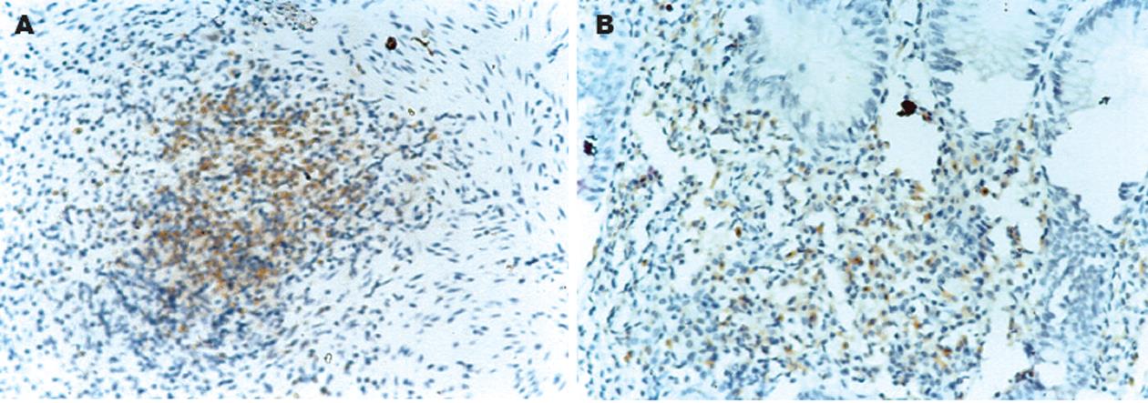
本组66例结肠癌标本中IL-8染色阳性44例(66.7%), 其中29例(43.9%)表达为++, 15例(22.7%)表达为+(图1); IL-15染色阳性40例(60.6%), 其中27例(40.9%)表达为++, 13例(19.7%)表达为+(图2).
IL-8与临床分期呈正相关(r = 0.437, P = 0.006)、与浸润深度呈正相关(r = 0.332, P = 0.003)、与淋巴结转移呈正相关(r = 0.316, P = 0.042)、与病理分级呈正相关(r = 0.826, P = 0.0001); IL-15与临床分期呈正相关(r = 0.317, P = 0.014)、与浸润深度呈正相关(r = 0.312, P = 0.015)、与淋巴结转移呈正相关(r = 0.236, P = 0.017)、与病理分级呈正相关(r = 0.368, P = 0.001). IL-8, IL-15与性别及年龄无相关性(P>0.05, 表1-2).
结肠癌是消化道常见的恶性肿瘤, 其发病率在我国仅次于胃癌和食道癌, 居消化道恶性肿瘤的第三位. 目前认为结肠癌的发病是多因素共同作用的结果, 随着研究的深入, 人们发现与结肠癌相关的细胞因子的检测可能对患者的早期诊断、手术及术后治疗方案的制订有重要的意义.
IL-8是1986年Kownazki首先发现的一种强有力的中性粒细胞趋化因子和活化因子[1], 其主要生物作用为趋化并激活中性粒细胞[2], 促进中性粒细胞的溶酶体活性和吞噬作用. 同时, IL-8能够趋化血管内皮细胞, 促进血管的形成, 此外他还具有免疫调节作用, 能够影响肿瘤的微环境. 有研究表明血管形成与肿瘤生长转移密切相关[3]. 国内研究发现, IL-8在直、结肠癌的肿瘤血管形成中起重要作用[4]. 另有研究发现IL-8能促进结肠癌细胞的增殖和转移[5]. Brew et al[6]利用免疫组化和RT-PCR等方法检测结肠癌患者结肠黏膜中IL-8的表达, 结果表明IL-8在结肠癌细胞系中扮演了自分泌细胞生长因子的角色. 还有国外学者认为, IL-8作为结肠癌细胞的自分泌或旁分泌生长因子, 具有调节肿瘤生长和转移的功能[7]. Ueda et al[8]检测结肠癌患者血清中的细胞因子, 其中IL-8的浓度明显升高. Hao et al[9]发现, 在有结肠癌家族史的正常人的结肠黏膜中, IL-8表达升高.
IL-15是1994年由Grabstein et al发现的分子量为14-15 kDa的糖蛋白细胞因子. IL-15与IL-2相似, 由不同类型的细胞产生, 他以IL-2rβ和γ链的成分作为其信号传导, 具有结合T细胞、B细胞、NK细胞以及上皮细胞的相应受体, 促进这些细胞的活化、增生, 抑制其凋亡以及促进促炎性细胞因子的合成等作用. 国外研究表明, IL-15通过影响细胞的生长、侵入和凋亡而在结肠癌的发生、发展过程中发挥着重要的作用[10]. IL-15能诱导结肠癌细胞增殖、侵入和产生前血管生长因子[11], 又能促进肠上皮细胞产生血管生长因子, 因此他能促进结肠黏膜增生和血管生成, 导致肿瘤生长和转移[12-13]. Baier et al利用RT-PCR方法检测结肠黏膜中细胞因子的表达, 结果IL-15的表达在结肠癌组和正常对照组间无显著差异. 国外学者发现: IL-15是一种NK细胞化学诱导剂[14], 因此他应间接抑制结肠癌和其他肿瘤[15-17]的发展, 而Cao et al[18]的研究证实, IL-15在结肠癌细胞和在宿主免疫系统中表现出相反的生物活性作用, 即促进结肠癌细胞的生长、转移, 这与Kuniyasu et al[19]的研究结果相一致. Baier et al[20]最新研究发现, IL-15通过消耗肿瘤相关性巨噬细胞而促进结肠癌细胞的生长. 总之, 多数研究表明, 结肠癌患者结肠黏膜中IL-8, IL-15的表达升高, 且与病情严重程度呈正相关. 我们的研究结果与之相符, 即IL-8, IL-15在结肠癌患者结肠黏膜中的表达水平与结肠癌临床分期、浸润深度、淋巴结转移、病理分级均呈显著正相关, 这表明IL-8和IL-15在结肠癌发病过程中起到了重要作用. 检测他们的表达情况可作为判断结肠癌恶性程度有价值的指标, 有助于临床诊断及治疗方案的选择.
结肠癌的发病原因尚未完全明确,细胞因子被认为在结肠癌的发病中具有极为重要的作用. 与结肠癌相关的细胞因子的检测可能对患者的早期诊断、手术及术后治疗方案的制订有重要的意义.
本研究结果与先前的多数研究相符, 即IL-8, IL-15在结肠癌患者结肠黏膜中的表达水平与结肠癌临床分期、浸润深度、淋巴结转移、病理分级均呈显著正相关, 这表明IL-8和IL-15在结肠癌发病过程中起到了重要作用. 检测他们的表达情况可做为判断结肠癌恶性程度有价值的指标, 有助于临床诊断及治疗方案的选择.
本文初步分析了IL-8, IL-15与结肠癌的关系, 具有一定的可读性, 和临床价值. 但后续研究需要进一步深入探讨细胞因子与肿瘤的关系.
电编: 张敏 编辑:张焕兰
| 1. | Yoshimura T, Matsushima K, Oppenheim JJ, Leonard EJ. Neutrophil chemotactic factor produced by lipopolysaccharide (LPS)-stimulated human blood mononuclear leukocytes: partial characterization and separation from interleukin 1 (IL 1). 1987. J Immunol. 2005;175:5569-5574. [PubMed] |
| 2. | Huber AR, Kunkel SL, Todd RF 3rd, Weiss SJ. Regulation of transendothelial neutrophil migration by endogenous interleukin-8. Science. 1991;254:99-102. [PubMed] [DOI] |
| 3. | Garcea G, Lloyd TD, Gescher A, Dennison AR, Steward WP, Berry DP. Angiogenesis of gastrointestinal tumours and their metastases--a target for intervention? Eur J Cancer. 2004;40:1302-1313. [DOI] |
| 5. | Itoh Y, Joh T, Tanida S, Sasaki M, Kataoka H, Itoh K, Oshima T, Ogasawara N, Togawa S, Wada T. IL-8 promotes cell proliferation and migration through metalloproteinase-cleavage proHB-EGF in human colon carcinoma cells. Cytokine. 2005;29:275-282. [PubMed] [DOI] |
| 6. | Brew R, Erikson JS, West DC, Kinsella AR, Slavin J, Christmas SE. Interleukin-8 as an autocrine growth factor for human colon carcinoma cells in vitro. Cytokine. 2000;12:78-85. [PubMed] [DOI] |
| 7. | Li A, Varney ML, Singh RK. Expression of interleukin 8 and its receptors in human colon carcinoma cells with different metastatic potentials. Clin Cancer Res. 2001;7:3298-3304. [PubMed] |
| 8. | Ueda T, Shimada E, Urakawa T. Serum levels of cytokines in patients with colorectal cancer: possible involvement of interleukin-6 and interleukin-8 in hematogenous metastasis. J Gastroenterol. 1994;29:423-429. [PubMed] [DOI] |
| 9. | Hao CY, Moore DH, Wong P, Bennington JL, Lee NM, Chen LC. Alteration of gene expression in macroscopically normal colonic mucosa from individuals with a family history of sporadic colon cancer. Clin Cancer Res. 2005;11:1400-1407. [PubMed] [DOI] |
| 10. | Kuniyasu H, Oue N, Tsutsumi M, Tahara E, Yasui W. Heparan sulfate enhances invasion by human colon carcinoma cell lines through expression of CD44 variant exon 3. Clin Cancer Res. 2001;7:4067-4072. [PubMed] |
| 11. | Kuniyasu H, Oue N, Nakae D, Tsutsumi M, Denda A, Tsujiuchi T, Yokozaki H, Yasui W. Interleukin-15 expression is associated with malignant potential in colon cancer cells. Pathobiology. 2001;69:86-95. [PubMed] [DOI] |
| 12. | Kuniyasu H, Oue N, Shigeishi H, Ito R, Kato Y, Yokozaki H, Yasui W. Prospective study of Ki-67 labeling index in the mucosa adjacent to cancer as a marker for colorectal cancer metastasis. J Exp Clin Cancer Res. 2001;20:543-548. [PubMed] |
| 13. | Ferrara N. Vascular endothelial growth factor and the regulation of angiogenesis. Recent Prog Horm Res. 2000;55:15-35. [PubMed] |
| 14. | Tasaki K, Yoshida Y, Miyauchi M, Maeda T, Takenaga K, Kouzu T, Asano T, Ochiai T, Sakiyamna S, Tagawa M. Transduction of murine colon carcinoma cells with interleukin-15 gene induces antitumor effects in immunocompetent and immunocompromised hosts. Cancer Gene Ther. 2000;7:255-261. [PubMed] [DOI] |
| 15. | Di Carlo E, Meazza R, Basso S, Rosso O, Comes A, Gaggero A, Musiani P, Santi L, Ferrini S. Dissimilar anti-tumour reactions induced by tumour cells engineered with the interleukin-2 or interleukin-15 gene in nude mice. J Pathol. 2000;191:193-201. [PubMed] [DOI] |
| 16. | Gri G, Chiodoni C, Gallo E, Stoppacciaro A, Liew FY, Colombo MP. Antitumor effect of interleukin (IL)-12 in the absence of endogenous IFN-gamma: a role for intrinsic tumor immunogenicity and IL-15. Cancer Res. 2002;62:4390-4397. [PubMed] |
| 17. | Fehniger TA, Cooper MA, Caligiuri MA. Interleukin-2 and interleukin-15: immunotherapy for cancer. Cytokine Growth Factor Rev. 2002;13:169-183. [PubMed] [DOI] |
| 18. | Cao S, Troutt AB, Rustum YM. Interleukin 15 protects against toxicity and potentiates antitumor activity of 5-fluorouracil alone and in combination with leucovorin in rats bearing colorectal cancer. Cancer Res. 1998;58:1695-1699. [PubMed] |
| 19. | Kuniyasu H, Ohmori H, Sasaki T, Sasahira T, Yoshida K, Kitadai Y, Fidler IJ. Production of interleukin 15 by human colon cancer cells is associated with induction of mucosal hyperplasia, angiogenesis, and metastasis. Clin Cancer Res. 2003;9:4802-4810. [PubMed] |
| 20. | Baier PK, Wolff-Vorbeck G, Eggstein S, Baumgartner U, Hopt UT. Cytokine expression in colon carcinoma. Anticancer Res. 2005;25:2135-2139. [PubMed] |


