修回日期: 2006-05-15
接受日期: 2006-05-22
在线出版日期: 2006-08-18
目的: 观察γ-氨基丁酸(GABA)对胰腺癌SW1990细胞生长的影响.
方法: 采用MTT法和流式细胞术检测GABA对胰腺癌SW1990细胞增殖、细胞凋亡、细胞周期的影响, 放射免疫分析法检测cAMP含量.
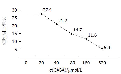
结果: 不同浓度的GABA (20-320 μmol/L)促进胰腺癌SW1990细胞的生长, 促进细胞周期比例的分布, 使G0/G1细胞减少, S期和G2/M细胞增多, 同时促进胰腺癌SW1990细胞内cAMP含量的增加, 且呈剂量依赖性(P<0.01). 显著抑制细胞凋亡, 凋亡率由27.4%降低到5.4%(χ2 = 10.19, P<0.01).
结论: GABA促进胰腺癌SW1990细胞生长, 可能通过受体后信息介导.
引文著录: 王营, 余胜利, 刘军权, 费素娟, 陈剑群, 许统俭, 王人灏, 刘伟. GABA对胰腺癌细胞株SW1990生长的影响. 世界华人消化杂志 2006; 14(23): 2337-2339
Revised: May 15, 2006
Accepted: May 22, 2006
Published online: August 18, 2006
AIM: To investigate the effect of gamma-aminobutyric acid (GABA) on the growth of pancreatic cancer cell line SW1990.
METHODS: Pancreatic cancer cell line SW1990 was cultured by routine method, and then treated with different concentrations of GABA (20-320 μmol/L). The proliferation, apoptosis, and cell cycle of SW1990 cells was investigated by MTT assay and flow cytometry, respectively. Radioimmunoassay was used to measure the intracellular cyclic adenosine monophosphate (cAMP) content.
RESULTS: The different concentrations of GABA promoted the growth of SW1990 cells and affected the distribution of cell cycle. The percentage of cells in G0/G1 phase was decreased while that in S and G2/M phase was increased. The content of intracellular cAMP was increased with the increase of GABA concentration in a dose-dependent manner (P < 0.01). The apoptosis rate of SW1990 cells was decreased from 27.5% to 5.4%, which had significant difference (χ2 = 10.19, P <0.01).
CONCLUSION: GABA can promote the proliferation of SW1990 cells by inhibiting apoptosis and influencing the distribution of cell cycle, which may be mediated by the information transition of post-receptor.
- Citation: Wang Y, Yu SL, Liu JQ, Fei SJ, Chen JQ, Xu TJ, Wang RH, Liu W. Effect of gamma-aminobutyric acid on the growth of pancreatic cancer cell line SW1990. Shijie Huaren Xiaohua Zazhi 2006; 14(23): 2337-2339
- URL: https://www.wjgnet.com/1009-3079/full/v14/i23/2337.htm
- DOI: https://dx.doi.org/10.11569/wcjd.v14.i23.2337
随着神经生物学研究的深入, 发现神经递质不仅参与支配局部组织的运动及感觉生理活动, 还可通过与细胞膜表面的特异性受体结合进行信号传导, 进而对靶细胞分化、生长、代谢、细胞骨架结构、基因表达产生影响. γ-氨基丁酸(gamma-aminobutyric acid, GABA)是体内重要的抑制性神经递质, 不仅存在于中枢神经系统, 也广泛存在于外周组织, 胰腺细胞含有GABA受体, 并能释放高活性的谷氨酸脱羧酶(glutamic acid decarboxylase, GAD)[1-3], 而GAD是GABA合成的关键酶. 近年研究显示, GABA含量和GAD活性在多种肿瘤组织中表达水平明显增加[4-8]. 我们观察体外GABA对胰腺癌细胞株SW1990增殖及凋亡的作用, 探讨神经递质在肿瘤发生、发展过程中的神经信号传导作用.
GABA和异丁基甲基黄嘌呤(IBMX)为Sigma公司产品, 125I-cAMP放免试剂盒为上海中医药大学产品, 噻唑氮蓝(MTT)为瑞士Fluka公司产品, 流式细胞检测仪(美国Becton Dickinson, BDFACSCalibur). 胰腺癌细胞株SW1990由上海交通大学附属第一人民医院王兴鹏教授惠赠. 胰腺癌细胞SW1990置含100 mL/L的小牛血清RMPI 1640培养液中, 37 ℃, 50 mL/L CO2条件培养. 将传代72 h的细胞消化脱壁, 再用含100 mL/L的小牛血清RPMI 1640培养液配成细胞悬液(109/L), 接种于96孔培养板(每孔0.1 mL), 待细胞贴壁生长后, 培养24 h.
实验组加入不同浓度的GABA, 对照组加入等量的培养液, 培养48 h后待测, 3孔重复. 应用甲基偶氮唑盐MTT比色法. 实验组与对照组加入MTT 20 μL, 培养4 h, 弃去上清, 加入二甲基亚砜100 μL, 待其沉淀产物完全溶解, 在酶标仪570 nm波长测定各孔吸光度(A). 另收集细胞, 1500 r/min离心5 min, 700 mL/L冷乙醇固定, 离心后每样品管中加碘化丙啶(PI)染液1 mL, 4 ℃避光染色30 min, 流式细胞仪检测细胞周期及细胞凋亡, 每个样本分析10000个细胞. 再取对数生长期细胞消化后, 在细胞悬液中(含细胞109/L), 加入10 mmol/L IBMX, 将细胞悬液分为实验组和对照组, 37 ℃培养. 取出悬液0.15 mL, 加入70.6 g/L三氯醋酸, 混匀, 4 ℃离心. 取上清洗涤后, 将水移入玻璃瓶内, 75 ℃水浴蒸干, 冰箱贮存待测. 放射免疫分析法检测cAMP.
统计学处理 实验数据以mean±SD表示, 计量资料采用t检验, 计数资料采用χ2检验.
胰腺癌细胞SW1990在给予20-320 μmol/L GABA培养48 h后, 不同浓度的GABA均能明显促进胰腺癌细胞SW1990的生长(表1).
与对照组相比, 不同浓度的GABA对胰腺癌SW1990细胞周期均有作用, 使G0/G1细胞减少, S期和G2/M细胞增多(表2). GABA抑制胰腺癌SW1990细胞凋亡, 随剂量的增加凋亡率下降, 凋亡率分别为27.4%, 21.2%, 14.7%, 11.6%, 5.4%, 组间比较差异有统计学意义(χ2 = 10.19, P<0.01, 图1).
GABA促进胰腺癌SW1990细胞内cAMP生成呈剂量依赖性(表1).
神经递质传统意义上被认为仅仅作为神经系统中细胞间的信号传递. 最新的肿瘤生物学研究结果显示, 神经递质和其受体结合与肿瘤生长、侵袭和转移有关[1,9]. 近年研究显示, GABA含量和GAD活性在结肠癌、乳腺癌、胃癌、甲状腺癌及前列腺癌中表达水平明显高于正常组织和良性病变, GABA含量和GAD活性与恶性肿瘤的发生及其生物学行为密切相关[4-8]. 国内有人发现, 胰腺癌GAD65表达明显增高, 且低分化腺癌表达高于高分化腺癌[10].
我们发现, 不同浓度的GABA均能促进SW1990增殖, 抑制细胞凋亡, 促进cAMP生成, 且呈剂量依赖性. GABA对SW1990细胞周期也有一定影响, 不同浓度GABA处理SW1990细胞48 h后, G1期细胞比例明显下降, S, G2期和M期细胞比例增加. 结果提示, GABA加快细胞周期进程. 最新研究发现, GABAA受体π亚单位在胰腺癌组织中过度表达[11]. GABA促进SW1990生长, 抑制细胞凋亡, 可能通过受体后信息传导途径, 使细胞内cAMP增加. GABA作为神经递质可能在肿瘤的生长过程中具有重要的作用.
随着神经生物学研究的深入, 作为神经系统中细胞间的信号传递物质--神经递质, 其神经系统以外的作用逐渐被认识. 神经递质与某些肿瘤生长、侵袭和转移有关. γ-氨基丁酸(gamma-aminobutyric acid, GABA)为氨基酸类神经递质之一, 有关GABA在肿瘤方面的研究国内仅有少数, 国外相对较多. 近年研究显示, GABA含量和GAD活性在多种进展期肿瘤组织中表达水平明显增加.
本文研究了体外GABA对胰腺癌细胞株SW1990具有促进生长的作用, 其作用机制有待进一步研究, 如其受体的表达, 与癌基因、抑癌基因的关系等等, 有助于揭示神经递质在肿瘤发生、发展过程中的作用, 将为肿瘤的诊断及治疗提供新的途径.
电编: 张敏 编辑:潘伯荣
| 1. | Sha L, Miller SM, Szurszewski JH. Electrophysio-logical effects of GABA on cat pancreatic neurons. Am J Physiol Gastrointest Liver Physiol. 2001;280:G324-331. [PubMed] |
| 2. | Watanabe M, Maemura K, Kanbara K, Tamayama T, Hayasaki H. GABA and GABA receptors in the central nervous system and other organs. Int Rev Cytol. 2002;213:1-47. [PubMed] |
| 4. | Kleinrok Z, Matuszek M, Jesipowicz J, Matuszek B, Opolski A, Radzikowski C. GABA content and GAD activity in colon tumors taken from patients with colon cancer or from xenografted human colon cancer cells growing as s.c. tumors in athymic nu/nu mice. J Physiol Pharmacol. 1998;49:303-310. [PubMed] |
| 5. | Mazurkiewicz M, Opolski A, Wietrzyk J, Radzikowski C, Kleinrok Z. GABA level and GAD activity in human and mouse normal and neoplastic mammary gland. J Exp Clin Cancer Res. 1999;18:247-253. [PubMed] |
| 6. | Matuszek M, Jesipowicz M, Kleinrok Z. GABA content and GAD activity in gastric cancer. Med Sci Monit. 2001;7:377-381. [PubMed] |
| 7. | Myshunina TM, Bogdanova TI, Tron'ko ND. On the possible role of gamma-aminobutyric acid in thyroid carcinogensis. Experimental Oncology. 2003;25:25-27. |
| 8. | Azuma H, Inamoto T, Sakamoto T, Kiyama S, Ubai T, Shinohara Y, Maemura K, Tsuji M, Segawa N, Masuda H. Gamma-aminobutyric acid as a promoting factor of cancer metastasis; induction of matrix metalloproteinase production is potentially its underlying mechanism. Cancer Res. 2003;63:8090-8096. [PubMed] |
| 9. | Entschladen F, Lang K, Drell TL, Joseph J, Zaenker KS. Neurotransmitters are regulators for the migration of tumor cells and leukocytes. Cancer Immunol Immunother. 2002;51:467-482. [PubMed] |
| 10. | Yang ZL, Wang QW, Deng XH, Li DQ, Lv F, Li YG. Enzymatic activities of ChAT, GAD65 and PKC in pancreatic carcinoma tissues. Shijie Huaren Xiaohua Zazhi. 2003;11:1554-1557. |


