修回日期: 2005-04-05
接受日期: 2005-04-09
在线出版日期: 2005-06-15
目的: 研究肝移植排斥反应中C4d沉积的部位和意义.
方法: 对肝移植患者的肝穿刺标本作C4d免疫组化染色, 观察C4d阳性沉积情况与肝脏病理改变的关系.
结果: 急性细胞性排斥反应中的69.2%肝移植标本, 在肝小叶汇管区小血管壁及肝血窦壁上有C4d的沉积.33.3%移植后乙肝复发和28.6%乙型肝炎的标本中, 仅汇管区小血管壁有C4d的沉积, 而无肝血窦壁上C4d的沉积.12例器官保存性损伤的肝标本中1例汇管区小血管壁及肝血窦壁上有C4d的沉积, 此例1 mo后再次穿刺发现急性细胞性排异反应.肝移植后胆管阻塞的标本中没有发现C4d的沉积.
结论: C4d在肝血窦壁的沉积可以作为肝移植急性排斥反应鉴别诊断一个比较特异的免疫组化指标.
引文著录: 卜宪敏, 郑智勇, 余英豪, 曾玲, 江艺. 肝移植排斥反应中C4d的表达意义. 世界华人消化杂志 2005; 13(11): 1314-1316
Revised: April 5, 2005
Accepted: April 9, 2005
Published online: June 15, 2005
AIM: To investigate the location and significance of C4d deposition in liver rejection.
METHODS: Immunohistological staining was performed to observe C4d deposition in the liver allografts biopsies as well as its relationship with the pathological alteration of liver.
RESULTS: C4d expression was 69.2% in the samples of liver cellular rejection, 33.3% in hepatitis B relapse after transplantation and 28.6% in hepatitis B. In the biopsies of liver rejection, C4d was found depositing on the vascular walls of portal areas and hepatic sinusoidal walls. In the biopsies of hepatitis B relapse after transplantation and hepatitis B, C4d deposited only in the vascular walls of portal area. In the biopsies of ischemia reperfusion damage, C4d deposition was observed on the vascular walls of portal areas and hepatic sinusoidal wall in 1 of 12 patients, and follow-up biopsy after 1 month revealed an acute cellular rejection. No C4d deposition was observed in bile duct occlusion after liver transplantation.
CONCLUSION: C4d deposition may be served as a sensitive marker in the diagnosis of liver rejection.
- Citation: Bu XM, Zheng ZY, Yu YH, Zeng L, Jiang Y. Signification of C4d deposition in liver rejection. Shijie Huaren Xiaohua Zazhi 2005; 13(11): 1314-1316
- URL: https://www.wjgnet.com/1009-3079/full/v13/i11/1314.htm
- DOI: https://dx.doi.org/10.11569/wcjd.v13.i11.1314
目前研究表明, 肝移植后排斥反应中体液排斥对细胞排斥起协同作用[1].肾移植病理研究中发现, C4d在移植肾间质毛细血管壁上弥漫沉积可以作为排异反应的一个特异性指标[2], 心脏移植病理中也有同样现象[3].肝移植排异反应中是否也有类似现象, 至今尚未见文献报道.我们对肝移植穿活检标本中C4d表达的情况进行研究.
我院2001-10/2004-11肝移植穿刺标本20例39次, 其中重复(1-4次)穿刺10例.男17例, 女3例, 年龄20-56(平均38)岁.受者主要原发病为: 乙肝肝硬化6例, 乙型慢性重型肝炎2例, Wilson病2例, 原发性肝癌9例, 原发性胆汁性肝硬化1例.供肝获取采用快速供肝切取法, UW液保存, 热缺血2-8 min, 冷缺血3.5-18 h.采用经典原位肝TX移植13例, 肝肾联合移植6例.术后免疫抑制剂采用FK506+激素+MMF三联方案或FK506+激素两联方案.
标本经40 g/L中性甲醛固定, 石蜡包埋, 连续切片2 μm厚, 分别作HE染色以及免疫组化抗HBsAg、HbcAg和C4d染色、部分选作CMV、EBV抗体染色.光镜下观察肝病理改变, 排异反应诊断及评分遵照Banff标准[4].另取乙型肝炎穿刺标本14例和移植肾急性细胞性排异反应穿刺标本10例作为免疫组化染色对照.兔抗人C4d采用Biomedica公司产品[5], 免疫组化染色采用Maixin.Bio公司EliVision TM plus广谱试剂盒.主要步骤如下: 石蜡切片脱蜡入水, 高压高温抗原修复, 浸入30 mL/L过氧化氢10 min, 1 g/L胰酶消化1 min, 滴加1∶40兔抗人C4d, 室温孵育2 h, 其余步骤按试剂盒说明书进行.各步骤间用0.01 mol/L pH7.2 PBS洗涤.
统计学处理 应用SPSS 12.0软件进行统计学分析, 采用χ2检验.
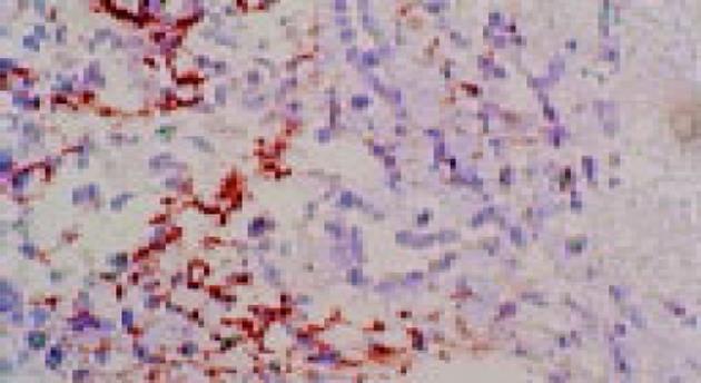
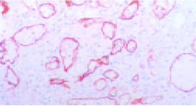
在移植肝急性细胞性排异反应中C4d阳性率(69.2%)较高, 阳性反应部位主要在肝血窦壁和汇管区小血管壁(图1).器官保存性损伤(8.3%)、移植后乙肝复发(33.3%)和乙型肝炎(28.6%)3组标本C4d阳性率均较低, P<0.05, 差异有显著意义.移植后乙肝复发和乙型肝炎C4d阳性部位主要为汇管区(图2), 而肝血窦壁没有C4d沉积; 器官保存性损伤只有1例C4d阳性, 阳性部位在肝血窦壁和汇管区小血管壁.此例1 mo后再次穿刺发现急性细胞性排异反应.对照组移植肾急性细胞性排异反应10例都有强阳性C4d沉积(表1), 沉积部位主要在肾小管周围的毛细血管壁和肾小球系膜区(图3).
| 病理类型 | n | 阳性 | 不同区域阳性 | ||
| n | % | 汇管区 | 血窦壁 | ||
| 急性细胞排异 | 13 | 9 | 69.2 | 9 | 8 |
| 慢性排异 | 1 | 0 | 0 | 0 | 0 |
| 器官保存性损伤 | 12 | 1 | 8.3 | 1 | 1 |
| 移植后乙肝复发 | 3 | 1 | 33.3 | 1 | 0 |
| 乙型肝炎 | 14 | 4 | 28.6 | 4 | 0 |
| 胆管阻塞 | 3 | 0 | 0 | 0 | 0 |
| 肾急性细胞排异 | 10 | 10 | 100 | ||
在抗体介导的体液性排异中, 补体起着协助和加强免疫的调节作用.体液性排斥反应发生时, 补体的经典途径即被激活, 被激活的CI蛋白水解为C4d, C4d与周围血管的内皮细胞共价结合, 所以能够比较持久存在, 从而可作为排异时存在体液成分激活的标志[6].移植肾肾小管周围的毛细血管壁C4d沉积是排异反应的一项较特异免疫组化指标[2,6,7-10].本组观察表明, 肝移植急性细胞性排斥反应时同样也会出现类似的现象, 即C4d在肝血窦壁上的沉积可以用来判别是否出现肝移植急性细胞排异反应.与移植肾急性排异反应相比, 肝移植急性排斥反应时肝血窦壁C4d沉积的阳性反应较弱, 但明确存在.肝移植急性细胞性排斥反应时C4d的阳性部位与乙型肝炎不同, 移植后乙肝复发和乙型肝炎病例C4d阳性部位主要在汇管区, 而肝血窦壁未见到C4d沉积.本组12例器官保存性损伤标本中只有1例同时存在汇管区和肝血窦壁C4d沉积, 而此例1 mo后再次穿刺发现急性细胞性排异反应.因此, C4d在肝血窦壁的沉积可能是肝移植急性排斥反应的一个比较特异的免疫组化指标.
从免疫学观点看, C4d沉积代表着经典途经的补体激活, 意味着体液免疫反应出现[6-7].以往的研究认为, 机体对移植物的急性排异反应, 主要是细胞排异反应[8].但从对移植肾急性排异反应的C4d沉积[9]和本观察结果表明, 急性细胞性排异反应过程中可以伴有体液排异反应出现.本研究也证实, 肝移植急性排异反应时也存在类似的情况; 在作为对照的肝炎病例中也有C4d的沉积, 提示乙肝的炎症也与体液免疫有关.在乙肝病例中, C4d的沉积主要在汇管区较多细胞浸润的区域, 在肝血窦中没有C4d的沉积.这种沉积并不是在所有的乙肝病例中出现, 其原因可能与肝炎的活动性有关.
编辑: 潘伯荣 审读: 张海宁
| 1. | Bohmig GA, Regele H, Exner M. C4d-positive acute humoral renal allograft rejection: effective treatment by immuno-adsorption. J Am Soc Nephrol. 2001;12:2482-2489. [PubMed] |
| 2. | 刘 志红, 陈 书芬, 陈 朝红, 曾 彩虹, 周 虹, 陈 劲松, 唐 政, 黎 磊石. 肾移植急性排斥患者肾组织C4d的沉积及其意义. 肾脏病与透析肾移植杂志. 2003;12:415-418. |
| 3. | Duong Van Huyen JP, Fornes P, Guillemain R, Amrein C, Chevalier P, Latremouille C, Creput C, Glotz D, Nochy D, Bruneval P. Acute vascular humoral rejection in a sensitized cardiac graft recipient: diagnostic value of C4d immunof-luorescence. Hum Pathol. 2004;35:385-388. [PubMed] [DOI] |
| 4. | Banff schema for grading liver allograft rejection: an international consensus document. Hepatology. 1997;25:658-663. [PubMed] [DOI] |
| 5. | Krukemeyer MG, Moeller J, Morawietz L, Rudolph B, Neumann U, Theruvath T, Neuhaus P, Krenn V. Description of B lymphocytes and plasma cells, complement, and chemokines/receptors in acute liver allograft rejection. Transplantation. 2004;78:65-70. [PubMed] [DOI] |
| 6. | Regele H, Bohmig GA, Habicht A, Gollowitzer D, Schillinger M, Rockenschaub S, Watschinger B, Kerjaschki D, Exner M. Capillary deposition of complement split product C4d in renal allografts is associated with basement membrane injury in peritubular and glomerular capillaries: a contribution of humoral immunity to chronic allograft rejection. J Am Soc Nephrol. 2002;13:2371-2380. [PubMed] [DOI] |
| 8. | Mauiyyedi S, Pelle PD, Saidman S, Collins AB, Pascual M, Tolkoff-Rubin NE, Williams WW, Cosimi AA, Schneeberger EE, Colvin RB. Chronic humoral rejection: identification of antibody-mediated chronic renal allograft rejection by C4d deposits in peritubular capillaries. J Am Soc Nephrol. 2001;12:574-582. [PubMed] |
| 9. | Herzenberg AM, Gill JS, Djurdjev O, Magil AB. C4d deposition in acute rejection: an independent long-term prognostic factor. J Am Soc Nephrol. 2002;13:234-241. [PubMed] |
| 10. | Collins AB, Schneeberger EE, Pascual MA, Saidman SL, Williams WW, Tolkoff-Rubin N, Cosimi AB, Colvin RB. Complement activation in acute humoral renal allograft rejection: diagnostic significance of C4d deposits in peritubular capillaries. J Am Soc Nephrol. 1999;10:2208-2214. [PubMed] |


