修回日期: 2004-03-25
接受日期: 2004-04-13
在线出版日期: 2004-07-15
目的: 探讨肝癌组织中脱--羧基凝血酶原的表达情况及临床意义.
方法: 用免疫组化法探讨了92例肝癌及癌旁组织, 7例转移性肝癌和19例慢性肝病组织中脱--羧基凝血酶原的表达水平, 并分析脱--羧基凝血酶原与肝癌的临床病理特征间的关系.
结果: 肝癌组织脱--羧基凝血酶原阳性率(73.9%)明显高于癌旁组织(26.1%, P<0.01), 但二者脱--羧基凝血酶原阳性率明显高于转移性肝癌和慢性肝病肝组织(3.5%, P<0.01). 浸润性生长的肝癌组织脱--羧基凝血酶原阳性率明显高于膨胀性生长的肝癌组织(P = 0.049), 无包膜形成的肝癌组织明显高于有包膜形成的肝癌组织(P = 0.037). 最大径>5 cm组的癌旁组织的脱--羧基凝血酶原阳性率明显高于直径≤5 cm组癌旁组织(P = 0.049), HBsAg和HCVAb均阴性或单HBsAg阳性的患者癌旁组织脱--羧基凝血酶原染色阳性率明显高于HCVAb阳性组患者(P<0.01). 肝硬化的癌旁组织脱--羧基凝血酶原阳性率明显低于慢性肝炎的癌旁组织(P<0.01). 肝癌组织、癌旁组织脱--羧基凝血酶原阳性率与肝癌其他临床病理特征无明显的关系(P>0.05).
结论: 脱--羧基凝血酶原可能是预测肝癌发生的重要标志物, 但不能作为肝癌的预后指标.
引文著录: 袁联文, 唐伟, 李永国, 幕内雅敏. 肝癌中脱--羧基凝血酶原的表达及意义. 世界华人消化杂志 2004; 12(7): 1543-1545
Revised: March 25, 2004
Accepted: April 13, 2004
Published online: July 15, 2004
AIM: To study the expression of des-gamma-carboxy-prothrombin (DCP) in hepatocellular carcinoma (HCC) tissues and its clinical significance.
METHODS: Cancerous and non-cancerous tissue samples prepared from 92 cases of HCCs, 7 metastatic HCCs and 19 chronic liver diseases were subjected to immunohistochemical staining for tissue DCP. Relation between DCP expression in cancerous and non-cancerous tissues and clinical parameters of HCC was analyzed.
RESULTS: The DCP expression in cancerous tissues (73.9%) was significantly higher than that in non-cancerous tissues (26.1%, P < 0.01). The DCP expression in cancerous and non-cancerous tissues of HCC was significantly higher than that in non-cancerous tissues of metastatic HCCs and chronic liver diseases (3.5%, P < 0.01). Positive DCP staining in cancerous tissues was more frequently in cases of infiltrative growth than in cases of expansive growth (P = 0.049), and was more frequently in cases where no capsule formation was noted than that in cases with capsule formation (P = 0.037). The DCP expression in non-cancerous tissues of HCC with size >5 cm was significantly higher than that of the size ≤ 5 cm (P = 0.049). Positive DCP staining in non-cancerous tissue was more frequently in cases of tumors larger than 5 cm than in cases of tumors that were 5 cm or smaller (P = 0.049). DCP expression in non-cancerous tissues of patients who were either negative for both hepatitis markers or positive for the HBsAg was significantly higher than the patients who were positive for the HCVAb (P < 0.01). The rate of positive DCP staining in non-cancerous tissues was also significantly lower in patients with liver cirrhosis than that in patients with chronic hepatitis (P < 0.05). No correlations were found between DCP expression in cancerous and non-cancerous tissues and other clinical parameters.
CONCLUSION: DCP may be an important marker in liver carcinogenesis, but DCP in tissues cannot be considered as a prognostic factor for HCC.
- Citation: Yuan LW, Tang W, Li YG, Makuuchi M. Des-gamma-carboxy-prothrombin expression in hepatocellular carcinoma and its clinical significance. Shijie Huaren Xiaohua Zazhi 2004; 12(7): 1543-1545
- URL: https://www.wjgnet.com/1009-3079/full/v12/i7/1543.htm
- DOI: https://dx.doi.org/10.11569/wcjd.v12.i7.1543
较多研究表明脱--羧基凝血酶原(Des-gamma-carboxy-prothrombin, DCP)是肝癌一种有用血清肿瘤标志物[1-11], 而有关肝癌组织中DCP表达和临床意义的研究甚少. 我们用免疫组化法探讨了92例肝癌组织中DCP的表达情况及意义.
原发性肝癌手术切除标本92例. 男68例, 女24例, 年龄16- 83(平均67.2±52.6岁); 伴肝硬化57例(62.0%); 有病毒性肝炎感染的血清学证据78例 (84.8%), 其中HBsAg阳性18例, HCV-Ab阳性58例, HBsAg和HCV-Ab均阳性2例, 其余14例均阴性. 肿瘤直径从1.0-19.0 (平均5.1±3.8 cm); 高分化癌20例, 中分化癌62例, 低分化癌10例. 伴血管浸润27例(29.3%), 存在肝内转移灶24例(26.1%). 另转移性肝癌7例, 原发部位为结肠; 慢性肝病患者肝穿刺活检标本19例, 其中肝硬化5例, 慢性肝炎14例. 这些组织经40 g/L甲醛固定, 石蜡包埋后, 制成4 m厚的切片. DCP单克隆抗体MU-3(东京Eisai 公司惠赠); Histofine SAB-PO (MULTI)试剂盒(100 mL/L牛血清; 生物素标识的抗鼠IgG的二抗; ABC试剂及DAB-HCl显色剂)(购自东京Nichirei 公司).
常规ABC免疫组化法, 免疫组化阳性物质定位于胞质. 用0.05 mol/LPBS液替代一抗作为染色阴性或替代对照. 随机选择10个高倍视野中DCP阳性细胞率, 以阳性细胞率≥15%为DCP阳性病例, <15%为阴性病例.
统计学处理 两组间的比较用为x2检验, 当P<0.05时为有显著性.
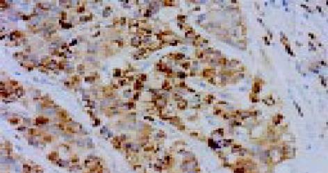
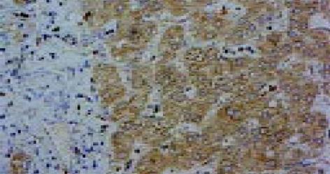
DCP染色阳性的细胞在其细胞质中出现大量棕褐色的颗粒. DCP染色阳性即可见于肝癌组织(图1)及癌旁组织(图2). 癌组织和癌旁组织DCP染色阳性率分别为73.9%(68/92)和26.1%(24/92). 7例转移性肝癌患者的非癌组织和19慢性肝病肝穿刺活检的肝组织中1例(1/26, 3.5%)DCP染色阳性. 统计分析显示肝癌组织阳性率明显高于癌旁组织(P<0.01), 二者的DCP阳性率又明显高于转移性肝癌和慢性肝病组(P<0.01).
肝癌组织DCP的表达与肿瘤直径无明显关系, 而直径>5 cm的肝癌癌旁组织的DCP表达明显高于直径≤5 cm组(P = 0.049, 表1). 浸润性生长的肝癌组织DCP染色阳性率明显高于膨胀性生长的肝癌组织(90.5% vs 69.0%, P = 0.049), 无包膜形成的肝癌组织明显高于有包膜形成的肝癌组织(90.9% vs 68.6%, P = 0.037). 肝癌组织DCP染色阳性率与肿瘤分化、血管浸润、肝内转移、TNM分期、肿瘤的直径、肿瘤复发和患者生存期无明显的关系. 也没发现肝癌组织DCP染色阳性率与肝炎病毒标志物、不同背景肝有明显关系. HBsAg和HCVAb均阴性或单HBsAg阳性的癌旁组织DCP染色阳性率明显高于HCVAb阳性组(二者阴性组 vs HCVAb阳性组: P = 0.014; HBsAg阳性组 vs HCVAb阳性组: P<0.01). 背景肝为肝硬化的癌旁组织DCP染色阳性率(11/57, 19.3%)明显低于背景肝存在慢性肝炎的癌旁组织(13/33, 39.4%; P<0.01). 但未发现癌旁组织DCP染色阳性率与其他临床病理因子有明显的关系.
| 因子 | n | 癌部组织 | 癌旁组织 | ||
| 阴性 | 阳性 | 阴性 | 阳性 | ||
| 直径≤5 cm | 61 | 14 (23.0) | 47 (77.0) | 49 (80.3) | 12 (19.7) |
| 直径> 5 cm | 31 | 10 (32.3) | 21 (67.7) | 19 (61.3) | 12 (38.7)a |
| HBV+HCV阴性 | 14 | 2 (14.3) | 12 (85.7) | 8 (57.1) | 6 (42.9)c |
| HBV 阳性 | 18 | 4 (22.2) | 14 (77.8) | 8 (44.4) | 10 (55.6)e |
| HCV阳性 | 58 | 18 (31.0) | 40 (69.0) | 50 (86.2) | 8 (13.8) |
| HBV+HCV阳性 | 2 | 0 | 2 (100) | 2 (100) | 0 |
| 肝硬化 | 57 | 14 (24.6) | 43 (75.4) | 46 (80.7) | 11 (19.3) |
| 慢性肝病 | 33 | 9 (27.3) | 24 (72.7) | 20 (60.6) | 13 (39.4)a |
| 正常 | 2 | 1 (50.0) | 1 (50.0) | 2 (100) | 0 |
| 膨胀性生长 | 71 | 22 (31.0) | 49 (69.0) | 54 (76.1) | 17 (23.9) |
| 浸润性生长 | 21 | 2 (9.5) | 19 (90.5)a | 14 (66.7) | 7 (33.3) |
| 有包膜浸润 | 70 | 22 (31.4) | 48 (68.6) | 51 (72.9) | 19 (27.1) |
| 无包膜浸润 | 22 | 2 (9.1) | 20 (90.9)a | 17 (77.3) | 5 (22.7) |
目前肝癌是世界最常见的恶性肿瘤之一, 其恶性程度高, 预后差[12-14]. 早期诊断、早期治疗仍是提高肝癌生存率最重要的手段. 目前肝癌的诊断主要依靠影像学的发现和血清肿瘤标志物的检测. 甲胎蛋白(AFP)作为肝癌代表性肿瘤标志物用于临床, 近年来血清DCP水平也被广泛应用于临床肝癌的诊断, 但关于肝癌组织DCP表达的研究不多. 慢性肝病患者肝结节性病灶可以从良性结节到肝癌, 其间按恶性程度存在有再生结节、腺瘤样增生、不典型性腺瘤样增生、高分化的早期肝癌、高分化肝癌、中分化肝癌至低分化肝癌多种组织学形态. 分化好的早期肝癌和不典型性腺瘤样增生间的组织学诊断标准至今仍有争议, 因此有必要寻找一种评价肝占位性病灶的组织标志物. Miskad et al研究了62例肝癌和9例腺瘤样增生的肝组织中DCP的表达的情况, 发现71%的肝癌组织DCP染色阳性, 而无1例腺瘤样增生组织表达DCP. 本研究也显示73.9%(68/92)肝癌癌组织DCP染色阳性, 而7例转移性肝癌患者的癌旁组织和19例肝穿刺活检的慢性肝病组织中仅1例DCP染色阳性. 因此以上结果提示DCP是肝癌一种有用的组织病理标志物.
我们发现肝癌癌组织染色阳性指数明显高于癌旁组织, 从组织学上证实肝癌癌组织可产生大量DCP, 是血清DCP的主要来源之一. 肝癌癌组织染色除了与肿瘤的生长类型和包膜形成与否存在明显的相关性外, 与其他的病理因子如血管浸润、肝内转移等无关. 我们也没发现肝癌组织染色与生存期之间有明显相关性, 上述证据提示肝癌组织DCP染色尚不能评价肝癌的进展, 不能作为肝癌的预后指标.
2001年Fujioka et al发现12%的小肝癌癌旁组织DCP染色阳性. 我们也发现26.1%(24/92)癌旁组织的DCP染色阳性, 而且一些癌旁组织的DCP染色阳性指数甚至高于癌组织, 这提示部分肝癌癌旁组织也可产生大量DCP, 这仍须组织中DCP定量测定研究加以验证. 统计分析显示癌旁组织DCP的染色高低与肿瘤的大小有明显关系, 癌旁组织的DCP染色阳性指数又明显高于转移性肝癌的癌旁组织和肝穿刺活检的组织, 说明肝癌肿瘤的存在影响着其周围非癌组织DCP的产生. 癌旁组织产生DCP增加的机制可能系肝癌癌细胞在快速增生过程中维生素K消耗增加造成癌旁局部维生素K缺乏或癌细胞分泌某种物质影响周围肝细胞产生DCP, 但具体的机制有待进一步研究.
编辑: N/A
| 1. | Cui R, Wang B, Ding H, Shen H, Li Y, Chen X. Usefulness of determining a protein induced by vitamin K absence in detection of hepatocellular carcinoma. Chin Med J (Engl). 2002;115:42-45. [PubMed] |
| 2. | Gotoh M, Nakatani T, Masuda T, Mizuguchi Y, Sakamoto M, Tsuchiya R, Kato H, Furuta K. Prediction of invasive activities in hepatocellular carcinomas with special reference to alpha-fetoprotein and des-gamma-carboxyprothrombin. Jpn J Clin Oncol. 2003;33:522-526. [PubMed] [DOI] |
| 3. | Ando E, Tanaka M, Yamashita F, Kuromatsu R, Takada A, Fukumori K, Yano Y, Sumie S, Okuda K, Kumashiro R. Diagnostic clues for recurrent hepatocellular carcinoma: comparison of tumour markers and imaging studies. Eur J Gastroenterol Hepatol. 2003;15:641-648. [PubMed] [DOI] |
| 4. | Nagaoka S, Yatsuhashi H, Hamada H, Yano K, Matsumoto T, Daikoku M, Arisawa K, Ishibashi H, Koga M, Sata M. The des-gamma-carboxy prothrombin index is a new prognostic indicator for hepatocellular carcinoma. Cancer. 2003;98:2671-2677. [PubMed] [DOI] |
| 5. | Ishii M, Gama H, Chida N, Ueno Y, Shinzawa H, Takagi T, Toyota T, Takahashi T, Kasukawa R. Simultaneous measurements of serum alpha-fetoprotein and protein induced by vitamin K absence for detecting hepatocellular carcinoma. South Tohoku District Study Group. Am J Gastroenterol. 2000;95:1036-1040. [PubMed] |
| 6. | Marrero JA, Su GL, Wei W, Emick D, Conjeevaram HS, Fontana RJ, Lok AS. Des-gamma carboxyprothrombin can differentiate hepatocellular carcinoma from nonmalignant chronic liver disease in american patients. Hepatology. 2003;37:1114-1121. [PubMed] [DOI] |
| 7. | Okuda H, Nakanishi T, Takatsu K, Saito A, Hayashi N, Yamamoto M, Takasaki K, Nakano M. Comparison of clinicopathological features of patients with hepatocellular carcinoma seropositive for alpha-fetoprotein alone and those seropositive for des-gamma-carboxy prothrombin alone. J Gastroenterol Hepatol. 2001;16:1290-1296. [PubMed] [DOI] |
| 8. | Omata M, Yoshida H. Resolution of liver cirrhosis and prevention of hepatocellular carcinoma by interferon therapy against chronic hepatitis C. Scand J Gastroenterol Suppl. 2003;47-51. [PubMed] [DOI] |
| 9. | Shimizu A, Shiraki K, Ito T, Sugimoto K, Sakai T, Ohmori S, Murata K, Takase K, Tameda Y, Nakano T. Sequential fluctuation pattern of serum des-gamma-carboxy prothrombin levels detected by high-sensitive electrochemiluminescence system as an early predictive marker for hepatocellular carcinoma in patients with cirrhosis. Int J Mol Med. 2002;9:245-250. [PubMed] |
| 10. | Nakagawa T, Seki T, Shiro T, Wakabayashi M, Imamura M, Itoh T, Tamai T, Nishimura A, Yamashiki N, Matsuzaki K. Clinicopathologic significance of protein induced vitamin K absence or antagonist II and alpha-fetoprotein in hepatocellular carcinoma. Int J Oncol. 1999;14:281-286. [PubMed] [DOI] |
| 11. | Okuda H, Nakanishi T, Takatsu K, Saito A, Hayashi N, Takasaki K, Takenami K, Yamamoto M, Nakano M. Serum levels of des-gamma-carboxy prothrombin measured using the revised enzyme immunoassay kit with increased sensitivity in relation to clinicopathologic features of solitary hepatocellular carcinoma. Cancer. 2000;88:544-549. [PubMed] [DOI] |


