修回日期: 2004-08-20
接受日期: 2004-08-25
在线出版日期: 2004-12-15
目的: 从基因蛋白表达层次探索胃癌中医证本质.
方法: 从第二军医大学长征医院普外科收集术前胃癌患者, 并按中医辨证分型标准将其归属; 取术后肿瘤标本, 用免疫组化方法检测胃癌肿瘤中E-Cadherin、C-erbB-2、P53、nm23、ICAM-1、VEGF、KDR、MMP-2、TIMP-29种基因蛋白表达情况.
结果: 每个证型中的9个基因蛋白之间均有不同表达, 统计学分析表明6证型的9个基因蛋白表达均有显著性差异P = 0.0 001, 进一步两两比较示同一证型内部多组基因都存在表达差异性; VEGF、E-cad、nm23表达率较高, 分别为94%、90%、92%. 痰湿凝结型中E-cad的蛋白表达最高, 以平均秩和表示为: 63.09, 肝胃不和、瘀毒内阻两型E-cad表达较低.
结论: 肝胃不和、瘀毒内阻两型胃癌发生转移可能主要与E-cad的缺失有关, 但内部既有癌基因的高表达亦有抑癌基因高表达.
引文著录: 许玲, 孙大志, 何金, 刘龙, 魏品康. 胃癌同证型中肿瘤转移相关基因蛋白的表达. 世界华人消化杂志 2004; 12(12): 2782-2787
Revised: August 20, 2004
Accepted: August 25, 2004
Published online: December 15, 2004
AIM: To explore the entity of gastric carcinoma under the same syndromic classification based on traditional Chinese medicine from the angle of gene protein expression.
METHODS: Patients with gastric carcinoma were collected and then classified before operation according to dialectic criterion of traditional Chinese medicine. Samples of gastric carcinoma from all the patient were collected after operation. EnVision two step immuhistochemical method was used to observe the expession of E-cadherin, C-erbB-2, P53, nm23, ICAM-1, VEGF, KDR, MMP-2, TIMP-2 in the cancerous samples.
RESULTS: The preceding 9 gene proteins were differently expressed in each type. There was significant difference (P = 0.0 001 < 0.01) in the expression of the 9 gene proteins between different syndromic types. Several genes had different expression in the same syndromic type. The rates of VEGF, E-cad, nm23 expression were higher (94%, 90% and 92% respectively) than other proteins. E-cad expression in the stagnation of phlegm-damp type is the highest expression (average rank = 63.09). E-cad expression was lower in the types of the stasis and endogenous toxics and the discordance of liver-stomach.
CONCLUSION: Metastasis of gastric carcinoma in the types of the stasis and endogenous toxics and the discordance of liver-stomach may relate to the loss of E-cadherin expression. However, both cancer gene and anti-oncogene are expressed at a high level in the two types.
- Citation: Xu L, Sun DZ, He J, Liu L, Wei PK. Expression of tumor metastasis-related gene protein in the same syndromic classification of gastric carcinoma on traditional Chinese medicine. Shijie Huaren Xiaohua Zazhi 2004; 12(12): 2782-2787
- URL: https://www.wjgnet.com/1009-3079/full/v12/i12/2782.htm
- DOI: https://dx.doi.org/10.11569/wcjd.v12.i12.2782
胃癌是常见的恶性肿瘤, 当前的诊治状况是一高三低, 即发病率高、早诊断率低, 手术切除率低、5年生存率低. 由于患者临床确诊时大多已为进展期胃癌, 故术后内科综合治疗对提高生存率, 减少转移复发, 改善生活质量显得尤为重要. 但许多西药或治疗方案在临床应用时也遇到不敏感人群, 或毒副作用过强患者; 因此包括国家相关医疗机构等开始呼吁, 希望重视"个体化医疗"这个问题[1]. 这正符合中医"辨证论治"原则. 然而目前中医对疾病的认识多是宏观的概括, 尚不能深入探求其本质内涵, 这从某种程度上限制了中医药的发展. 此研究项目即从肿瘤相关基因E-cadherin, C-erbB-2, P53, nm23, ICAM-1, VEGF, KDR, MMP-2, TIMP-2的蛋白表达上探求中医证本质内涵.
胃癌100例来源于第二军医大学长征医院普外科. 我院病理科联系, 收集胃癌标本, 蜡块备用. 辨证标准按照1978年全国第一届胃癌学术会议北京市胃癌协作组制定的六型分法辨证. (1)肝胃不和型: 主症为胃脘胀满, 时时隐痛, 窜及两胁, 呃逆呕吐, 脉沉或弦细, 舌质淡红, 苔薄白或薄黄; (2)脾胃虚寒型: 主症为胃脘隐痛, 喜按喜温, 或朝食暮吐, 暮食朝吐, 面色苍白, 肢冷神疲, 便溏浮肿, 舌淡而胖, 苔白滑润, 脉沉缓; (3)瘀毒内阻型: 主症为胃脘刺痛, 心下痞硬, 吐血便黑, 皮肤甲错, 舌质暗紫, 脉沉细涩; (4)胃热伤阴型: 主症为胃内灼热, 口干欲饮, 胃脘嘈杂, 食后脘痛, 五心烦热, 大便干燥, 食欲不振, 脉弦细数, 舌红少苔, 或苔黄少津; (5)痰湿凝结型: 主症为胸闷膈满, 面黄虚肿, 呕吐痰涎, 腹胀便溏, 痰核累累, 舌淡红, 苔滑腻; (6)气血双亏型: 主症为全身乏力, 心悸气短, 头晕目眩, 面色无华, 虚烦不寐, 自汗盗汗, 甚至阴阳两虚, 脉沉细无力, 舌淡苔薄. 本组胃热伤阴、瘀毒内阻各16例, 痰湿凝结、气血双亏、肝胃不和、脾胃虚寒各17例. 男69例, 女31例; 年龄<40岁7例, >40岁93例. 胃热伤阴型中有1例不能明确是否有远处转移, 临床分期亦无法确定(表1). E-cad: 小鼠抗人E-cad蛋白一抗, C-erbB-2: 兔抗人C-erbB-2蛋白一抗, MMP-2: 鼠抗人MMP-2蛋白一抗, TIMP-2: 鼠抗人TIMP-2蛋白一抗, KDR: 兔抗人蛋白一抗和p53: 兔抗人p53蛋白一抗购自福州迈新生物技术开发公司; nm23: 鼠抗人nm23蛋白一抗, 购自上海长岛生物技术有限公司; 6.0 mL, 编号: MAB-0244, 购自福州迈新生物技术开发公司; 6.0 mL, 编号: MAB-0244, 购自福州迈新生物技术开发公司; ICAM-1: 鼠抗人ICAM-1蛋白一抗, 购自上海长岛生物技术有限公司; VEGF: 鼠抗人VEGF蛋白一抗, 购自上海长岛生物技术有限公司. 免疫组化EnVision试剂盒购自DAKO公司.
| 分析因素 | 胃癌辨证分型 | 合计 | ||||||
| 肝胃不和 | 脾胃虚寒 | 瘀毒内阻 | 胃热伤阴 | 痰湿凝结 | 气血双亏 | |||
| 年龄 | <40 | 1 | 1 | 2 | 0 | 3 | 0 | 7 |
| ≥40 | 16 | 16 | 14 | 16 | 14 | 17 | 93 | |
| 性别 | 男 | 12 | 12 | 12 | 9 | 13 | 11 | 69 |
| 女 | 5 | 5 | 4 | 7 | 4 | 6 | 31 | |
| 病理 | 腺癌 | 12 | 13 | 11 | 15 | 14 | 14 | 79 |
| 黏液腺癌 | 3 | 2 | 3 | 1 | 0 | 3 | 12 | |
| 分型 | 印戒细胞癌 | 1 | 2 | 1 | 0 | 3 | 0 | 7 |
| 间质瘤细胞瘤 | 1 | 0 | 1 | 0 | 0 | 0 | 2 | |
| 恶性淋巴瘤 | 0 | 0 | 0 | 0 | 0 | 0 | 0 | |
| 腺癌 | Ⅰ | 1 | 0 | 1 | 1 | 2 | 0 | 5 |
| 分级 | Ⅱ | 10 | 11 | 3 | 11 | 2 | 8 | 45 |
| Ⅱ-Ⅲ | 1 | 1 | 1 | 1 | 2 | 1 | 7 | |
| Ⅲ | 5 | 5 | 11 | 3 | 9 | 7 | 40 | |
| Ⅳ | 0 | 0 | 0 | 0 | 2 | 1 | 3 | |
| 浸润 | 1 | 5 | 0 | 1 | 1 | 2 | 0 | 9 |
| 程度 | 2 | 3 | 7 | 4 | 5 | 4 | 3 | 26 |
| 3 | 6 | 9 | 8 | 8 | 5 | 7 | 43 | |
| 4 | 3 | 1 | 3 | 2 | 6 | 7 | 22 | |
| 淋巴结转 | 0 | 7 | 8 | 6 | 5 | 8 | 7 | 41 |
| 移情况 | 1 | 5 | 5 | 7 | 4 | 4 | 3 | 28 |
| 2 | 5 | 4 | 3 | 7 | 5 | 7 | 31 | |
| 远处 | 0 | 13 | 15 | 14 | 13 | 14 | 15 | 84 |
| 转移 | 1 | 4 | 2 | 2 | 2 | 3 | 2 | 15 |
| 临床 | Ⅰ | 6 | 5 | 4 | 3 | 5 | 3 | 26 |
| 分期 | Ⅱ | 4 | 4 | 3 | 4 | 3 | 1 | 19 |
| Ⅲa | 1 | 3 | 4 | 3 | 2 | 5 | 18 | |
| Ⅲb | 3 | 4 | 2 | 4 | 2 | 4 | 19 | |
| Ⅳ | 3 | 1 | 3 | 2 | 5 | 4 | 18 | |
| 肿瘤 | 肿块形 | 2 | 4 | 2 | 0 | 1 | 6 | 15 |
| 形状 | 溃疡形 | 10 | 5 | 7 | 7 | 11 | 6 | 46 |
| 溃疡型肿块 | 4 | 8 | 6 | 7 | 3 | 5 | 33 | |
| 局部黏膜病变 | 1 | 0 | 1 | 1 | 2 | 0 | 5 | |
| 革状胃 | 0 | 0 | 0 | 1 | 0 | 0 | 1 | |
免疫组化采用EnVision二步法. 具体流程如下: 人胃癌肿瘤标本石蜡连续切片4 μm厚, 30 mL/L过氧化氢甲醇溶液浸泡15 min以封闭内源性过氧化物酶, 放入高压锅中进行抗原修复. 加入非免疫性小牛血清37 ℃ 20 min, 甩去血清, 加入浓缩型第一抗体E-cadherin, ICAM-1(稀释50倍)50 μL; C-erbB-2, P53, nm23, VEGF, KDR, MMP-2, TIMP-2 50 μL 4 ℃过夜; PBS液冲洗3次, 每次2 min; 加入EnVision 50 μL, 37 ℃ 2 h; PBS液冲洗3次, 每次2 min; 加DAB显色液50 μL, 显微镜下观察3-10 min; 苏木素复染2 min; 自来水冲洗; 10 mL/L盐酸酒精分化、温水中浸洗返蓝、逐级酒精脱水、二甲苯透明、中性树胶封片、读结果. 在做VEGF时抗原修复用EDTA 40 mL加入2 000 mL蒸馏水中, 水煮开后将片子放入开水中(不用高压)煮20 min, 余与其他8个免疫组化实验流程同. 阳性为棕色, 以任意5个高倍镜视野中阳性显色作为评定依据. 浅棕色(+), 棕黄色(++), 深棕色(+++). 免疫组化着色按其强弱分为(-), (+), (++), (+++)分别给予分值为0, 1, 2, 3分; 细胞表达数0, 1-25%, 26-50%, 51-75%, 76-100%, 分别给予分值为0, 1, 2, 3, 4分; 将每一标本的两个分值相乘, 作为结果.
统计学处理 各证型组之间比较采用SAS统计软件包, 非正态分布资料用完全随机化设计资料的非参数kruskal-Wallis H检验进行数据处理和分析, 两两比较采用Nemenyi test.
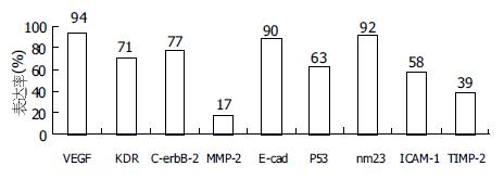
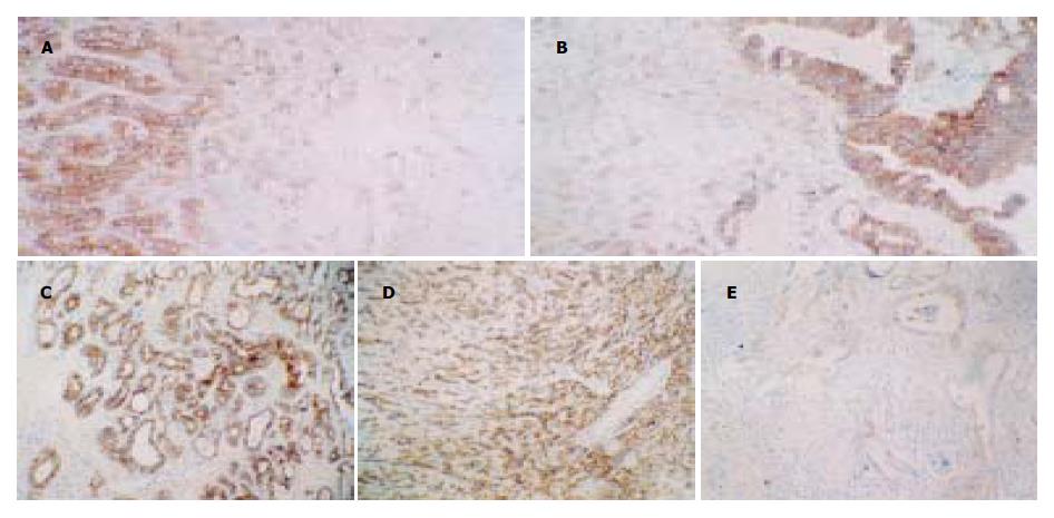
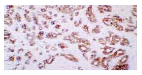
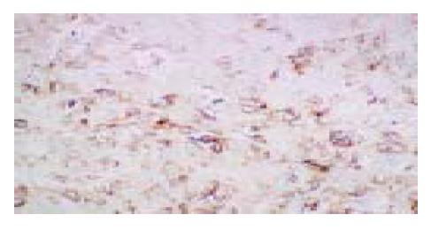
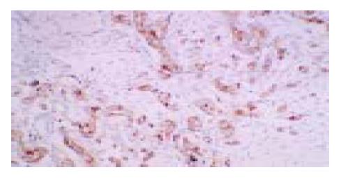
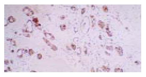
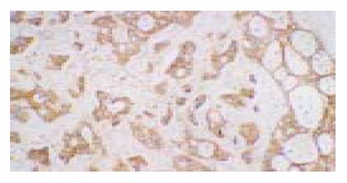
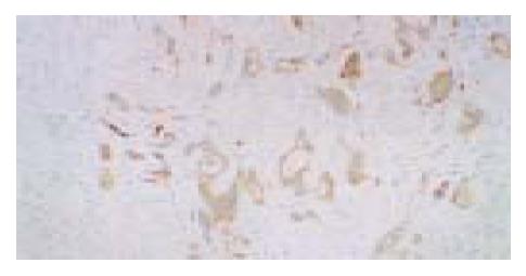
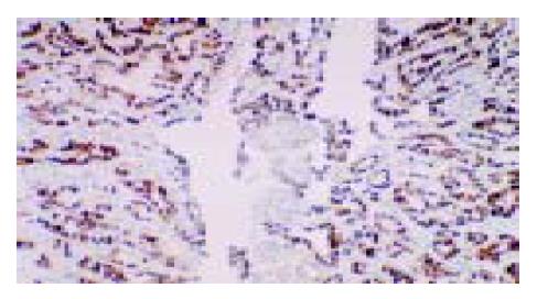
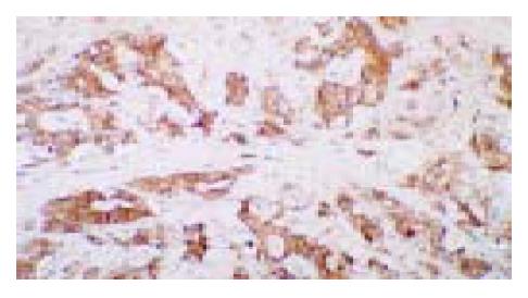
在100例标本中9种肿瘤相关基因蛋白皆有表达(图2-10), 其中VEGF, E-cad, nm23表达率较高, 分别为94%, 90%, 92%; 而MMP-2, TIMP-2及ICAM-1的表达相对较低, 分别为17%, 39%, 58%(图1). 同证型内整体上皆存在显著性差异, P = 0.0 001(P<0.01), 进一步两两比较发现同一证型内普遍存在基因蛋白表达差异性(表3), 尤其重要的是发现痰湿凝结型中E-cad的表达最高, 以平均秩和表示为: 63.09, 而肝胃不和(图2D-E)、瘀毒内阻(图1-2)两型E-cad表达较低, 以平均秩和表示分别为: 34.09, 35.50(表2).
| 基因 | 痰湿凝结 | 胃热伤阴 | 瘀毒内阻 | 气血双亏 | 脾胃虚寒 | 肝胃不和 |
| E-cad | 63.09 | 47.19 | 35.50 | 63.06 | 59.00 | 34.09 |
| P53 | 51.53 | 46.09 | 53.94 | 56.24 | 56.24 | 38.91 |
| nm23 | 43.91 | 49.59 | 53.00 | 66.91 | 46.71 | 42.97 |
| ICAM-1 | 56.00 | 56.53 | 62.16 | 44.71 | 47.44 | 37.21 |
| VEGF | 59.52 | 44.16 | 47.13 | 52.41 | 48.62 | 50.59 |
| KDR | 49.65 | 45.34 | 47.91 | 61.94 | 54.03 | 43.68 |
| MMP-2 | 50.47 | 48.00 | 58.50 | 53.88 | 47.65 | 44.82 |
| TIMP-2 | 43.21 | 55.72 | 45.69 | 60.47 | 53.12 | 44.82 |
| C-erbB-2 | 52.24 | 52.31 | 35.16 | 53.85 | 56.79 | 51.85 |
| 中医证型 | 肿瘤相关基因表达差异情况 |
| 痰湿凝结型 | E-cad>ICAM-1; E-cad>MMP-2; E-cad>TIMP-2; nm23>MMP-2, nm23>TIMP-2; VEGF>MMP-2, VEGF>TIMP-2; |
| 胃热伤阴型 | E-cad>MMP-2; E-cad>TIMP-2; nm23>MMP-2; VEGF>MMP-2; |
| 瘀毒内阻型 | nm23>MMP-2; nm23>TIMP-2; |
| 气血双亏型 | E-cad>ICAM-1; E-cad>MMP-2; E-cad>TIMP-2; nm23>ICAM-1; nm23>MMP-2, nm23>TIMP-2; VEGF>MMP-2, |
| 脾胃虚寒型 | E-cad>ICAM-1; E-cad>MMP-2; E-cad>TIMP-2; C-erbB-2>MMP-2, nm23>MMP-2, nm23>TIMP-2; VEGF>MMP-2, VEGF>TIMP-2; |
| 肝胃不和型 | E-cad>MMP-2; nm23>MMP-2; VEGF>ICAM-1; VEGF>MMP-2, VEGF>TIMP-2 |
中医证候的重要性早在唐朝就有所论述[2]. 中医治病必须按照辨证论治的基本原则, 同病异治、异病同治, 提倡个体化治疗, 真正地体现中医治疗的精髓. 胃癌是常见病、多发病, 当今生物学研究已经进入了在生物大分子整体水平上将不同研究技术与手段有机地结合, 以攻克生物学难题的时代. 这一点突出体现在基因组学(genomics)以及蛋白质组学(proteomics)等新学科领域的研究中[3]. 我们思考, 同样是胃癌, 但出现的疾病临床表现并不相同, 有的患者以泛酸、嗳气为主, 即中医辨证为肝胃不和, 而有些患者则以泛吐黏液清水为主, 并无泛酸、嗳气, 伴有舌苔白厚腻辨证应属痰湿凝结表现. 这使我们进一步想到, 这些表型是否能从基因学方面来阐释胃癌症候本质.
随着现代科技与医学的结合, 肿瘤的侵袭与转移在肿瘤分子机制的研究方面取得了令人鼓舞的进展. 肿瘤转移的产生是多步骤的连续过程: 肿瘤细胞首先脱离原发瘤, 侵入周围组织, 进入血管, 然后瘤细胞随血流或淋巴道运行至某些器官或组织, 并受阻停留在小血管中, 穿出血管, 直至在新部位分裂繁殖形成转移瘤或称继发瘤. 肿瘤浸润是肿瘤细胞和细胞外基质在宿主多因素调节下相互作用的结果, 也是肿瘤远处转移的前奏. 肿瘤的浸润和转移共同构成肿瘤播散, 是成为大多肿瘤患者死亡的重要原因. 可见转移是一个涉及肿瘤细胞-宿主细胞-细胞外基质(ECM)之间相互作用的多步骤分子生物化学变化的过程. 其中分子生物学机制研究已经较为成熟, 尤其是在胃癌肿瘤细胞同质性黏附上起重要作用的E-cadherin[4-7], ICAM-1[8-9]; 在胃癌细胞对基底膜的侵袭上起平衡作用[10-13]的MMP-2, TIMP-2; 在肿瘤细胞发生侵袭、转移的整个过程起作用的肿瘤血管生成因子及其受体VEGF, KDR[14-16]; 以及与胃癌发生侵袭、转移行为密切相关的调控基因[17-27]C-erbB-2, P53, nm23等皆已成为普遍共识.因此我们采用这9项指标, 用免疫组化方法进行研究, 并试以进一步从基因蛋白表达上探索胃癌证本质.
目前从事临床工作的学者, 逐渐倾向于个体化治疗, 认为疾病的发生、发展以及预后皆存在着差异性, 胃癌同样如此. 在治疗上个体对各种药物的敏感程度及其疗效亦不同, 因此倡导个体化治疗. 这正应中医临床思维诊疗特点"辨证论治", "有诸内必形诸外", 相同的疾病, 中医可辩为不同的证, 针对证候的不同应进行不同治疗. 但中医"辨证论治"的内在本质何在, 即中医证本质, 为此我们从基因水平探讨胃癌中医"证"本质. 本研究结果发现VEGF, E-cad, nm23表达率较高, 分别为94%, 90%, 92%. 尤其重要的发现痰湿凝结型中E-cad的表达最高, 以平均秩和表示为: 63.09, 其值最高, 而肝胃不和、瘀毒内阻两型E-cad表达较低, 我们考虑此两型胃癌发生的转移主要E-cad的缺失, 即肿瘤细胞间同质性黏附降低促进了肿瘤的侵袭、转移的发生. 同时研究亦表明在胃癌每个证型内部均有基因蛋白表达的差异性存在(P = 0.0 001), 同证型内部既有抑癌基因的表达, 亦有促癌基因的表达; 而且癌基因与促癌基因的表达可出现在同一证型中. 因此确定患者的预后情况及判断某一证型的整体预后情况还有待于结合随访进行观察.
编辑: N/A
| 3. | 李 林. 蛋白质组学的进展. 生物化学与生物物理进展. 2000;27:227-231. |
| 4. | Frixen UH, Behrens J, Sachs M, Eberle G, Voss B, Warda A, Löchner D, Birchmeier W. E-cadherin-mediated cell-cell adhesion prevents invasiveness of human carcinoma cells. J Cell Biol. 1991;113:173-185. [PubMed] [DOI] |
| 5. | Becker KF, Keller G, Hoefler H. The use of molecular biology in diagnosis and prognosis of gastric cancer. Surg Oncol. 2000;9:5-11. [DOI] |
| 6. | Lee JH, Koh JT, Shin BA, Ahn KY, Roh JH, Kim YJ, Kim KK. Comparative study of angiostatic and anti-invasive gene expressions as prognostic factors in gastric cancer. Int J Oncol. 2001;18:355-361. [PubMed] [DOI] |
| 8. | Tanaka H, Yashiro M, Sunami T, Ohira M, Hirakawa-Y S Chung K. Lipid-mediated gene transfection of intercellular adhesion molecule-1 suppresses the peritoneal metastasis of gastric carcinoma. Int J Mol Med. 2002;10:613-617. [PubMed] |
| 10. | Okada Y. Tumor cell-matrix interaction: pericellular matrix degradation and metastasis. Verh Dtsch Ges Pathol. 2000;84:33-42. [PubMed] |
| 11. | Chen BQ, Yang YM, Gao YH, Liu JR, Xue YB, Wang XL, Zheng YM, Zhang JS, Liu RH. Inhibitory effects of c9, t11-conjugated linoleic acid on invasion of human gastric carcinoma cell line SGC-7901. World J Gastroenterol. 2003;9:1909-1914. [PubMed] [DOI] |
| 12. | Sier CF, Kubben FJ, Ganesh S, Heerding MM, Griffioen G, Hanemaaijer R, van Krieken JH, Lamers CB, Verspaget HW. Tissue levels of matrix metalloproteinases MMP-2 and MMP-9 are related to the overall survival of patients with gastric carcinoma. Br J Cancer. 1996;74:413-417. [DOI] |
| 14. | Zhang H, Wu J, Meng L, Shou CC. Expression of vascular endothelial growth factor and its receptors KDR and Flt-1 in gastric cancer cells. World J Gastroenterol. 2002;8:994-998. [PubMed] [DOI] |
| 16. | Ren J, Dong L, Xu CB, Pan BR. The role of KDR in the interactions between human gastric carcinoma cell and vascular endothelial cell. World J Gastroenterol. 2002;8:596-601. [DOI] |
| 17. | Aoyagi K, Kohfuji K, Yano S, Murakami N, Miyagi M, Takeda J, Shirouzu K. Evaluation of the epidermal growth factor receptor (EGFR) and c-erbB-2 in superspreading-type and penetrating-type gastric carcinoma. Kurume Med J. 2001;48:197-200. [PubMed] [DOI] |
| 19. | García I, Vizoso F, Martín A, Sanz L, Abdel-Lah O, Raigoso P, García-Muñiz JL. Clinical significance of the epidermal growth factor receptor and HER2 receptor in resectable gastric cancer. Ann Surg Oncol. 2003;10:234-241. [PubMed] [DOI] |
| 20. | Yeung P, Lee CS, Marr P, Sarris M, Fenton-Lee D. Nm23 gene expression in gastric carcinoma: an immunohistochemical study. Aust N Z J Surg. 1998;68:180-182. [DOI] |
| 21. | Huang SP, Wu MS, Shun CT, Wang HP, Lin JT. Tumor angiogenesis increases with nuclear p53 accumulation in gastric carcinoma. Hepatogastroenterology. 2002;49:1453-1456. [PubMed] |
| 22. | Kimura M, Tsuda H, Morita D, Ichikura T, Ogata S, Aida S, Yoshizumi Y, Maehara T, Mochizuki H, Matsubara O. A proposal for diagnostically meaningful criteria to classify increased epidermal growth factor receptor and c-erbB-2 gene copy numbers in gastric carcinoma, based on correlation of fluorescence in situ hybridization and immunohistochemical measurements. Virchows Arch. 2004;445:255-262. [PubMed] [DOI] |
| 23. | Maeta Y, Tsujitani S, Matsumoto S, Yamaguchi K, Tatebe S, Kondo A, Ikeguchi M, Kaibara N. Expression of Mcl-1 and p53 proteins predicts the survival of patients with T3 gastric carcinoma. Gastric Cancer. 2004;7:78-84. [PubMed] [DOI] |
| 24. | Pinto-de-Sousa J, Silva F, David L, Leitão D, Seixas M, Pimenta A, Cardoso-de-Oliveira M. Clinicopathological significance and survival influence of p53 protein expression in gastric carcinoma. Histopathology. 2004;44:323-331. [PubMed] [DOI] |
| 25. | Nishikura K, Watanabe H, Iwafuchi M, Fujiwara T, Kojima K, Ajioka Y. Carcinogenesis of gastric endocrine cell carcinoma: analysis of histopathology and p53 gene alteration. Gastric Cancer. 2003;6:203-209. [PubMed] [DOI] |
| 26. | Lu C, Xu HM, Ren Q, Ao Y, Wang ZN, Ao X, Jiang L, Luo Y, Zhang X. Somatic mutation analysis of p53 and ST7 tumor suppressor genes in gastric carcinoma by DHPLC. World J Gastroenterol. 2003;9:2662-2665. [PubMed] [DOI] |
| 27. | Ishikawa M, Kitayama J, Kazama S, Nagawa H. Expression of vascular endothelial growth factor C and D (VEGF-C and -D) is an important risk factor for lymphatic metastasis in undifferentiated early gastric carcinoma. Jpn J Clin Oncol. 2003;33:21-27. [PubMed] [DOI] |


