修回日期: 2002-09-20
接受日期: 2002-10-03
在线出版日期: 2003-06-15
介绍并评价脾静脉结扎诱导的继发性脾功能亢进犬动物模型.
18只健康成年杂种狗随机分为Ⅰ组(对照组n = 4)、Ⅱ组(脾静脉结扎n = 10)和Ⅲ组(脾静脉结扎+脾切除n = 4), 通过结扎狗的脾静脉主干和脾静脉属支引起淤血性脾肿大; 脾静脉结扎后第3周第Ⅲ组行脾切除术. 定期观察动物外周血细胞变化以及影像学、组织病理学改变.
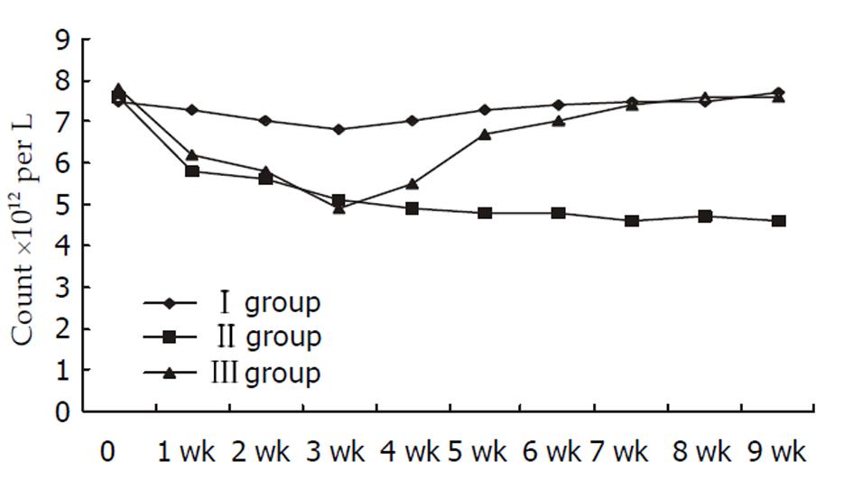
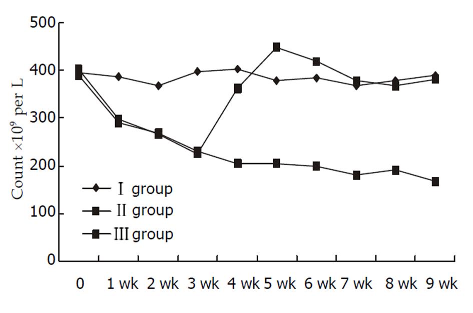
脾静脉结扎后1 wk内外周血红细胞、血小板开始下降, 第3周末二者下降明显(Ⅰ组红细胞和血小板计数分别为(6.8±1.2)×1012/L、(398±58)×109/L, Ⅱ组为(5.1±0.7)×1012/L、(230±86)×109/L, 分别P<0.01和P<0.05); 红细胞和血小板减少、脾脏肿大可持续9 wk以上; 但脾静脉结扎后白细胞水平无显著改变. 脾切除术后2 wk红细胞和血小板逐渐恢复正常. 脾静脉结扎3 wk后脾脏组织病理学改变逐渐符合慢性脾脏淤血改变.
脾静脉结扎方法简单, 可以建立确切的继发性脾功能亢进, 可以作为脾功能亢进外科或介入治疗的较理想模型.
引文著录: 刘全达, 马宽生, 何振平, 丁钧, 董家鸿. 脾静脉结扎诱导继发性脾功能亢进犬动物模型的评价. 世界华人消化杂志 2003; 11(6): 749-752
Revised: September 20, 2002
Accepted: October 3, 2002
Published online: June 15, 2003
To introduce and evaluate a canine model of secondary hypersplenism induced by splenic vein ligation.
Eighteen healthy mongrel dogs were randomly divided into three groups. The first group (n = 4) underwent laparotomy, the second (n = 10) and third groups (n = 4) underwent laparotomy plus ligation of splenic vein and its collateral branches to induce congestive splenomegaly. At the end of the third week, splenectomy was performed in the third group. The blood cell counts for peripheral venous blood were determined weekly, and the radiographic and histopathological changes of spleen also obtained regularly.
The erythrocyte and platelet counts decreased in the first week, and were significantly lowered (erythrocyte count of (6.8 ± 1.2)×1012/L in control vs (5.1± 0.7)×1012/L in second group, P<0.01; and platelet counts of (398 ± 58)×109/L vs (230 ± 86)109/L, P<0.05 respectively) at the end of 3rd week after splenic vein ligation thereafter sustained. The splenomegaly, erythrocytopenia and thrombocytopenia had remained over 9 weeks. No significant changes of the leukocyte counts were observed after splenic vein ligation throughout the experiment (P>0.05). The abnormal status of erythrocytopenia and thrombocytopenia was ameliolated by splenectomy, and the erythrocyte and platelet counts were similarly to the levels of the control group in the second week after splenectomy. After the end of 3rd week after splenic vein ligation, the splenic histopathological changes conformed to the changes of chronic congestive splenomagely.
The method of splenic vein ligation to induce experimental secondary hypersplenism is simple and effective. This is a relative ideal model for surgical or interventional therapy on hypersplenism.
- Citation: Liu QD, Ma KS, He ZP, Ding J, Dong JH. Evaluation of a canine model of secondary hypersplenism induced by splenic vein ligation. Shijie Huaren Xiaohua Zazhi 2003; 11(6): 749-752
- URL: https://www.wjgnet.com/1009-3079/full/v11/i6/749.htm
- DOI: https://dx.doi.org/10.11569/wcjd.v11.i6.749
继发性脾功能亢进(脾亢)是肝硬化的常见表现, 其直接后果是出凝血机制紊乱, 引发或加重曲张静脉出血[1-4]. 脾切除术是外科最常用的方法, 且疗效确切[5-7]. 但随着对脾脏免疫功能认识的深入[8-12], 保留部分脾脏已得到公认[13-18]. 临床也逐渐应用选择性脾动脉栓塞术、脾内无水酒精注射等微创方法[19-22]治疗继发性脾亢. 但在继发性脾亢的微创治疗的实验研究中, 尚缺乏脾脏肿大的大动物模型[23-28]. 我们在对脾脏射频消融治疗脾亢的实验研究中, 建立了一种快速诱导的继发性脾亢的犬动物模型, 现报告如下.
健康成年杂种狗18只(第三军医大学实验动物中心提供), 雌雄不拘, 质量12-17 kg, 随机分为3组: Ⅰ组(对照组, n = 4); Ⅱ组(脾静脉结扎, n = 10); Ⅲ组(脾静脉结扎+脾切除, n = 4). 脾静脉结扎参照Sahin et al [28]加以改良. 动物空腹过夜, 腹部备皮后, 以速眠新(长春市军需大学兽医研究所提供)肌肉注射(0.1 mL/kg)全身麻醉, 建立静脉通道, 术中快速输入平衡液500 ml, 无菌条件下手术: Ⅰ组剖腹术; Ⅱ组剖腹术+脾静脉结扎; Ⅲ组剖腹术+脾静脉结扎. 为避免脾静脉侧枝循环建立, 在脾静脉汇入门静脉处结扎脾静脉主干后20 min, 再在脾门处分别结扎扩张的脾静脉属支. 术后归圈饲养, 温度242 ℃, 食水不限. 术后2 wk再手术, Ⅰ组: 剖腹术; Ⅱ, Ⅲ组: 剖腹术+脾静脉侧枝血管结扎. 术后3 wk再次手术, Ⅰ, Ⅱ组: 剖腹术; Ⅲ组: 剖腹术+脾切除术.
所有动物术后每周行腹部CT或超声检查, 了解脾脏大小(脾脏厚度和长径)改变; 实验4 wk起第Ⅱ组每周处死1只, 实验9 wk动物全部处死, 切除脾脏均经40 g/L甲醛固定, 石蜡包埋, 连续5 μm切片, HE染色后镜检, 了解脾脏组织病理学改变. 实验当天术前和术后每周采动物外周血行血细胞计数(白细胞、红细胞和血小板). 血标本立即送我院门诊检验科行自动血细胞计数仪Sysmex(K-4500)检测, 每个标本重复2次, 取其均值.
统计学处理 计量数据以均数北曜疾(mean±SD)表示, 均与对照Ⅰ组行t检验, P<0.05为相差显著.
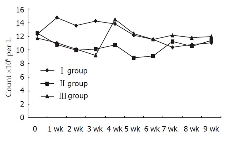
与作对照组Ⅰ组的外周血的血小板计数[(398±58)×109/L]和红细胞计数红细胞[(6.8±1.2)×1012/L]相比, Ⅱ、Ⅲ组动物在脾静脉结扎后1 wk即出现红细胞和血小板下降. 在3 wk末Ⅱ组[(230±86)×109/L](t = 3.553, P<0.01)和Ⅲ组的血小板计数[(225±96)×109/L](t = 3.085, P<0.05)显著下降; 3 wk末Ⅱ组[(5.1±0.7)×1012/L](t = 3.369, P<0.05)和Ⅲ组红细胞计数[(4.9±0.6)×1012/L](t = 2.832, P<0.05)亦显著降低; 第Ⅲ组脾切除术后红细胞和血小板逐渐恢复, 脾切术后2 wk基本恢复至正常; Ⅱ组到9 wk实验结束血小板和红细胞一直保持在低水平. 整个实验过程中三组白细胞水平变化均不显著(P>0.05, 图1-3).
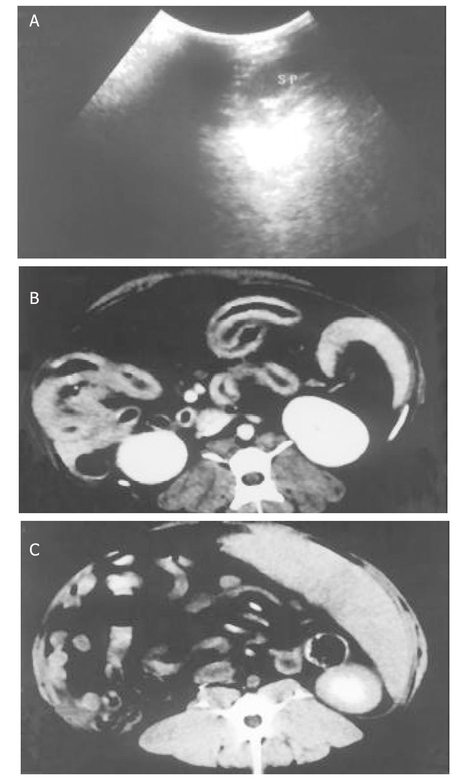
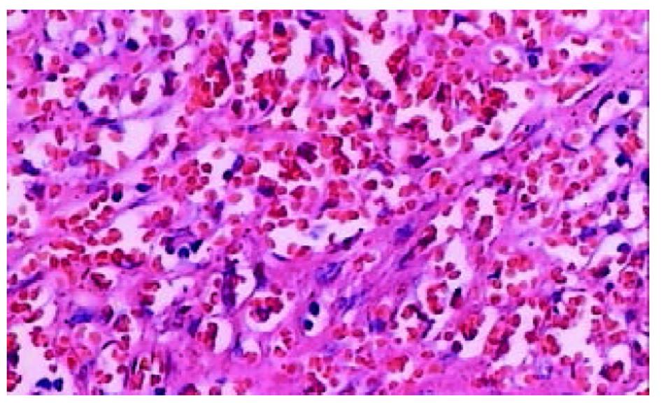
采用Doppler超声(图4A)和CT检查(图4B)显示, 脾脏结扎后体积明显增大(图4C), 与术中探查和影像学结果一致. 2 wk剖腹时发现脾脏略有缩小, 脾门周围又出现数支扩张的脾静脉属支; 予再次结扎后, 此后探查脾门仅见网状迂曲的小静脉网, 无明显扩张的粗大静脉. 术中组织病理学检查提示, 与正常脾脏结构比较, 脾静脉结扎组3 wk末脾脏明显充血, 脾窦扩张, 4 wk以后脾脏内可见纤维组织增生, 脾窦内有较多微血栓, 脾小梁明显增粗(图5). 上述组织病理学表现符合慢性脾淤血改变.
我们采用脾静脉结扎均成功诱导继发性脾亢, 该方法类似于临床的脾静脉血栓形成引起区域性门静脉高压, 但后者仅50%左右出现脾脏肿大, 脾亢者更少[29-33]. 其根本原因是脾静脉慢性闭塞过程中血流逐渐通过扩张的胃短静脉、胃网膜静脉等侧枝血管代偿, 因而脾大、脾亢并不明显. 为了防止上述侧枝形成, Sahin et al [28]采用在脾门处分别结扎脾静脉属支的方法, 但该方法诱导的脾亢仅能维持5 wk左右. 我们发现, 即使在脾门处结扎脾静脉, 术后1 wk探查时仍可见到代偿的粗大脾静脉属支, 脾脏缩小, 提示1次手术无法保证彻底的阻断脾静脉流出道. 因此我们设计结扎脾静脉主干基础上, 结合快速液体输入, 待脾静脉属支扩张后, 再在脾门处分别结扎扩张的脾静脉属支, 2 wk后再次结扎代偿的粗大脾静脉属支, 这就尽可能地完成脾静脉流出道的阻断. 该方法极大地提高了脾亢动物模型的成功率. 由于潜在侧枝循环被阻断, 而高压的动脉血流仍持续向脾内灌注, 从而确保了巨脾和脾亢的长时间维持. 研究表明, 我们的脾亢模型持续时间达9 wk以上.
脾脏长时间淤血状态下, 除脾门周围出现细小网状血管网外, 脾脏组织学结构出现一些特征性改变, 即脾脏被膜增厚、脾窦淤血扩张、白髓萎缩、纤维化, 本实验脾静脉结扎后的组织病理学改变符合上述脾淤血改变. Ⅱ, Ⅲ组动物脾静脉结扎后红细胞和血小板计数在1 wk即出现下降, 在3 wk末与对照组相比下降显著, 而且脾静脉结扎引起的红细胞和血小板下降可以被脾切除术纠正. 这些结果证实, 脾静脉结扎可以诱导红细胞减少和血小板减少. 外周血细胞计数和脾脏组织学改变均提示, 动物脾静脉结扎可以成功诱导继发性脾亢模型. 整个实验过程中, 白细胞下降并不显著, 脾静脉结扎后引起的急性脾脏肿大是否能刺激并动员全身炎症反应从而代偿白细胞减少?本实验未进行相关研究, 但脾静脉结扎可能不是脾亢性白细胞减少症的理想模型.
随着脾脏免疫功能认识的深入, 尽可能地保留良性脾脏疾病的部分脾脏功能的认识已经被临床医师所接受. 临床许多脾亢患者由于全身情况差、高龄或者伴有其他不适合手术的疾病, 如何处理脾亢相对棘手. 目前有选择性脾动脉栓塞、脾脏酒精注射等微创措施[19-22], 但临床疗效均不太满意, 前者较多出现脾脓肿, 后者栓塞范围不易控制. 我们正在探索射频消融[34-37]治疗脾亢的动物研究, 发现狗的脾肿大模型比兔、猪更适合射频消融疗效的需要, 能够满足射频针展开直径2.0-3.5 cm的工作需要.
总之, 脾静脉结扎建立继发性脾亢方法简单、有效, 脾静脉属支的彻底结扎是保证模型成功的关键. 该模型可以作为脾功能亢进外科或介入治疗的较理想模型.
梁天学、朱斌、马新、吴政谦等医师提供手术协助和周代全副主任技师的帮助.
| 1. | Okudaira M, Ohbu M, Okuda K. Idiopathic portal hypertension and its pathology. Semin Liver Dis. 2002;22:59-72. [PubMed] [DOI] |
| 2. | Jiao YF, Okumiya T, Saibara T, Kudo Y, Sugiura T. Erythrocyte creatine as a marker of excessive erythrocyte destruction due to hypersplenism in patients with liver cirrhosis. Clin Biochem. 2001;34:395-398. [DOI] |
| 3. | Bajaj JS, Bhattacharjee J, Sarin SK. Coagulation profile and platelet function in patients with extrahepatic portal vein obstruction and non-cirrhotic portal fibrosis. J Gastroenterol Hepatol. 2001;16:641-646. [DOI] |
| 4. | Bashour FN, Teran JC, Mullen KD. Prevalence of peripheral blood cytopenias (hypersplenism) in patients with nonalcoholic chronic liver disease. Am JGastroenterol. 2000;95:2936-2939. [PubMed] [DOI] |
| 5. | Lin MC, Wu CC, Ho WL, Yeh DC, Liu TJ, Peng FK. Concomitant splenectomy for hypersplenic thrombocytopenia in hepatic resection for hepatocellular carcinoma. Hepatogastroenterology. 1999;46:630-634. |
| 6. | Carr JA, Shurafa M, Velanovich V. Surgical indications in idiopathic splenomegaly. Arch Surg. 2002;137:64-68. [PubMed] [DOI] |
| 7. | McCormick PA, Murphy KM. Splenomegaly, hypersplenism and coagulation abnormalities in liver disease. Baillieres Best Prac Res Clin Gastroenterol. 2000;14:1009-1031. [PubMed] [DOI] |
| 9. | Hansen K, Singer DB. Asplenic-hyposplenic overwhelming sepsis: postsplenectomy sepsis revisited. Pediatr Dev Pathol. 2001;4:105-121. [PubMed] [DOI] |
| 10. | Kyriazanos ID, Tachibana M, Yoshimura H, Kinugasa S, Dhar DK, Nagasue N. Impact of splenectomy on the early outcome after oesophagectomy for squamous cell carcinoma of the oesophagus. Eu J Surg Onco. 2002;28:113-119. [PubMed] [DOI] |
| 11. | Altamura M, Caradonna L, Amati L, Pellegrino NM, Urgesi G, Miniello S. Splenectomy and sepsis: the role of the spleen in the immune-mediated bacterial clearance. Immunopharmacol Immunotoxicol. 2001;23:153-161. [PubMed] [DOI] |
| 12. | Seabrook TJ, Hein WR, Dudler L, Young AJ. Splenectomy selectively affects the distribution and mobility of the recirculating lymphocyte pool. Blood. 2000;96:1180-1183. |
| 13. | de Buys Roessingh AS, de Lagausie P, Rohrlich P, Berrebi D, Aigrain Y. Follow-up of partial splenectomy in children with hereditary spherocytosis. J Pediatr Surg. 2002;37:1459-1463. [PubMed] [DOI] |
| 14. | Jiang HC, Sun B, Qiao HQ, Xu J, Piao DX, Yin H. Clinical application o f serial operations with preserving spleen. World J Gastroenterol. 2001;7:876-879. [DOI] |
| 15. | Sarkar PK, Bhattacharya DK. Splenectomy and splenic slice grafting in the management of thalassemia. Pediatr Surg Int. 2001;17:369-372. [PubMed] [DOI] |
| 16. | Zhang H, Chen J, Kaiser GM, Mapudengo O, Zhang J, Exton MS, Song E. The value of partial splenic autotransplantation in patients with portal hypertension: a prospective randomized study. Arch Surg. 2002;137:89-93. [DOI] |
| 18. | Sockrider CS, Boykin KN, Green J, Marsala A, Mladenka M, Zibari GB. Partial splenic embolization for hypersplenism after liver transplantation. Transplant Proc. 2001;33:3472-3473. [DOI] |
| 19. | Sakai T, Shiraki K, Inoue H, Sugimoto K, Ohmori S, Murata K, Takase K, Nakano T. Complications of partial splenic embolization in cirrhotic patients. Dig Dis Sci. 2002;47:388-391. [DOI] |
| 20. | Obatake M, Muraji T, Kanegawa K, Satoh S, Nishijima E, Tsugawa C. A new volumetric evaluation of partial splenic embolization for hypersplenism in biliary astresia. J Pediatr Surg. 2001;36:1615-1616. [PubMed] [DOI] |
| 21. | Kimura F, Itoh H, Ambiru S, Shimizu H, Togawa A, Yoshidome H, Ohtsuka M, Shimizu Y, Shimamura F, Miyazaki M. Long-term results of initial and repeated partial splenic embolization for the treatment of chronic idiopathic thrombocytopenic purpura. Am J Roentgenol. 2002;179:1323-1326. [PubMed] [DOI] |
| 22. | Shimizu T, Onda M, Tajiri T, Yoshida H, Mamada Y, Taniai N, Aramaki T, Kumazaki T. Bleeding portal-hypertensive gastropathy managed successfully by partial splenic embolization. Hepatogastroenterology. 2002;49:947-949. |
| 27. | Lei DX, Peng CH, Peng SY, Jiang XC, Wu YL, Shen HW. Safe upper limit of intermit tent hepatic inflow occlusion for liver resection in cirrhotic rats. World J Gastroenterol. 2001;7:713-717. [DOI] |
| 28. | Sahin M, Tekin S, Aksoy F, Vatansev H, Seker M, Avunduk MC, Kartal A. The effects of splenic artery ligation in an experimental model of secondary hypersplenism. J R Coll Surg Edinb. 2000;45:148-152. |
| 29. | Olakowski M, Lampe P, Boldys H, Slota J, Olakowska E. Neuroendocrine pancreatic carcinoma causing sinistral portal hypertension. Med Sci Monit. 2001;7:1326-1328. |
| 30. | Jaroszewski DE, Schlinkert RT, Gray RJ. Laparoscopic splenectomy for the treatment of gastric varices secondary to sinistral portal hypertension. Surg Endosc. 2000;14:87. |
| 31. | Sakorafas GH, Tsiotou AG. Splenic-vein thrombosis complicating chronic pancreatitis. Scand J Gastroenterol. 1999;34:1171-1177. [PubMed] [DOI] |
| 32. | Sakorafas GH, Sarr MG, Farnell MB. The significance of sinistral portal hypertension complicating chronic pancreatitis. Am J Surg. 2000;179:129-133. [DOI] |
| 33. | Suhocki PV, Berend KR, Trotter JF. Idiopathic splenic vein stenosis: a cause of gastric variceal hemorrhage. South Med J. 2000;93:812-814. [DOI] |
| 34. | Nishida T, Inoue K, Kawata Y, Izumi N, Nishiyama N, Kinoshita H, Matsuoka T, Toyoshima M. Percutaneous radiofrequency ablation of lung neoplasms: a minimally invasive strategy for inoperable patients. J Am Coll Surg. 2002;195:426-430. [DOI] |
| 35. | Liu LX, Jiang HC, Piao DX. Radiofrequence ablation of liver cancers. World J Gastroenterol. 2002;8:393-399. [DOI] |
| 36. | Jiang HC, Liu LX, Piao DX, Xu J, Zheng M, Zhu AL, Qi SY, Zhang WH, Wu LF. Clinical short-term results of radiofrequency ablation in liver cancers. World J Gastroenterol. 2002;8:624-630. [DOI] |
| 37. | Liu QD, Ma KS, He ZP, Ding J, Huang XQ, Dong JH. Experimental study on the feasibility and safety of radiofrequency ablation for secondary splenomagely and hypersplenism. World J Gastroenterol. 2003;9:813-817. [DOI] |


