修回日期: 2002-08-20
接受日期: 2002-08-23
在线出版日期: 2003-01-15
目的: 探讨人胃溃疡愈合过程中碱性成纤维细胞生长因子(basic fibroblast growth factor, bFGF)的变化及其作用.
方法: 采用免疫组织化学法, 对正常胃黏膜(20例)、胃溃疡活动期(24例)、愈合期(26例)和瘢痕期(20例)组织的bFGF 的表达进行定位观察和图像半定量分析.
结果: bFGF在正常胃黏膜呈弱阳性表达, 在胃溃疡急性期呈阳性表达, 在愈合期和瘢痕期呈强阳性表达. 阳性信号主要位于腺上皮、细胞外基质、成纤维细胞和内皮细胞中. bFGF平均光密度值和面密度值在胃溃疡活动期、愈合期、瘢痕期与正常胃黏膜相比均有显著性差异(0.247±0.042, 0.321±0.096, 0.296±0.048 vs 0.125±0.062, P<0.05; 0.131±0.024, 0.165±0.031, 0.162±0.028 vs 0.081±0.008, P<0.05), 在愈合期及瘢痕期组织与活动期溃疡相比也有显著性差异(0.321±0.096, 0.296±0.048 vs 0.247±0.042, P<0.05; 0.165±0.031, 0.162±0.028 vs 0.131±0.024, P<0.05).
结论: 在人胃溃疡愈合过程中, 存在着内源性bFGF由弱到强的演变, 说明bFGF与胃溃疡愈合密切相关. 临床上合理应用bFGF有助于消化性溃疡愈合.
引文著录: 贺建华, 罗和生. 碱性成纤维细胞生长因子在胃溃疡愈合中的表达及意义. 世界华人消化杂志 2003; 11(1): 61-64
Revised: August 20, 2002
Accepted: August 23, 2002
Published online: January 15, 2003
AIM: To investigate changes of basic fibroblast growth factor (bFGF) in the healing of human gastric ulcer.
METHODS: The expression of basic fibroblast growth factor (bFGF) in human gastric mucosa: normal (20 cases), active stage of gastric ulcer (24 cases), healing stage of gastric ulcer (26 cases) and scarring stage of gastric ulcer (20 cases) was detected by immunohistochemical methods and computerized image analysis.
RESULTS: The expression intensity of bFGF was different among normal gastric mucosa and mucosa at different stages of gastric ulcer. The expression in normal gastric mucosa was weakly positive, at the active stage of gastric ulcer was positive, at the healing stage and scarring stage was strongly positive. The positive signal of bFGF was localized in granulation tissues, extracellular matrix (ECM), fibroblasts and endothelial cells. There were significant differences in average integral optic density and area density among different stages of gastric ulcer and normal gastric mucosa (0.247±0.042, 0.321±0.096, 0.296±0.048 vs 0.125±0.062 P < 0.05; 0.131±0.024, 0.165±0.031, 0.162±0.028 vs 0.081±0.008, P < 0.05) They were significantly higher at the healing stage and scarring stage than that at the active stage in average integral optic density and area density (0.321±0.096, 0.296±0.048 vs 0.247±0.042, P < 0.05; 0.165±0.031, 0.162±0.028 vs 0.131±0.024, P < 0.05).
CONCLUSION: The changes of bFGF were closely related to the healing of human gastric ulcer, and rational use of bFGF can promote the healing of gastric ulcer.
- Citation: He JH, Luo HS. Expression of basic fibroblast growth factor(bFGF) in healing human gastric ulcer. Shijie Huaren Xiaohua Zazhi 2003; 11(1): 61-64
- URL: https://www.wjgnet.com/1009-3079/full/v11/i1/61.htm
- DOI: https://dx.doi.org/10.11569/wcjd.v11.i1.61
消化性溃疡愈合是一个十分复杂的过程, 包括上皮、黏膜肌层和结缔组织的重建. 其中会涉及到一系列细胞和分子机制的参与. 许多生长因子参与了其愈合过程[1,2]. 碱性成纤维细胞生长因子(basic fibroblast growth factor, bFGF)即为其中一种. 有研究认为外源性bFGF可促进实验性胃、十二指肠溃疡愈合[3-5], 但有关人胃溃疡愈合过程中内源性bFGF的变化研究较少. 我们采用免疫组织化学方法, 对bFGF在正常胃黏膜、胃溃疡活动期、愈合期和瘢痕期的表达情况进行了研究, 探讨bFGF在胃溃疡愈合中的变化及作用.
全部胃溃疡标本均来源于胃镜取材. 胃溃疡分期采用日本学者畸田隆夫倡导的分期法, 将溃疡分为活动期(active stage)、愈合期(healing stage)和瘢痕期(scarring stage). 其中活动期(GA组)24例, 愈合期(GH组)26例, 瘢痕期(GS期)20例, 标本均取自溃疡边缘, 每例取两块, 一块用于免疫组化, 一块用于HE染色. 另取20例完整胃窦黏膜组织作对照. 标本经10%中性甲醛固定, 脱水, 石蜡包埋, 切片后备用.
(1)组织学观察采用HE染色, 光镜下观察溃疡各期组织结构.(2)组织碱性成纤维细胞生长因子(bFGF)表达采用免疫组织化学SABC法. bFGF免疫组织化学染色试剂盒购于武汉博士德生物工程有限公司, 操作按试剂盒说明书进行. 将石蜡切片脱蜡并进行抗原热修复后系列染色, 其中一抗用抗体稀释液按1: 100稀释. 光镜观察, 结果以胞质或/和胞膜着棕色者为阳性. 另用PBS代替一抗为阴性对照. 在400倍光镜下观察, 每个视野观察100个细胞, 按染色强度或细胞阳性数确定表达强度. 不染色, 表达强度即为阴性. 染色较弱, 每个视野阳性细胞数少于10个为弱阳性; 中度染色, 每个视野阳性细胞数10-30个为阳性; 强染色, 每个视野阳性细胞数>30个为强阳性.(3)图像半定量分析 应用H. PYLORIIAS-1000型全自动医学图像彩色分析系统, 分别测定正常胃黏膜、胃溃疡活动期、愈合期和瘢痕期中胃组织bFGF的平均积分光密度值和面密度值. 每张切片随机选取5个视野, 然后取其均值.
统计学处理 数据以表示, 组间比较采用t 检验.
溃疡活动期: 主要表现为黏膜变性、坏死, 大量中性粒细胞为主的炎性细胞浸润和少量新生肉芽组织; 愈合期: 各种炎性细胞减少, 可见再生的腺体组织、毛细血管、纤维结缔组织; 瘢痕期: 肉芽组织逐渐纤维化和胶原形成, 腺体重构.
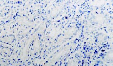
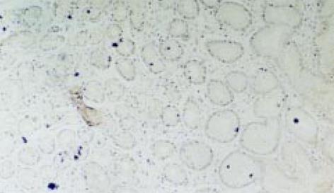
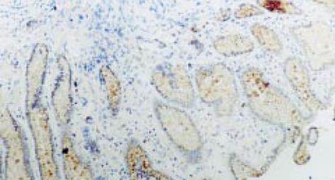
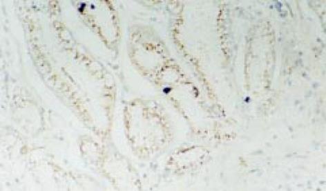
在正常胃窦黏膜, bFGF有弱阳性表达, 主要位于腺体组织(图1), 在溃疡活动期bFGF呈阳性表达, 主要位于腺上皮, 细胞外基质及炎性细胞中(图2); 在溃疡愈合期和瘢痕期均有强阳性表达, 主要位于再生腺体组织, 成纤维细胞和内皮细胞中(图3, 4).
结果见表1 bFGF平均积分光密度值在胃溃疡各期与正常胃黏膜组相比均有显著差异(P<0.05). 在胃溃疡愈合期和瘢痕期与活动期相比均有显著差异(P<0.05). bFGF平均面密度值在胃溃疡各期与正常胃黏膜组均有显著差异(P<0.05), 在愈合期和瘢痕期与活动期相比均有显著性差异(P<0.05).
损伤修复是多种细胞, 生长因子和细胞外基质相互作用的复杂过程, 生长因子在损伤修复过程中起着重要作用[6,7]. 碱性成纤维细胞生长因子(basic fibroblast growth factor, bFGF)是一种对创伤修复有重要调控作用的细胞因子[8-12], 其参与了调控组织修复的全过程, 包括调控炎性反应、诱导毛细胞血管胚芽形成, 促进上皮及肉芽组织生长. 早期生长反应素-1(Erg-1)对组织修复有重要作用, 而Erg-1的许多作用均依赖于bFGF在组织中的释放和旁分泌. 成纤维细胞生长因子主要通过三条途径发挥其促分裂效应: (1)通过受体介导. 流式细胞仪检测发现, 成纤维细胞生长因子促进细胞增生的作用发生在与受体结合后, 可能会促使细胞周期中的G0期与G1期细胞减少, S期加速; (2)通过限制性的酶解作用, 在酶的作用下使与肝素结合的无活性的FGF变成可溶性的、有活性的FGF; (3)通过FGF结构修饰而发挥作用. 此外, 成纤维细胞生长因子还具有非促分裂激素样活性, 可以趋化炎性细胞与组织修复细胞向创面聚集, 发挥抗感染作用和产生生长因子释放的级联效应.
由于胃溃疡是穿透黏膜肌层或其以下的坏死病灶, 因而他的愈合过程也十分复杂, 包括坏死物的清除、基底部长出肉芽组织, 进而形成纤维组织和瘢痕组织及血管的生成, 上皮重构等过程. 溃疡愈合在形态、发生、衍化和分期上与皮肤伤口愈合特别相似, 所以同样也会涉及到一系列细胞和分子机制参与[13-24]. 有研究认为外源性bFGF可加速实验性胃、十二指肠溃疡愈合. Szabo et al利用bFGF可促进慢性伤口血管形成并加速其愈合的特性, 把具有酸稳定特性的bFGF(bFGF-CS23)用于治疗鼠十二指肠球部溃疡, 结果显示bFGF虽不影响胃酸和胃蛋白酶的分泌, 却比西米替丁更有效地促进溃疡面血管形成, 加速溃疡愈合. 王军志et al[3]利用rh-bFGF治疗实验性大、小鼠胃溃疡, 发现溃疡边缘再生上皮宽度, 肉芽组织内毛细血管密度及瘢痕组织内胶原含量提高, 并促进再生腺体成熟与溃疡边缘组织RNA合成. Ernst et al[4]应用bFGF局部注射治疗小鼠胃溃疡, 可加速溃疡愈合. 而应用抗bFGF抗体则会使溃疡愈合延迟. 许志华et al[5]应用rh-bFGF局部注射治疗162例胃、十二指溃疡患者, 证实其确实能明显加快溃疡愈合.
Satoh et al[25]曾研究发现在小鼠胃溃疡愈合过程中内源性bFGF起很重要的作用. 但有关人胃溃疡愈合过程中内源性bFGF变化研究较少. 我们采用免疫组织化学方法检测bFGF在人胃溃疡中的表达情况. 结果显示: 正常胃黏膜中bFGF呈弱阳性表达, 活动期胃溃疡呈阳性表达; 在愈合期和瘢痕期均呈强阳性表达. 阳性信号主要位于腺体组织、成纤维细胞、细胞外基质、内皮细胞中. bFGF平均积分光密度和面密度值, 在胃溃疡各期与正常胃黏膜相比均有显著性差异(P<0.05), 在胃溃疡愈合期和瘢痕期与胃溃疡活动期相比有显著性差异(P<0.05). 从实验结果观察, bFGF存在于正常胃黏膜, 在胃溃疡愈合过程中有从弱至强的演变过程, 推测可能与溃疡愈合有关. 胃黏膜损伤或(和)溃疡可激活编码bFGF及其受体的基因, 导致bFGF增加. bFGF增多, 可刺激上皮移行、增生, 刺激成纤维细胞增生和细胞外基质形成, 促进结缔组织形成和新生血管生成[26,27]. 瘢痕期bFGF表达较愈合期有所减少, 但仍呈高表达状态, 二者无显著差别(P>0.05), 可能与瘢痕期组织结构仍需改造, 还需要bFGF的作用. 因为高质量的溃疡愈合应有良好的绒毛结构、完整的腺体和丰富的Goblet细胞, 即使溃疡面有肉眼观愈合和上皮形成, 但如果缺乏腺体结构或绒毛, 标志组织学恢复不良[28,29]. 故推测bFGF在影响溃疡愈合质量方面起重要的调控作用. 一些溃疡愈合不良或难治性溃疡可能存在一些生长因子减少或缺乏[30]. 国外学者曾研究认为胃溃疡组织中bFGF表达高于正常胃黏膜[31]. 我们的研究更详细地阐明了溃疡愈合过程中内源性bFGF的变化规律, 说明bFGF参与了胃溃疡的修复. 为bFGF用于临床治疗消化性溃疡提供了一定的理论依据, 将为消化性溃疡的治疗开辟一条新途径.
编辑: N/A
| 1. | Milani S, Calabrò A. Role of growth factors and their receptors in gastric ulcer healing. Microsc Res Tech. 2001;53:360-371. [PubMed] [DOI] |
| 2. | Szabo S, Vincze A. Growth factors in ulcer healing: lessons from recent studies. J Physiol Paris. 2000;94:77-81. [PubMed] [DOI] |
| 3. | Wang JZ, Wu YJ, Rao CM, Gao MT, Li WG. Effect of recombinant human basic fibroblast growth factor on stomach ulcers in rats and mice. Zhongguo Yao Li Xue Bao. 1999;20:763-768. [PubMed] |
| 4. | Ernst H, Konturek PC, Hahn EG, Stosiek HP, Brzozowski T, Konturek SJ. Effect of local injection with basic fibroblast growth factor (BFGF) and neutralizing antibody to BFGF on gastric ulcer healing, gastric secretion, angiogenesis and gastric blood flow. J Physiol Pharmacol. 2001;52:377-390. [PubMed] |
| 7. | Kunimoto BT. Growth factors in wound healing: the next great innovation? Ostomy Wound Manage. 1999;45:56-64; quiz 65-6. [PubMed] |
| 8. | Fu XB, Yang YH, Sun TZ, Gu XM, Jiang LX, Sun XQ, Sheng ZY. Effect of intestinal ischemia-reperfusion on expressions of endogenous basic fibroblast growth factor and transforming growth factor betain lung and its relation with lung repair. World J Gastroenterol. 2000;6:353-355. [PubMed] [DOI] |
| 11. | Fu X, Shen Z, Chen Y, Xie J, Guo Z, Zhang M, Sheng Z. Randomised placebo-controlled trial of use of topical recombinant bovine basic fibroblast growth factor for second-degree burns. Lancet. 1998;352:1661-1664. [PubMed] [DOI] |
| 12. | Tarnawski A, Szabo IL, Husain SS, Soreghan B. Regeneration of gastric mucosa during ulcer healing is triggered by growth factors and signal transduction pathways. J Physiol Paris. 2001;95:337-344. [PubMed] [DOI] |
| 13. | Konturek PC, Brzozowski T, Konturek SJ, Ernst H, Drozdowicz D, Pajdo R, Hahn EG. Expression of epidermal growth factor and transforming growth factor alpha during ulcer healing. Time sequence study. Scand J Gastroenterol. 1997;32:6-15. [PubMed] [DOI] |
| 14. | Palomino A, Hernández-Bernal F, Haedo W, Franco S, Más JA, Fernández JA, Soto G, Alonso A, González T, López-Saura P. A multicenter, randomized, double-blind clinical trial examining the effect of oral human recombinant epidermal growth factor on the healing of duodenal ulcers. Scand J Gastroenterol. 2000;35:1016-1022. [PubMed] [DOI] |
| 15. | Niki S, Matsubayashi H, Mizoue T, Mizuguchi Y, Sanada J, Takei K, Miwa K, Horibe T, Niido T, Seki T. [Study of transforming growth factor-alpha expression in duodenal ulcer]. Nihon Shokakibyo Gakkai Zasshi. 1999;96:385-391. [PubMed] |
| 16. | Longman RJ, Douthwaite J, Sylvester PA, Poulsom R, Corfield AP, Thomas MG, Wright NA. Coordinated localisation of mucins and trefoil peptides in the ulcer associated cell lineage and the gastrointestinal mucosa. Gut. 2000;47:792-800. [PubMed] [DOI] |
| 17. | Tominaga K, Arakawa T, Kim S, Iwao H, Kobayashi K. Increased expression of transforming growth factor-beta1 during gastric ulcer healing in rats. Dig Dis Sci. 1997;42:616-625. [PubMed] [DOI] |
| 18. | Hori K, Shiota G, Kawasaki H. Expression of hepatocyte growth factor and c-met receptor in gastric mucosa during gastric ulcer healing. Scand J Gastroenterol. 2000;35:23-31. [PubMed] [DOI] |
| 19. | Kinoshita Y, Kishi K, Asahara M, Matasushima Y, Wang HY, Miyazawa K, Kitamura N, Chiba T. Production and activation of hepatocyte growth factor during the healing of rat gastric ulcers. Digestion. 1997;58:225-231. [PubMed] [DOI] |
| 20. | Szabo S, Vincze A, Sandor Z, Jadus M, Gombos Z, Pedram A, Levin E, Hagar J, Iaquinto G. Vascular approach to gastroduodenal ulceration: new studies with endothelins and VEGF. Dig Dis Sci. 1998;43:40S-45S. [PubMed] |
| 21. | Takahashi M, Kawabe T, Ogura K, Maeda S, Mikami Y, Kaneko N, Terano A, Omata M. Expression of vascular endothelial growth factor at the human gastric ulcer margin and in cultured gastric fibroblasts: a new angiogenic factor for gastric ulcer healing. Biochem Biophys Res Commun. 1997;234:493-498. [PubMed] [DOI] |
| 22. | Jones MK, Kawanaka H, Baatar D, Szabó IL, Tsugawa K, Pai R, Koh GY, Kim I, Sarfeh IJ, Tarnawski AS. Gene therapy for gastric ulcers with single local injection of naked DNA encoding VEGF and angiopoietin-1. Gastroenterology. 2001;121:1040-1047. [PubMed] [DOI] |
| 24. | 胡 义亭, 甄 承恩, 邢 国章, 张 曼利, 张 建生, 王 鼎鑫, 卢 亚敏. 消化性溃疡患者转化生长因子a 、表皮生长因子和前列腺素E2的关系. 世界华人消化杂志. 2002;10:43-47. [DOI] |
| 25. | Satoh H, Shino A, Sato F, Asano S, Murakami I, Inatomi N, Nagaya H, Kato K, Szabo S, Folkman J. Role of endogenous basic fibroblast growth factor in the healing of gastric ulcers in rats. Jpn J Pharmacol. 1997;73:59-71. [PubMed] [DOI] |
| 26. | Jones MK, Tomikawa M, Mohajer B, Tarnawski AS. Gastrointestinal mucosal regeneration: role of growth factors. Front Biosci. 1999;4:D303-D309. [PubMed] |
| 27. | Akiba Y, Nakamura M, Oda M, Kimura H, Miura S, Tsuchiya M, Ishii H. Basic fibroblast growth factor increases constitutive nitric oxide synthase during healing of rat gastric ulcers. J Clin Gastroenterol. 1997;25 Suppl 1:S122-S128. [PubMed] [DOI] |
| 30. | Shih SC, Chien CL, Tseng KW, Lin SC, Kao CR. Immunohistochemical studies of transforming growth factor-beta and its receptors in the gastric mucosa of patients with refractory gastric ulcer. J Formos Med Assoc. 1999;98:613-620. [PubMed] |


