Published online Aug 15, 1999. doi: 10.3748/wjg.v5.i4.351
Revised: December 3, 1998
Accepted: December 19, 1998
Published online: August 15, 1999
- Citation: Guo SP, Ma ZS, Wang WL. Construction of eukaryotic expression vector of HBV x gene. World J Gastroenterol 1999; 5(4): 351-352
- URL: https://www.wjgnet.com/1007-9327/full/v5/i4/351.htm
- DOI: https://dx.doi.org/10.3748/wjg.v5.i4.351
Chronic infection with hepatitis B virus is closely related to liver diseases, including hepatocellular carcinoma. Hepatitis B virus x gene and its product HBx Ag possibly play an important role in carcinogenesis of hepatocellular carcinoma. HBV x gene can integrate into cellular DNA during chronic infection[1]. HBx Ag overexpression may alter signal transduction pathways of hepatocyte[2]. HBx Ag can bind to and inactivate negative growth-regulatory molecules such as tumor suppressor p53[3], suggesting its role in hepatocarcinogenesis.
We have constructed the HBV x gene eukaryotic expression vector in order to study its contribution to chronic hepatitis and hepatocellular carcinoma.
Plasmids pTTHK containing HBV DNA and pT7Blue were provided by Dr. ZHANG Huai-Zhong. PCR primers were synthesized by Shanghai Shengon Biology Coporation. Plasmid pCDNA3.1 was obtained from our department.
All restriction enzymes and T4 DNA ligase were purchased from Hua Mei Coporation. E. coli JM109 and DH5 α were obtained from our department.
Polymerase chain reaction (PCR) was used to obtain HBV x gene from the plasmid pTTHK. The sequence of primers was as follows. The restriction enzyme sites of EcoR I and Kpn I were added at 5′ end of upper and lower primers respectively.
5′ATCGGTACCATGGCTGCTAGGCTG 3′
5′GGAGAATTCATGATTAGGCAGAGGTG 3′
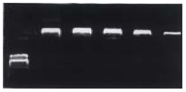
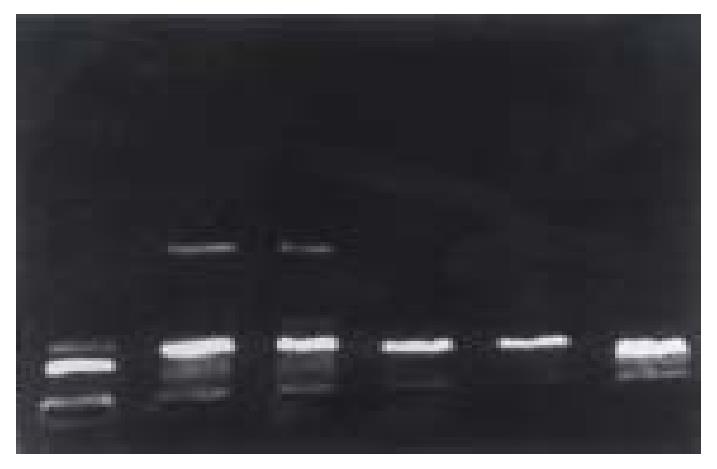
The condition of PCR is 94 °C 5 min, 59 °C 1 min, 74 °C 1 min, 30 cycles and 74 °C 10 min. The PCR results are shown in Figure 1. The PCR product was recovered from the low melting agarose gel and was digested by endonuclease EcoR I and Kpn I simultaneously and was ligated to plasmid pT7Blue with T4 DNA ligase. After recombinant plasmid pT7Blue-HBX- was introduced into DH5 α, seven ampicillin resistant clones were selected and the plasmid DNA was extracted. The correct plasmids identified by the restriction analysis were sequenced.
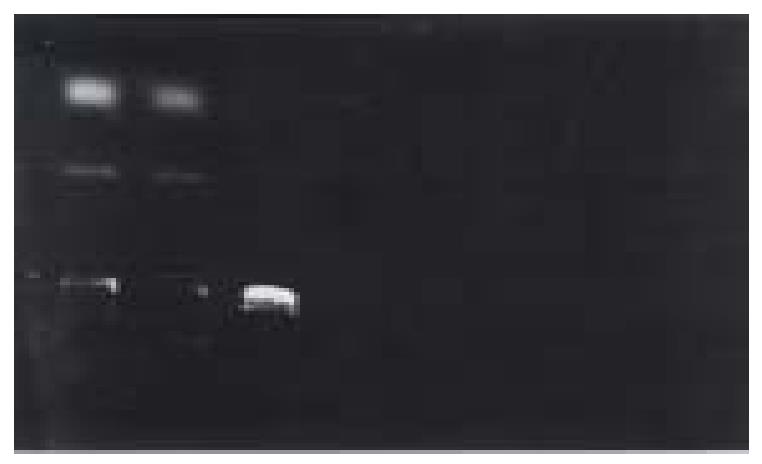
Plasmid pCDNA3.1 and pT7Blue-HBX were digested by endonuclease EcoR I and Kpn I. 480 bp fragment of HBX and 5.4 kb fragment of pCDNA3.1 were recovered from the low melting point agarose gel re spectively. They were ligated by T4 DNA and named pCDNA3.1-HBX. The recombinant DNA was introduced into DH5 α. Eight clones were selected an d correct plasmids were identified by combinative digestion of EcoR I and Kpn I. One colony exhibited 480 bp and 5.4 kb was in right junction named pCDAN3.1-HBX.
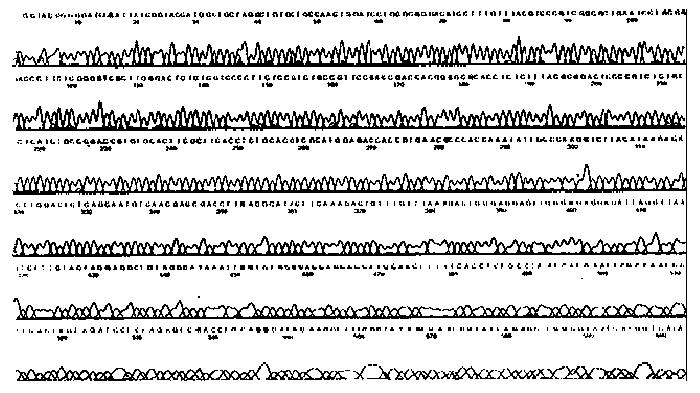
In order to construct the eukaryotic expression vector of HBV x gene, we first constructed the vector pT7Blue-HBX. The PCR result and the gene structure of recombinant plasmid are shown in Figure 1 and Figure 2. The result of the sequence is shown in Figure 3.
On the basis of correct sequence result of pT7Blue-HBX, we inserted HBV x gene at the sites of EcoR I and Kpn I of pCDNA3.1 to construct the eukaryotic expression vector pCDNA3.1-HBX. The recombinant plasmid gene structure is shown in Figure 4.
Hepatitis B virus (HBV) is a formidable threat to public health. HBV associated HCC is one of the 10 commonly encountered cancers in the world. The relative risk of HBV carriers developing HCC approaches 200:1, which is one of the highest relative risks known for a human cancer[4]. HBV x gene and x antigen may play an important role in the development of chronic infections and chronic liver disease. HBV can integrate into cell DNA and the HBV x region is the most frequently integrated sequence[1]. These integrated fragments make HBV encode x antigen that is capable of trans activation both in vitro and in vivo[5]. HBx Ag can stimulate the cell growth directly[6], and inactivate the negative growth regulators, such as tumor suppressor p53 [2]. Furthermore, HBx Ag can increase the resistance of HBx Ag-positive cells to apoptosis mediated by cytotoxic cytokine through altering signal transduction pathway of hepatocyte[2].
In conclusion, the topic of HBV is an old but important one, and is worthy of further studies. We have constructed the eukaryotic expression vector of HBV x gene for this study.
Dr. Shuang-Ping Guo, female, born on December 17, 1967 in Gansu Province, graduated from Department of Medicine in Lanzhou Medical College, engaged in molecular pathology of neoplasm.
Edited by WANG Xian-Lin
| 1. | Feitelson MA, Duan LX. Hepatitis B virus X antigen in the pathogenesis of chronic infections and the development of hepatocellular carcinoma. Am J Pathol. 1997;150:1141-1157. [PubMed] |
| 2. | Wang XW, Forrester K, Yeh H, Feitelson MA, Gu JR, Harris CC. Hepatitis B virus X protein inhibits p53 sequence-specific DNA binding, transcriptional activity, and association with transcription factor ERCC3. Proc Natl Acad Sci USA. 1994;91:2230-2234. [RCA] [PubMed] [DOI] [Full Text] [Cited by in Crossref: 469] [Cited by in RCA: 481] [Article Influence: 15.5] [Reference Citation Analysis (1)] |
| 3. | Beasly RP. Hepatitis B virus. The major etiology of hepatocellular carcinoma. Cancer. 1988;61:1942-1948. [RCA] [DOI] [Full Text] [Cited by in RCA: 19] [Reference Citation Analysis (0)] |
| 4. | Balsano C, Billet O, Bennoun M, Cavard C, Zider A, Grimber G, Natoli G, Briand P, Levrero M. Hepatitis B virus X gene product acts as a transactivator in vivo. J Hepatol. 1994;21:103-109. [RCA] [PubMed] [DOI] [Full Text] [Cited by in Crossref: 37] [Cited by in RCA: 35] [Article Influence: 1.1] [Reference Citation Analysis (0)] |
| 5. | Benn J, Schneider RJ. Hepatitis B virus HBx protein deregulates cell cycle checkpoint controls. Proc Natl Acad Sci USA. 1995;92:11215-11219. [RCA] [PubMed] [DOI] [Full Text] [Cited by in Crossref: 226] [Cited by in RCA: 236] [Article Influence: 7.9] [Reference Citation Analysis (0)] |


