Published online Aug 15, 1999. doi: 10.3748/wjg.v5.i4.301
Revised: July 3, 1999
Accepted: July 19, 1999
Published online: August 15, 1999
AIM: To study the complications and the risk factors of percutaneous liver biopsy, and to compare the complication rate between the periods o f 1987-1993 and 1994-1996.
METHODS: Medical records of all patients undergoing percutaneous liver biopsy between January 1, 1987 to September 31, 1996 in Songklanagarind Hospital were reviewed retrospectively.
RESULTS: There were 484 percutaneous liver biopsies performed. The total complication rate was 6.4%, of which 4.5% were due to major bleeding; the death rate was 1.6%. The important risk factors correlated with bleeding complications and deaths were a platelet count of 70 × 109/L or less, a prolonged prothrombin time of > 3 s over control, or a prolonged activated partial thromboplastin time of > 10 s over control. Although physician inexperience was not statistically significantly associated with bleeding complications and deaths, there was a reduction of death rate from 2.2% in 1987-1993 to 0% in 1993-1996. This reduction is thought to result from both increased experience o f senior staff and increased supervision of residents.
CONCLUSIONS: Screening of platelet count, prothrombin time, and activated partial thromboplastin time should be done and need to be corrected in case of abnormality before liver biopsy. Percutaneous liver biopsy should be performed or supervised by an expert in gastrointestinal diseases, especially in high risk cases.
- Citation: Thampanitchawong P, Piratvisuth T. Liver biopsy: complications and risk factors. World J Gastroenterol 1999; 5(4): 301-304
- URL: https://www.wjgnet.com/1007-9327/full/v5/i4/301.htm
- DOI: https://dx.doi.org/10.3748/wjg.v5.i4.301
Percutaneous liver biopsy is a helpful diagnostic procedure which has been used for 100 years[1-3]. Although ultrasonography, computed tomography, and magnetic resonant imaging are useful in investigation of liver disease, liver biopsy is still essential for diagnosis[4] in the majority of patients. To avoid fatal complications, biopsy must be performed in patients with indications for and no contraindications against biopsy[5]. In our hospital, this procedure has been practised for several years. In 1992 there was an unofficial report of an unusual high complication rate, but this is the first investigative study. After 1994, all liver biopsies were performed by well-trained gastroente rologists or under their supervision; this seemed to lead to a decrease in the complication rate. Thus this study reviewed the incidence of complications in unsupervised biopsy performed between 1987-1993, and compare it with the incidence of complications in supervised biopsy performed between 1994-1996. Risk factors and additional procedures required for complications were evaluated.
We retrospectively collected all percutaneous liver biopsies performed in inpatients between January 1, 1987 and September 31, 1996. Eligible patients were aged 15 years or older and underwent biopsy performed by medical staff or residents using Menghini needle. Recorded data included: demographic data (age and gender); indication for biopsy; post biopsy notes, both immediate and follow-up; hematocrit (Hct) result within 48 h pre-biopsy and 5-48 h post-biopsy (if several Hct were reported, the median was calculated); prothrombin time (PT), p artial thromboplastin time (PTT), platelet count and liver function test within 10 d pre-biopsy; history of FFP and/or platelet transfusion prior to biopsy; ultrasonography report and pathological diagnosis.
Complications were defined as follows: Major bleeding: a decrease in Hct of 4% or more requiring packed red cell (PRC) transfusion and/or surgical intervention. Minor bleeding: a decrease in Hct of 4% or more not requiring intervention (PRC- transfusion or surgery). Transient hypotension: blood pressure of lower than 90/60 mmHg, a decrease in systolic pressure of 20 mmHg or more or a decrease in diastolic pressure of 15 mmHg or more.
Data analysis. A Chi-square and two-tailed Fisher exact test was used to comp are proportions; a two-tailed Student s t test was used to compare means; and a logistic regression analysis was used to determine the best predictors among several independent variables. Probability (P) value of less than 0.05 w as considered to be statistically significant.
Of 484 percutaneous liver biopsies, 50 biopsies were performed in 25 patients (2 biopsies/1 patient). The distribution of biopsies in each time period is summarized in Table 1.
| 1987-1993 | 1994-1996 | P value | |
| Liver biopsies (%) | 367 (75.8) | 117 (24.2) | NS |
| Mean age (years) | 51 | 48 | NS |
| Gender (%) | NS | ||
| Male (n = 372) | 74.5 | 80.4 | |
| Female (n = 112) | 25.5 | 19.6 | |
| Hct (%) | 34.40 ± 8.33 | 33.35 ± 8.39 | NS |
| PT (sec) | 14.15 ± 2.34 | 13.61 ± 1.63 | NS |
| PTT (sec) | 32.61 ± 6.67 | 33.19 ± 5.88 | NS |
| Platelet (× 109/L) | 298 ± 172 | 332 ± 162 | NS |
| TB+ (g/dl) | 1.33 ± 1.22 | 1.30 ± 1.30 | NS |
| AST+ (U/L) | 125 ± 117.64 | 148.44 ± 134.36 | NS |
| ALT+ (U/L) | 82.03 ± 80.37 | 90.86 ± 78.72 | NS |
| ALP+ (U/L) | 355.69 ± 278.32 | 347.10 ± 303.76 | NS |
| Alb+ (g/dl) | 3.54 ± 0.75 | 3.58 ± 0.81 | NS |
| Hepatic malignancy | 49.3 | 36.8 | 0.019 |
Demographic data. The mean age of patients was 50 years (range 15-85 years), 76.9% (372 of 484) of patients were men and 23.1% (112 of 484) were women. There was no difference of age and gender between the periods of 1987-1993 and 1994-1996 (Table 1).
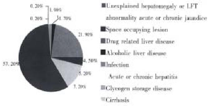
Indications for biopsy and pre-biopsy data. Indications for liver biopsy are shown in Figure 1. The most frequent indication was presence of a space occupying lesion, 57.2% (277 of 484), other indications were unexplained hepatomegaly or liver function test abnormality, 21.9% (106 of 484), acute or chronic hepatitis, 7.2% (35 of 484), acute or chronic jaundice, 5.2% (25 of 484), infection, 4.5% (22 of 484), cirrhosis 1.9% (9 of 484), alcoholic liver disease, 1.7% (9 of 484), drug related liver disease, 0.2% (1 of 484), and glycogen storage disease, 0.2% (1 of 484). All pre-biopsy data comparing between 1987-1993 and 1994-1996 showed no statistically significant difference (Table 1).
Blood transfusion. FFP was transfused in 11.2% (54 of 484) of all liver biopsies, of which 69.8% had PT prolonged > 3 s over control, 41.2% had PTT prolonged > 10 s and 25.9% had abnormal PT and PTT. The average amount of FFP given was 1.8 × 0.3 units (range 2-32 units). Platelet concentrates were transfused in 1.9% (9 of 484) of all liver biopsies, of which 55.6% had a platelet count 70 × 109/L or less and the average amount of platelet concentrate was 1.9 × 0.1 units (range 4-21 units).
Physicians involved in liver biopsy. Between 1987-1993, most percutaneous liver biopsies were performed by non-gastroenterologist without supervision; after 1994 all liver biopsies were performed by well-trained gastroenterologists or by residents under their supervision.
Pathological results. There was a significant decrease of hepatic malignancy from 49.3% (181 of 367) in 1987-1993 to 36.8% (43 of 117) in 1994-1996 (P = 0.019, Table 1).
Complications. The overall complication rate was 6.4% (31 of 484); 45% (17 of 484) were due to major bleeding, 2.7% (13 of 484) due to transient hypotension, and 1.0% (5 of 484) due to minor bleeding. The death rate was 1.6% (8 of 484) but when patients who died from other causes were excluded, it was 1.2%. There was no association between age or gender and complication (P > 0.05). There was no significant difference between major bleeding between 1987-1993 and 1994-1996 (4.72%, 17 of 360) vs 3.4% (5 of 124, P = 0.617). The death rate was reduced from 2.2% (8 of 360) during first period to 0% (0 of 124) in the later period; but this was not statistically significant (P = 00208). Bleeding complication of hepatic malignancy (3.5%, 9 of 260) and non-hepatic malignancy (5.8%, 13 of 224) were not different. 38.7% of pathological findings of complicated cases were hepatic malignancy. Coagulopathy was related to both bleeding complications and death. In patients with a normal PT the bleeding complication rate was 3.6% (15 of 416) and the death rate was 1.0% (4 of 416). The rates increased to 10.5% (6 of 57) and 7% (4 of 57), respectively, when PT was prolonged by > 3 s over control, (P = 0.017 and P = 0.009). Patients with a PTT prolonged by > 10 s over control also had increased rates of bleeding complications (10.3% vs 3.8%) among patients with normal PTT (P = 0.079). The death rate in patients with prolonged PTT, on the other hand, was significantly higher than that in patients with a normal PTT (7.7% vs 102%, P = 0.023). There was also a significant difference in both bleeding and death rates between patients with platelet counts ≤ 70 × 109/L and those with platelet counts greater than 70 × 109/L (bleeding rate: 25.0% vs 4.0%, P = 0.014; death rate: 33.3% vs 0.8%, P = 0.001).
Logistic regression analysis of the independent variables demonstrated that the most important predictors for bleeding (P = 0.000) and death(P = 0.003) was only platelet count 70 × 109/L or less.
Eight patients (1.6%) died after liver biopsy between 1987-1993. Only five of these deaths could be definitely attributed to liver biopsy and one additional death was probably related to biopsy procedures. Therefore, the actual death rate should be 1.2% (6 of 484). In three of the attributable deaths, bleeding and puncture wounds were observed during laparotomy; in two other cases, Hct was markedly decreased both 3 and 24 h post-biopsy. One patient developed sudden dyspnea and death in which pneumothorax could not be excluded; thus, this death is probably related to biopsy. Two of the deaths may not have been associated with liver biopsy; the first patient died of pulmonary hemorrhage and the second of pneumonia with sepsis.
In this patient population, the overall complication rate was 6.4% and the over all death rate was 1.6%. Our complication and death rates were unusually high a s compared with other studies which have shown a complication rate of 0.13%-3.2%, and a mortality rate 0%-0.33%[1,6,7]. However, MaVay and associates also reported a high rate of major bleeding (4.5%)[6].
We found the most important independent risk factors for bleeding complications and death were platelet count of 70 × 109/L or less. However, prolonged PT > 3 s over control and prolonged PTT > 10 s over control also contributed to the increasing bleeding complications. Our study support those of commonly considered contraindications for liver biopsy that is a platelet count of 50.80 × 10⁹/L or less or a PT > 3 s[1,8]. Shama et al[9] also found no bleeding in 16 patients with platelet counts of 61.90 × 10⁹/L, but 3 of 13 patients with platelet counts of 3060 × 109/L bled. Ewe, while investigating laparoscopic liver bleeding time, found bleeding from the biopsy site occurred in cases with a platelet count of 50 × 109/L or less, but this bleeding could be stopped with compression[10]. Although these studies demonstrated no increased risk of bleeding with a platelet count greater than 60 × 109/L, one patient in our study with platelet count of 63 × 109/L had post liver biopsy bleeding. Therefore, we chose a platelet count of greater than 70 × 109/L as the safety margin for doing liver biopsy. Sherlock also recommended that liver biopsy should not be performed when platelet count was less than 80 × 109/L[1]. In this study, PT being greater than 3 s is a risk for bleeding complication, but McVay et al[6] have reported that mild prolongation of PT and PTT 1.1-1.5 times that of normal is not associated with increased bleeding complications. Other studies have also shown no increased risk for bleeding in open surgical procedures if PT and PTT were prolonged by 1.5 times or less[3,11]. Therefore, it has been suggested that bleeding after liver biopsy is a random event that cannot be predicted by currently available methods[9,11], local intrahepatic clotting and elastic factors may prevent bleeding[10].
In this study, the death rate decreased from 2.2% in 1987-1993 to 0% in 1994- 1996. This decrease was not statistically significant probably due to the small sample size of patients biopsied between 1994-1996. Gilmore et al[3] reported a frequency of bleeding complications of 3.25% when biopsies were performed by inexperienced physicians (less than 20 previous biopsies performed), which was higher than the complication rate of 1.1% when biopsies were performed by physicians who had done more than 100 biopsies before. A survey in Switzerland also showed the complication rate was markedly higher in physicians with less experience (0.88% in physicians who performed biopsies less than 12 times per year vs 0.06% in physicians who performed biopsies 50 or more times per year)[12]. This may suggest that liver biopsy complication rate is determined by both the experience and training of those performing the procedure.
As in other studies, the most frequent indication for liver biopsy was the presence of a space occupying lesion and the most frequent pathological diagnosis was hepatic malignancy[6,9]. Our study showed no association between pathological findings and bleeding complications. Hepatic malignancy and cirrhosis have previously been identified as risk factors by Gonalez-Vallina, Mc Gill, and Fisher et al[6,8,13]. In these reports, Vim-Silverman and Tru-cut needles were frequently used and may have caused increased bleeding. Malignancy may also predispose to bleeding because of hypervascularity and capillary fragility, cirrhosis is thought to increase the risk of bleeding due to large subcapsular venous pooling[9]. However, Paisal showed no significant increase in bleeding due to cirrhosis[14].
Bleeding complications of percutaneous liver biopsy may occur in the setting of a platelet counts of 70 × 109/L or less, a prolonged PT > 3 s over control, or a PTT > 10 s over control. In order to minimize complications, all pre biopsy patients should be screened with platelet counts, PT, and PTT. If any of these value is abnormal, it should be corrected with FFP or platelet concentrate before liver biopsy. This study showed that physician experience is not a statistically significant risk factor for complications of liver biopsy. However other reports have suggested that performance of liver biopsy by untrained or inexperienced physician increase complications. Therefore, percutaneous liver biopsy should be performed or supervised by an expert in gastroenterology; this is especially important in high risk cases.
The authors are grateful to Ms. Nualta Arpakoppakul of the Department of Epidemiology for her help in statistical analysis, and to Ms. Anne Kasmar, and Mr. Ben Worley for their help in English language advice.
Edited by MA Jing-Yun
| 1. | Sherlock S. Diseases of the liver and biliary system: needle biopsy of the liver. 8th ed. Boston, Melbourne: Blackwell Scientific Publications 1989; 36-48. |
| 2. | Valori R, Elias E. How to perform a percutaneous liver biopsy. Br J Hosp Med. 1989;42:408-410. [PubMed] |
| 3. | Gilmore IT, Burroughs A, Murray-Lyon IM, Williams R, Jenkins D, Hopkins A. Indications, methods, and outcomes of percutaneous liver biopsy in England and Wales: an audit by the British Society of Gastroenterology and the Royal College of Physicians of London. Gut. 1995;36:437-441. [PubMed] |
| 4. | Stotland BR, Lichtenstein GR. Liver biopsy complications and routine ultrasound. Am J Gastroenterol. 1996;91:1295-1296. [PubMed] |
| 5. | McGill DB. Predicting hemorrhage after liver biopsy. DigDis Sci. 1981;26:235-237. |
| 6. | McVay PA, Toy PT. Lack of increased bleeding after liver biopsy in patients with mild hemostatic abnormalities. Am J Clin Pathol. 1990;94:747-753. [PubMed] |
| 7. | Hegarty JE, Williams R. Liver biopsy: techniques, clinical applications, and complications. Br Med J (Clin Res Ed). 1984;288:1254-1256. [PubMed] |
| 8. | Schiff ER, Schiff L. Disease of the liver: needle biopsy of the liver. 7th editor. Philadelphia: Lippincott. 1993;216-225. |
| 9. | Sharma P, McDonald GB, Banaji M. The risk of bleeding after percutaneous liver biopsy: relation to platelet count. J Clin Gastroenterol. 1982;4:451-453. [PubMed] |
| 10. | Ewe K. Bleeding after liver biopsy does not correlate with indices of peripheral coagulation. Dig Dis Sci. 1981;26:388-393. [PubMed] |
| 11. | Pasha T, Gabriel S, Therneau T, Dickson ER, Lindor KD. Cost-effectiveness of ultrasound-guided liver biopsy. Hepatology. 1998;27:1220-1226. [PubMed] |
| 12. | Froehlich F, Lamy O, Fried M, Gonvers JJ. Practice and complications of liver biopsy. Results of a nationwide survey in Switzerland. Dig Dis Sci. 1993;38:1480-1484. [RCA] [PubMed] [DOI] [Full Text] [Cited by in Crossref: 116] [Cited by in RCA: 102] [Article Influence: 3.2] [Reference Citation Analysis (1)] |
| 13. | Gonzalez-Vallina R, Alonso EM, Rand E, Black DD, Whitington PF. Outpatient percutaneous liver biopsy in children. J Pediatr Gastroenterol Nutr. 1993;17:370-375. [PubMed] |
| 14. | Pongchairerks P. Ultrasound-guided liver biopsy: accuracy, safety and sonographic findings. J Med Assoc Thai. 1993;76:597-600. [PubMed] |


