Letter to the Editor Open Access
Copyright ©The Author(s) 2025. Published by Baishideng Publishing Group Inc. All rights reserved.
World J Gastroenterol. Mar 7, 2025; 31(9): 100221
Published online Mar 7, 2025. doi: 10.3748/wjg.v31.i9.100221
Perianal fistulizing Crohn’s disease: Mechanisms and treatment options focusing on cellular therapy
Payal Bhatnagar, Department of Pharmaceutical Technology, School of Pharmacy, IMU University, Kuala Lumpur 57000, Malaysia
Sherreen Elhariri, Ismail A S Burud, Department of Surgery, IMU University, Clinical Campus, Seremban 70300, Negeri Sembilan, Malaysia
Nabil Eid, Department of Anatomy, Division of Human Biology, School of Medicine, IMU University, Kuala Lumpur 57000, Malaysia
ORCID number: Sherreen Elhariri (0000-0001-7604-6708); Ismail A S Burud (0000-0003-0326-424X); Nabil Eid (0000-0002-2938-2618).
Author contributions: Bhatnagar P designed the figure; Burud IAS revised the manuscript; Bhatnagar P, Elhariri S, and Eid N wrote the manuscript; Eid N approved the final draft; and all authors have read and approved the final manuscript.
Conflict-of-interest statement: All the authors report no relevant conflicts of interest for this article.
Open Access: This article is an open-access article that was selected by an in-house editor and fully peer-reviewed by external reviewers. It is distributed in accordance with the Creative Commons Attribution NonCommercial (CC BY-NC 4.0) license, which permits others to distribute, remix, adapt, build upon this work non-commercially, and license their derivative works on different terms, provided the original work is properly cited and the use is non-commercial. See: https://creativecommons.org/Licenses/by-nc/4.0/
Corresponding author: Nabil Eid, Associate Professor, Academic Editor, Senior Lecturer, MD, PhD, Department of Anatomy, Division of Human Biology, School of Medicine, IMU University, No. 126 Jalan Jalil Perkasa 19, Kuala Lumpur 57000, Malaysia. nabilsaleheid@imu.edu.my
Received: August 10, 2024
Revised: January 6, 2025
Accepted: January 21, 2025
Published online: March 7, 2025
Processing time: 192 Days and 2.9 Hours

Abstract

Perianal fistulizing Crohn’s disease (PFCD) is a common presentation of CD, which affects the patients’ quality of life, including social and sexual function. The management of PFCD remains a critical challenge in inflammatory bowel disease, primarily due to limited understanding of the mechanisms involved in its pathogenesis, complicating medical treatment. Increased production of inflammatory cytokines such as tumor necrosis factor and interleukin-13 by infiltrating macrophages and other inflammatory cells stimulate the epithelial-to-mesenchymal transition, resulting in activation of myofibroblasts and elevation of matrix metalloproteinases, leading to fistula formation. Given the potential for malignant transformation, PFCD screening is critical. Cytokine and inflammation-targeted therapies can help control this disease, but recurrence is a common complication. Surgical interventions such as fistulotomy represent viable therapeutic options, with magnetic resonance imaging serving as an important diagnostic tool for delineating fistula tract anatomy. Animal models and clinical trials demonstrate that injection of mesenchymal stem cells (MSCs) into the fistula results in suppression of the inflammatory cells and cytokines and complete resolution of PFCD. Recently, MSC-derived extracellular vesicles were found to stimulate fistula healing, with encouraging results. In this article, we comment on the review article by Pacheco et al, summarizing the various lines of PFCD treatment and highlighting the role of screening for this disease. Importantly, we focus on the various mechanisms involved in the pathogenesis of PFCD, the therapeutic roles of MSCs and related extracellular vesicles, and explore the potential role of autophagy in enhancing the therapeutic efficacy of these cells, which may help in the treatment of this disease.

Key Words: Stem cell therapy; Crohn’s disease; Perianal fistulas; Regenerative medicine; Treatment outcomes; Extracellular vesicles; Autophagy; Magnetic resonance imaging

Core Tip: Perianal fistulizing Crohn’s disease (PFCD) is a common complication of CD, characterized by complex pathogenic mechanisms involving inflammatory cell accumulation, particularly macrophages, and enhanced cytokine production. These processes drive epithelial-to-mesenchymal transition, myofibroblast activation, upregulation of matrix metalloproteinases, ultimately leading to fistula formation. While various medical and surgical options target inflammatory processes in this disease, recurrence remains a substantial challenge. Local administration of mesenchymal stem cells or their derived extracellular vesicles has emerged as a promising therapeutic strategy through suppression of the inflammatory cells and promotion of fistula healing in PFCD. Here we summarize the various mechanisms involved in PFCD pathogenesis, with particular emphasis on mesenchymal stem cell therapy and their derived extracellular vesicles.


TO THE EDITOR

One of the most challenging complications of Crohn’s disease (CD) is perianal fistula, with an estimated lifetime risk of 14%-38% in population-based estimates[1,2]. This condition significantly impacts the quality of life through its presentation of pain, perianal discharge, and faecal incontinence, impacting the patients’ physical, social, and sexual well-being. Management of perianal fistulizing CD (PFCD) requires a multidisciplinary approach, encompassing medical interventions, surgical procedures, and other newer forms of treatment such as stem cell therapy. The review article by Pacheco et al[1] presents various medical interventions for PFCD, including antibiotics, thiopurines, tacrolimus, anti-tumor necrosis factor (TNF) agents, vedolizumab, ustekinumab, and hyperbaric oxygen therapy. Endoscopic approaches and innovative surgical techniques such as ligation of the inter-sphincteric fistula tract, advancement flaps, fistula plugs, fibrin glue injection, fistula laser closure, and video-assisted anal fistula treatment, are discussed as well. In addition, the authors highlighted the role of stem cells in the treatment of this disease and the importance of screening to prevent complications such as cancer.

A review of the various modalities of therapeutic approaches revealed that antibiotics, mainly ciprofloxacin in combination with infliximab, demonstrated superior healing outcomes when compared to anti-TNF monotherapy[2]. Notably, higher infliximab drug levels (median level at 2 weeks: 20 μg/mL vs 5.6 μg/mL) correlated with better response rates in terms of fistula healing and closure[3]. The healing process can be monitored through magnetic resonance imaging (MRI), with successful healing characterized by the resolution of T2-weighted image hyperintensity and absence of enhancement 12 months post-treatment[4]. Disease recurrence and maintenance of remission remain significant challenges, hence the decision to continue or stop anti-TNF treatment remains unanswered. Current evidence suggests that disease relapse is associated with discontinuation of treatment, supporting the continuation of anti-TNF therapy to achieve clinical remission[5]. Therapeutic drug monitoring is important to improve fistula healing rates, as higher dosing regimens are required for treating PFCD. While anti-TNF therapy using infliximab and adalimumab remains the preferred medical intervention for perianal CD, some patients demonstrate non-response to anti-TNF therapy[6].

Where PFCD is associated with perianal abscess, adequate drainage of the abscess must precede any definite surgery. The standard surgical procedures in the management of perianal fistula include seton placement and fistulectomy[7]. However, surgical interventions for PFCD demonstrate relatively moderate success rates: For example, ligation of the inter-sphincteric fistula tract achieves 53% success, advancement flaps 61%, fistula plugs 50%-60% and fibrin glue injection 38%. Importantly, surgical fistula closure should only be attempted after endoscopic remission of the proctitis[1]. Recent surgical innovations for PFCD include ablative procedures targeting the fistula tract epithelium, such as fistula laser closure and video-assisted anal fistula treatment[7].

Long-standing CD carries a significant risk of cancer development, particularly squamous cell carcinoma and adenocarcinoma. Patients with CD have a 4- to 20-fold increased risk of colorectal carcinoma[8]. The incidence of cancer arising from CD-associated fistulas is 0.2/1000 person-year, and the overall incidence of fistula-associated anal cancer is 0.3% to 0.7%[9]. The latency period between initial diagnosis of CD and detection of carcinoma varied between 18 and 30 years[10]. Detection of carcinoma in perianal CD is difficult, with malignancy often discovered at the advanced stage during surgical intervention, resulting in poor prognosis. Definitive diagnosis typically requires biopsy or curettage performed during examination under anaesthesia[11].

MRI: The primary radiological modality for PFCD investigation

MRI demonstrates utility not only for evaluation of fistulizing disease, but also in the diagnosis of perianal carcinoma. Characteristic MRI findings suggestive of perianal neoplasia in patients include irregular inner wall morphology, delayed enhancement of internal tissue, and mucinous patterns. While the fistula wall is frequently observed, this finding alone is insufficient to differentiate cancer from inflammation. Additionally, a distinct double-layered enhancement pattern characterized by an early bright external wall enhancement contrasting with a darker delayed-enhancing internal wall may be observed[12].

Mechanisms of PFCD: Roles of inflammatory cells, cytokines, and epithelial-to-mesenchymal transition

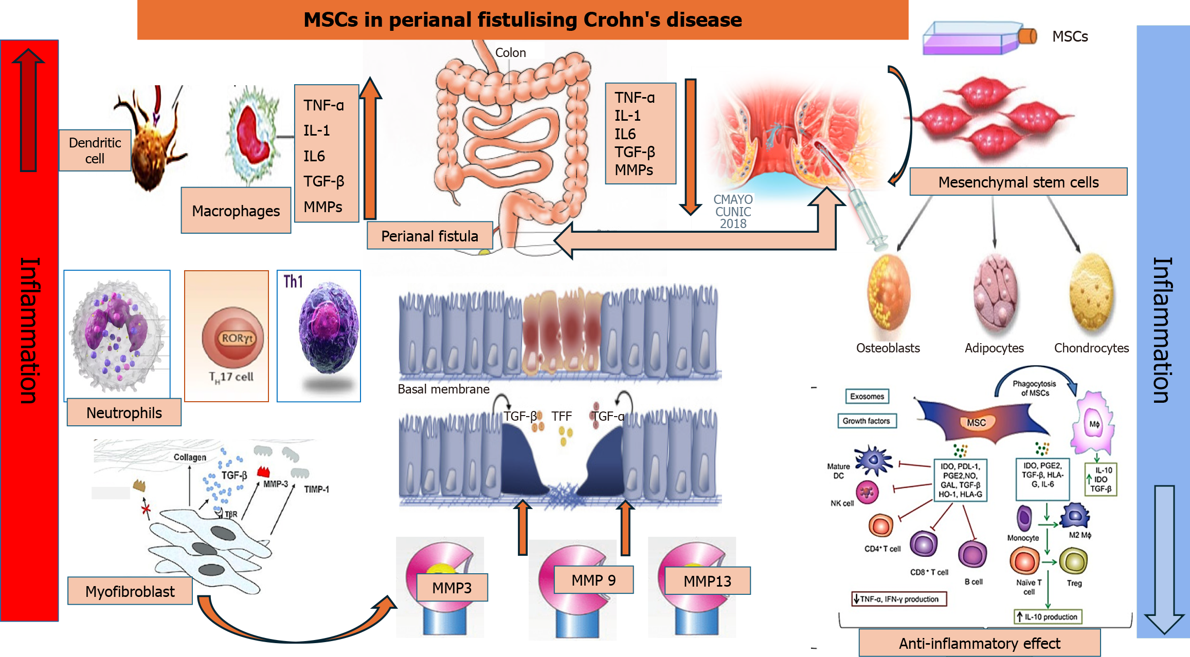
Perianal fistulas in CD, which can affect the small and large intestines, are characterized by narrow squamous epithelial cells. These cells undergo epithelial-to-mesenchymal transition to become transition cells in response to immunological triggers or bacterial invasion. As a result, the cells lose their phenotypic and functional properties such as polarity and adhesiveness, while acquiring mesenchymal cell traits. This leads to reduced cell-to-cell adhesion and enhanced migratory activity[13]. This anomaly results in a cascade of cellular events, such as infiltration of inflammatory cells, i.e., neutrophils, T helper (Th) cells, M1 macrophages, dendritic cells, and myofibroblasts, as well as the release of inflammatory cytokines and regulatory proteins (Figure 1). Consequently, the peculiar channel of transition cells appears to be the outcome of chronic inflammation spreading through the mucosal wall, which in turn leads to the development of fistulas[14].

Figure 1
Figure 1 Mechanisms of perianal fistulizing Crohn’s disease and therapeutic roles of mesenchymal stem cells. Enhanced production of inflammatory cytokines by infiltrating macrophages and other inflammatory cells stimulates the epithelial-to-mesenchymal transition, resulting in activation of myofibroblasts and elevation of matrix metalloproteinases, leading to fistula formation. Injection of mesenchymal stem cells into the fistula results in suppression of the inflammatory cells and cytokines and complete resolution of perianal fistulizing Crohn’s disease. MSCs: Mesenchymal stem cells; TNF: Tumor necrosis factor; IL: Interleukin; TGF: Transforming growth factor; MMP: Matrix metalloproteinase; Th: T helper cells; TFF: Trefoil factor family; DC: Dendritic cell; NK: Natural killer; Treg: Regulatory T cell; IDO: Indolamine 2,3-dioxygenase; PDL-1: Programmed death ligand 1; PGE2: Prostaglandin E2; NO: Nitrogen oxide; GAL: Gallic acid; HO-1: Heme oxygenase-1; HLA: Human leukocyte antigen.

Immunohistochemical analyses of CD-related perianal fistulas indicate acute inflammation in 56% of cases[13], characterized by significant infiltration of T cells into the inner wall of the fistula, along with pronounced activation of inflammatory M1 macrophages. Investigations into the phenotypic and functional characteristics of T cells derived from individuals with PFCD revealed significant infiltration of the fistula by T cells with Th17, Th1, and Th17/1 phenotypes[14]. The inflammatory cascade underlying PFCD is predominantly mediated by inflammatory cytokine activation. Severe intestinal inflammation triggers the release of TNF-α, interleukin-6 (IL-6), IL-13, and transforming growth factor-beta (TGF-β). The abundance of these cytokines in the lining of fistula tracts, adjacent tissue, and peripheral blood suggests their involvement in the pathogenesis of PFCD[15].

Matrix metalloproteinases (MMPs) play crucial roles in tissue degradation and remodeling. Excessive MMP activity can cause cancer or inflammatory bowel disease through abnormal degradation of the extracellular matrix[16]. Kiesler et al[17] and Washburn et al[18] identified elevated expression of MMP-3 in CD-associated fistula tissue regardless of the inflammatory state and increased goblet cell presence when compared to controls. Additionally, they found that MMP-3 and MMP-9 were produced by mononuclear cells and fibroblasts, especially in fistulas with active inflammation. Furthermore, MMP-13 protein expression was detected in CD-associated fistulas, but was almost absent in non-fistulizing CD tissue[13]. This provides evidence for the role of MMPs as mediators in the pathogenesis of perianal fistula in CD through abnormal degradation of the extracellular matrix in the bowel wall.

Mesenchymal stem cell therapy in PFCD

Mesenchymal stem cells (MSCs) are multipotent stromal cells with self-renewal and differentiation ability. They are acquired from the umbilical cord, bone marrow, adipose tissue, and various other sources, and can be obtained as autologous or allogeneic, depending on clinical requirements[19,20]. MSCs exhibit well-documented immunomodulatory effects, tissue regeneration properties, and antiaging and anti-inflammatory potential via regulation of function of T cells, B cells, natural killer cells, macrophages, neutrophils and dendritic cells. This regulation occurs via changing of the local inflammatory cell environment, migration to inflammation sites, and reduction of immune response (Figure 1)[21,22]. The precise regulatory mechanism of MSCs in perianal fistula is not well-established and suggested to be via immunosuppression, i.e., controlling the hyperactivity of T-cells, release of inflammatory cytokines such as TGF-β, TNF-α, IL-7, IL-6 and IL-13, and secretion of anti-inflammatory molecules such as IL-10. Through these mechanisms, MSCs maintain an anti-inflammatory environment that is conducive to the repair of eroded tissue in the perianal area, particularly in the context of PFCD[23].

The application of MSCs in phase II and phase III clinical trials has demonstrated significant therapeutic efficacy in PFCD. MSCs, commonly derived from adipose tissue or bone marrow, release anti-inflammatory cytokines through autocrine or paracrine mechanisms to promote blood vessel repair and re-epithelialization[24]. Swaroop et al[25] conducted a trial to examine the therapeutic potential of locally administered human allogeneic bone marrow-derived mesenchymal stromal cells in PFCD patients non-responsive to conventional treatments. In an open-trial, phase I/II, single-arm study, clinical severity and biomarkers were analyzed at baseline through week 104, incorporating MRI scans at weeks 24 and 104. The authors found that across various clinical trials, local administration of MSCs demonstrated favorable safety profile with no significant adverse effects compared to the placebo group[25]. Intraperitoneal injection of human MSCs into SAMP-1/YitFc mice models for CD achieved long-term therapeutic efficacy through sustained anti-inflammatory macrophage programming via efferocytosis[26]. Stem cells have emerged as a promising therapeutic option for Crohn’s fistula, based on its high efficacy and lower incidence of adverse events. Clinical studies have shown that a 3 × 10⁷ cells/mL dosage yields superior outcomes in patients with Crohn’s fistula[27]. However, further clinical and pre-clinical studies are needed to strengthen the evidence base for this approach in the future.

MSC-derived extracellular vesicles therapy for PFCD

Mammalian cells secrete extracellular vesicles (EVs), which serve as vehicles for functional nucleic acids (mRNA, miRNA, and other RNA species) to facilitate cellular communication and uptake both in vitro and in vivo. EVs are categorized based on size into two primary subsets: Exosomes (50–100 nm in diameter), which have an endosomal origin, and microvesicles (100–1000 nm in diameter), derived from cytoplasmic budding[28]. Recently, MSC-derived EVs have emerged as an effective and safe therapeutic modality for the treatment of various autoimmune conditions, including CD[29]. In the dextran sodium sulfate-induced mouse model of colitis, intravenous injection of human umbilical cord-derived exosomes resulted in reduced macrophage infiltration and inflammatory cytokines in colonic tissue, with improvement of colitis[30]. Moreover, a recent phase I clinical trial demonstrated complete tract resolution in 5 of 11 patients with non-CD fistulizing disease following injection of human placental MSCs-derived EVs over a 6-month period[31]. These findings underscore the need for further investigation of both local and systemic administration of EVs in the treatment of PFCD through targeted animal models and expanded clinical trials.

Autophagy modulation in MSCs and EVs for treatment of PFCD

Macroautophagy (hereafter referred to as autophagy) is a prosurvival mechanism for the clearance of damaged cellular components in response to various stressors, such as oxidative stress, inflammation, and hypoxia[32,33]. Therefore, in the context of inflammatory conditions such as CD, autophagy activation in MSCs may serve as a protective mechanism. Recent in vitro investigations reported that autophagy upregulation in MSCs by rapamycin enhanced the immunosuppression of inflammatory CD4+ T cells via secretion of TGF-β1[34]. Moreover, autophagy plays an important role in the differentiation of MSCs at the cellular and molecular levels, thereby influencing their therapeutic potential across a wide range of diseases, including PFCD[35]. Notably, autophagy-induced MSC-derived EVs have demonstrated therapeutic efficiency in attenuating renal fibrosis in vitro via reduction of pro-inflammatory gene expression, including IL-1 and TNF-α[36]. Taken together, these findings suggest that autophagy upregulation in MSCs may enhance their immunosuppressive functions and survival, in addition to the production of effective EVs, potentially advancing the treatment of autoimmune diseases such as PFCD.

Conclusions

PFCD is a common presentation of CD. Various mechanisms are involved in fistula formation, mainly related to the accumulation of inflammatory cells such as M1 macrophages and the production of inflammatory cytokines. This results in epithelial-to-mesenchymal transition, myofibroblast activation, and upregulation of MMPs, culminating in PFCD. While various medical and surgical options target the underlying inflammation, disease recurrence remains a frequent complication. The potential progression to anal carcinoma necessitates screening protocols. MSC therapy has emerged as a relatively novel local therapeutic approach for PFCD, with MSC-derived EVs demonstrating enhanced fistula-healing properties. Further research in vitro and in vivo using specific animal models for PFCD is needed to explore the therapeutic roles of MSCs and EVs in both luminal and fistulizing CD. Moreover, additional studies, including larger randomized controlled trials, long-term follow-up studies, and investigations into patient selection criteria, are required. Additionally, investigation into the potential roles of autophagy modulation in MSCs is needed to enhance their immunosuppressive properties and optimize production of high-quality EVs for treatment of PFCD.

Footnotes

Provenance and peer review: Invited article; Externally peer reviewed.

Peer-review model: Single blind

Specialty type: Gastroenterology and hepatology

Country of origin: Malaysia

Peer-review report’s classification

Scientific Quality: Grade B, Grade C, Grade D

Novelty: Grade B, Grade B, Grade B

Creativity or Innovation: Grade B, Grade B, Grade B

Scientific Significance: Grade B, Grade B, Grade C

P-Reviewer: Huang C; Wang S S-Editor: Wei YF L-Editor: A P-Editor: Zheng XM

References
1.  Pacheco T, Monteiro S, Barros L, Silva J. Perianal disease in inflammatory bowel disease: Broadening treatment and surveillance strategies for anal cancer. World J Gastroenterol. 2024;30:3373-3385.  [PubMed]  [DOI]  [Cited in This Article: ]  [Reference Citation Analysis (2)]
2.  Lee MJ, Parker CE, Taylor SR, Guizzetti L, Feagan BG, Lobo AJ, Jairath V. Efficacy of Medical Therapies for Fistulizing Crohn's Disease: Systematic Review and Meta-analysis. Clin Gastroenterol Hepatol. 2018;16:1879-1892.  [PubMed]  [DOI]  [Cited in This Article: ]  [Cited by in Crossref: 76]  [Cited by in RCA: 87]  [Article Influence: 12.4]  [Reference Citation Analysis (0)]
3.  Yarur AJ, Kanagala V, Stein DJ, Czul F, Quintero MA, Agrawal D, Patel A, Best K, Fox C, Idstein K, Abreu MT. Higher infliximab trough levels are associated with perianal fistula healing in patients with Crohn's disease. Aliment Pharmacol Ther. 2017;45:933-940.  [PubMed]  [DOI]  [Cited in This Article: ]  [Cited by in Crossref: 184]  [Cited by in RCA: 223]  [Article Influence: 27.9]  [Reference Citation Analysis (1)]
4.  Richard N, Derinck A, Bridoux V, Charpentier C, Savoye Collet C, Savoye G. Which magnetic resonance imaging feature is associated with treatment response in perianal fistulizing Crohn's disease? Abdom Radiol (NY). 2024;49:2913-2920.  [PubMed]  [DOI]  [Cited in This Article: ]  [Reference Citation Analysis (0)]
5.  Mak JWY, Tang W, Yip TCF, Ran ZH, Wei SC, Ahuja V, Kumar S, Leung WK, Hilmi I, Limsrivilai J, Aniwan S, Lam BCY, Chan KH, Ng KM, Leung CM, Li MKK, Lo FH, Sze ASF, Tsang SWC, Hui AJ, Hartono JL, Ng SC. Stopping anti-tumour necrosis factor therapy in patients with perianal Crohn's disease. Aliment Pharmacol Ther. 2019;50:1195-1203.  [PubMed]  [DOI]  [Cited in This Article: ]  [Cited by in Crossref: 6]  [Cited by in RCA: 5]  [Article Influence: 0.8]  [Reference Citation Analysis (0)]
6.  Vasudevan A, Bruining DH, Loftus EV Jr, Faubion W, Ehman EC, Raffals L. Approach to medical therapy in perianal Crohn's disease. World J Gastroenterol. 2021;27:3693-3704.  [PubMed]  [DOI]  [Cited in This Article: ]  [Cited by in CrossRef: 8]  [Cited by in RCA: 4]  [Article Influence: 1.0]  [Reference Citation Analysis (3)]
7.  Adegbola SO, Sahnan K, Twum-Barima C, Iqbal N, Reza L, Lung P, Warusavitarne J, Tozer P, Hart A. Current review of the management of fistulising perianal Crohn's disease. Frontline Gastroenterol. 2021;12:515-523.  [PubMed]  [DOI]  [Cited in This Article: ]  [Cited by in Crossref: 8]  [Cited by in RCA: 5]  [Article Influence: 1.0]  [Reference Citation Analysis (0)]
8.  Fiorillo C, Schena CA, Quero G, Laterza V, Pugliese D, Privitera G, Rosa F, Schepis T, Salvatore L, Di Stefano B, Larosa L, Minordi LM, Natale L, Tortora G, Armuzzi A, Alfieri S. Challenges in Crohn's Disease Management after Gastrointestinal Cancer Diagnosis. Cancers (Basel). 2021;13:574.  [PubMed]  [DOI]  [Cited in This Article: ]  [Cited by in Crossref: 2]  [Cited by in RCA: 3]  [Article Influence: 0.8]  [Reference Citation Analysis (0)]
9.  Sharma P, Putambekar V, Kumar P, Thomas DM, Vuyyuru SK, Kante B, Mundhra SK, Sharma R, Dash NR, Makharia G, Kedia S, Ahuja V. Incidence of intestinal & extra-intestinal cancers among individuals with Crohn's disease in northern India. Indian J Med Res. 2024;160:61-69.  [PubMed]  [DOI]  [Cited in This Article: ]  [Reference Citation Analysis (0)]
10.  Burke KE, Nayor J, Campbell EJ, Ananthakrishnan AN, Khalili H, Richter JM. Interval Colorectal Cancer in Inflammatory Bowel Disease: The Role of Guideline Adherence. Dig Dis Sci. 2020;65:111-118.  [PubMed]  [DOI]  [Cited in This Article: ]  [Cited by in Crossref: 19]  [Cited by in RCA: 18]  [Article Influence: 3.6]  [Reference Citation Analysis (0)]
11.  Shwaartz C, Munger JA, Deliz JR, Bornstein JE, Gorfine SR, Chessin DB, Popowich DA, Bauer JJ. Fistula-Associated Anorectal Cancer in the Setting of Crohn's Disease. Dis Colon Rectum. 2016;59:1168-1173.  [PubMed]  [DOI]  [Cited in This Article: ]  [Cited by in Crossref: 21]  [Cited by in RCA: 27]  [Article Influence: 3.0]  [Reference Citation Analysis (0)]
12.  Horio Y, Ikeda J, Matsumoto K, Okada S, Nagano K, Kusunoki K, Kuwahara R, Kimura K, Kataoka K, Beppu N, Uchino M, Ikeda M, Okadome T, Yamakado K, Ikeuchi H. Machine learningbased radiomics models accurately predict Crohn's diseaserelated anorectal cancer. Oncol Lett. 2024;28:421.  [PubMed]  [DOI]  [Cited in This Article: ]  [Reference Citation Analysis (0)]
13.  McGregor CGC, Tandon R, Simmons A. Pathogenesis of Fistulating Crohn's Disease: A Review. Cell Mol Gastroenterol Hepatol. 2023;15:1-11.  [PubMed]  [DOI]  [Cited in This Article: ]  [Cited by in Crossref: 1]  [Reference Citation Analysis (0)]
14.  Rizzo G, Rubbino F, Elangovan S, Sammarco G, Lovisa S, Restelli S, Pineda Chavez SE, Massimino L, Lamparelli L, Paulis M, Maroli A, Roda G, Shalaby M, Carvello M, Foppa C, Drummond SP, Spaggiari P, Ungaro F, Spinelli A, Malesci A, Repici A, Day AJ, Armuzzi A, Danese S, Vetrano S. Dysfunctional Extracellular Matrix Remodeling Supports Perianal Fistulizing Crohn's Disease by a Mechanoregulated Activation of the Epithelial-to-Mesenchymal Transition. Cell Mol Gastroenterol Hepatol. 2023;15:741-764.  [PubMed]  [DOI]  [Cited in This Article: ]  [Cited by in Crossref: 8]  [Reference Citation Analysis (0)]
15.  Lightner AL, Ashburn JH, Brar MS, Carvello M, Chandrasinghe P, van Overstraeten AB, Fleshner PR, Gallo G, Kotze PG, Holubar SD, Reza LM, Spinelli A, Strong SA, Tozer PJ, Truong A, Warusavitarne J, Yamamoto T, Zaghiyan K. Fistulizing Crohn's disease. Curr Probl Surg. 2020;57:100808.  [PubMed]  [DOI]  [Cited in This Article: ]  [Cited by in Crossref: 8]  [Cited by in RCA: 9]  [Article Influence: 1.8]  [Reference Citation Analysis (0)]
16.  Anandabaskaran S, Hanna L, Iqbal N, Constable L, Tozer P, Hart A. Where Are We and Where to Next?-The Future of Perianal Crohn's Disease Management. J Clin Med. 2023;12:6379.  [PubMed]  [DOI]  [Cited in This Article: ]  [Cited by in Crossref: 3]  [Reference Citation Analysis (0)]
17.  Kiesler P, Fuss IJ, Strober W. Experimental Models of Inflammatory Bowel Diseases. Cell Mol Gastroenterol Hepatol. 2015;1:154-170.  [PubMed]  [DOI]  [Cited in This Article: ]  [Cited by in Crossref: 385]  [Cited by in RCA: 461]  [Article Influence: 46.1]  [Reference Citation Analysis (1)]
18.  Washburn S, Maddipatla SC, Murthy S, Dodd A, Pelia RS, Kolachala VL, Geem D, Matthews JD, Gibson G, Kugathasan S. Persistent Inflammation of the Rectum in Perianal Fistulizing Crohn's Disease Is Associated With Goblet Cell Function. Gastro Hep Adv. 2024;3:131-133.  [PubMed]  [DOI]  [Cited in This Article: ]  [Reference Citation Analysis (0)]
19.  Zhou J, Shi Y. Mesenchymal stem/stromal cells (MSCs): origin, immune regulation, and clinical applications. Cell Mol Immunol. 2023;20:555-557.  [PubMed]  [DOI]  [Cited in This Article: ]  [Cited by in Crossref: 12]  [Cited by in RCA: 28]  [Article Influence: 14.0]  [Reference Citation Analysis (0)]
20.  El-Akabawy G, El-Kersh SOF, El-Kersh AOFO, Amin SN, Rashed LA, Abdel Latif N, Elshamey A, Abdallah MAAEM, Saleh IG, Hein ZM, El-Serafi I, Eid N. Dental pulp stem cells ameliorate D-galactose-induced cardiac ageing in rats. PeerJ. 2024;12:e17299.  [PubMed]  [DOI]  [Cited in This Article: ]  [Reference Citation Analysis (0)]
21.  Song N, Scholtemeijer M, Shah K. Mesenchymal Stem Cell Immunomodulation: Mechanisms and Therapeutic Potential. Trends Pharmacol Sci. 2020;41:653-664.  [PubMed]  [DOI]  [Cited in This Article: ]  [Cited by in Crossref: 372]  [Cited by in RCA: 487]  [Article Influence: 97.4]  [Reference Citation Analysis (0)]
22.  Li X, Li C, Zhang W, Wang Y, Qian P, Huang H. Inflammation and aging: signaling pathways and intervention therapies. Signal Transduct Target Ther. 2023;8:239.  [PubMed]  [DOI]  [Cited in This Article: ]  [Cited by in Crossref: 27]  [Cited by in RCA: 306]  [Article Influence: 153.0]  [Reference Citation Analysis (0)]
23.  Sarsenova M, Kim Y, Raziyeva K, Kazybay B, Ogay V, Saparov A. Recent advances to enhance the immunomodulatory potential of mesenchymal stem cells. Front Immunol. 2022;13:1010399.  [PubMed]  [DOI]  [Cited in This Article: ]  [Cited by in RCA: 37]  [Reference Citation Analysis (0)]
24.  Regmi S, Pathak S, Kim JO, Yong CS, Jeong JH. Mesenchymal stem cell therapy for the treatment of inflammatory diseases: Challenges, opportunities, and future perspectives. Eur J Cell Biol. 2019;98:151041.  [PubMed]  [DOI]  [Cited in This Article: ]  [Cited by in Crossref: 117]  [Cited by in RCA: 172]  [Article Influence: 28.7]  [Reference Citation Analysis (0)]
25.  Swaroop S, Vuyyuru SK, Kante B, Kumar P, Mundhra SK, Arora U, Goyal A, Kandasamy D, Sharma R, Kabilan K, Kedia S, Dash NR, Ahuja V. A phase I/II clinical trial of ex-vivo expanded human bone marrow derived allogeneic mesenchymal stromal cells in adult patients with perianal fistulizing Crohn's Disease. Stem Cell Res Ther. 2024;15:140.  [PubMed]  [DOI]  [Cited in This Article: ]  [Reference Citation Analysis (0)]
26.  Dave M, Dev A, Somoza RA, Zhao N, Viswanath S, Mina PR, Chirra P, Obmann VC, Mahabeleshwar GH, Menghini P, Durbin-Johnson B, Nolta J, Soto C, Osme A, Khuat LT, Murphy WJ, Caplan AI, Cominelli F. MSCs mediate long-term efficacy in a Crohn's disease model by sustained anti-inflammatory macrophage programming via efferocytosis. NPJ Regen Med. 2024;9:6.  [PubMed]  [DOI]  [Cited in This Article: ]  [Cited by in Crossref: 3]  [Cited by in RCA: 4]  [Article Influence: 4.0]  [Reference Citation Analysis (0)]
27.  Cao Y, Su Q, Zhang B, Shen F, Li S. Efficacy of stem cells therapy for Crohn's fistula: a meta-analysis and systematic review. Stem Cell Res Ther. 2021;12:32.  [PubMed]  [DOI]  [Cited in This Article: ]  [Cited by in Crossref: 21]  [Cited by in RCA: 29]  [Article Influence: 7.3]  [Reference Citation Analysis (0)]
28.  Ito Y, Taniguchi K, Kuranaga Y, Eid N, Inomata Y, Lee SW, Uchiyama K. Uptake of MicroRNAs from Exosome-Like Nanovesicles of Edible Plant Juice by Rat Enterocytes. Int J Mol Sci. 2021;22:3749.  [PubMed]  [DOI]  [Cited in This Article: ]  [Cited by in Crossref: 16]  [Cited by in RCA: 26]  [Article Influence: 6.5]  [Reference Citation Analysis (0)]
29.  González-González A, García-Sánchez D, Dotta M, Rodríguez-Rey JC, Pérez-Campo FM. Mesenchymal stem cells secretome: The cornerstone of cell-free regenerative medicine. World J Stem Cells. 2020;12:1529-1552.  [PubMed]  [DOI]  [Cited in This Article: ]  [Cited by in Crossref: 55]  [Cited by in RCA: 74]  [Article Influence: 14.8]  [Reference Citation Analysis (4)]
30.  Mao F, Wu Y, Tang X, Kang J, Zhang B, Yan Y, Qian H, Zhang X, Xu W. Exosomes Derived from Human Umbilical Cord Mesenchymal Stem Cells Relieve Inflammatory Bowel Disease in Mice. Biomed Res Int. 2017;2017:5356760.  [PubMed]  [DOI]  [Cited in This Article: ]  [Cited by in Crossref: 103]  [Cited by in RCA: 163]  [Article Influence: 20.4]  [Reference Citation Analysis (0)]
31.  Pak H, Hadizadeh A, Heirani-Tabasi A, Soleimani M, Asbagh RA, Fazeli MS, Kazemeini A, Keshvari A, Keramati MR, Salahshour F, Nazari H, Tafti SMA, Behboudi B. Safety and efficacy of injection of human placenta mesenchymal stem cells derived exosomes for treatment of complex perianal fistula in non-Crohn's cases: Clinical trial phase I. J Gastroenterol Hepatol. 2023;38:539-547.  [PubMed]  [DOI]  [Cited in This Article: ]  [Cited by in RCA: 19]  [Reference Citation Analysis (0)]
32.  Eid N, Davamani F. Human β-defensin-1 activates autophagy in human colon cancer cells via regulation of long non-coding RNA TCONS_00014506. World J Gastrointest Oncol. 2024;16:2894-2901.  [PubMed]  [DOI]  [Cited in This Article: ]  [Reference Citation Analysis (5)]
33.  Horibe A, Eid N, Ito Y, Hamaoka H, Tanaka Y, Kondo Y. Upregulated Autophagy in Sertoli Cells of Ethanol-Treated Rats Is Associated with Induction of Inducible Nitric Oxide Synthase (iNOS), Androgen Receptor Suppression and Germ Cell Apoptosis. Int J Mol Sci. 2017;18:1061.  [PubMed]  [DOI]  [Cited in This Article: ]  [Cited by in Crossref: 19]  [Cited by in RCA: 19]  [Article Influence: 2.4]  [Reference Citation Analysis (0)]
34.  Gao L, Cen S, Wang P, Xie Z, Liu Z, Deng W, Su H, Wu X, Wang S, Li J, Ouyang Y, Wu Y, Shen H. Autophagy Improves the Immunosuppression of CD4+ T Cells by Mesenchymal Stem Cells Through Transforming Growth Factor-β1. Stem Cells Transl Med. 2016;5:1496-1505.  [PubMed]  [DOI]  [Cited in This Article: ]  [Cited by in Crossref: 36]  [Cited by in RCA: 45]  [Article Influence: 5.0]  [Reference Citation Analysis (0)]
35.  Wei Y, Zheng Z, Zhang Y, Sun J, Xu S, Di X, Ding X, Ding G. Regulation of mesenchymal stem cell differentiation by autophagy. Open Med (Wars). 2024;19:20240968.  [PubMed]  [DOI]  [Cited in This Article: ]  [Reference Citation Analysis (0)]
36.  Ahrabi B, Abbaszadeh HA, Piryaei A, Shekari F, Ahmady Roozbahany N, Rouhollahi M, Azam Sayahpour F, Ahrabi M, Azimi H, Moghadasali R. Autophagy-induced mesenchymal stem cell-derived extracellular vesicles ameliorated renal fibrosis in an in vitro model. Bioimpacts. 2023;13:359-372.  [PubMed]  [DOI]  [Cited in This Article: ]  [Reference Citation Analysis (0)]