Published online Jun 15, 1997. doi: 10.3748/wjg.v3.i2.95
Revised: January 31, 1997
Accepted: March 1, 1997
Published online: June 15, 1997
AIM: To evaluate the application of magnetic resonance imaging (MRI) to the preoperative staging of advanced gastric cancer.
METHODS: An MRI (SE sequence) was preoperatively performed on 34 patients with advanced gastric cancer. The tumors were located at the cardia fundus in 11 patients, the corpus in 14, the antrum in ten and throughout the entire stomach in two. The images were analyzed and staged on the basis of the criteria proposed by Matsushita M. The results were compared with the corresponding histopathologic findings to analyze the rate of diagnostic accordance.
RESULTS: The diagnostic rate accuracy by MRI was 77.8% (seven out of nince) for T2 tumors; 77.3% (17 out of 22) for T3 tumors and 100% (three out of three) for T4 tumors, with the overall accuracy equaling 79.4%. When grades T3 and T4 tumors were considered as a single group to determine the presence or absence of extraserosal invasion using MRI technology, the diagnostic accuracy was 88.3%. Statistically, MRI staging showed a significant correlation with the corresponding histopathologic staging using the Spearman correlation test (rs′ = 0.743, P < 0.01). When the concordance between MRI and histopathologic staging results were studied according to tumor location, the staging accuracy was highest (90.9%) in tumors located in gastric cardia fundus.
CONCLUSION: MR imaging is moderately valuable when staging advanced gastric cancer, especially for tumors located in gastric cardia fundus.
- Citation: Tang GY, Guo QL, Xin PP. Evaluation of preoperative staging in advanced gastric cancer with MRI. World J Gastroenterol 1997; 3(2): 95-97
- URL: https://www.wjgnet.com/1007-9327/full/v3/i2/95.htm
- DOI: https://dx.doi.org/10.3748/wjg.v3.i2.95
Endoscopy and barium studies are two main methods for clinical diagnosis of gastric cancer. Nevertheless, computed tomography and ultrasonography are used for diagnosis in patients with known gastric cancer as well as in complementary studies for the evaluation of extraserosal invasion, metastasis to lymph nodes and other organs[1-3]. However, there have only been a few studies that have utilized magnetic resonance imaging (MRI) for gastric cancer. The purpose of this study was to evaluate the utility of MR imaging in the preoperative staging of gastric cancer by analyzing the MR images from 34 patients with advanced gastric cancer.
MR imaging was performed on 34 patients (26 men and eight women; mean age 55.6 years) with advanced gastric cancer. The lesions of all the patients were diagnosed pathologically following surgery. The interval between MRI and surgery ranged from three to 21 d. Gastrectomy was performed on 29 patients and exploratory laparotomy was performed on five patients. The tumors were located at the cardia fundus in 11 patients, the corpus in 14, the antrum in ten and the entir estomach in two. The degree of tumor invasiveness was evaluated according to the criteria[4] of the Tumor Node Metastasis (TNM) classification formulated by the China National Cooperative Group of Gastric Cancer. Nine patients had stage T2 cancer, 19 had stage T3, and six had stage T4.
The patients fasted for 10-12 h. Before undergoing the MRI examination, the patients drank 600-1000 mL of water and were administered 10 mg of 654-2 intramuscularly to relax their stomachs. All patients were imaged in the supine position. MR imaging was performed on a 0.15 tesla MRI unit (ASM-015P Analogic Scientific Inc.) using body coil, a T1-weighted spin echo (SE) sequence (TR 700 ms/TE 30 ms), and a T2-weighted SE sequence (TR 1800 ms/TE 90 ms). The slice thickness was 8-10 mm. Axial and coronal images were routinely obtained. When necessary, sagittal imaging was also conducted.
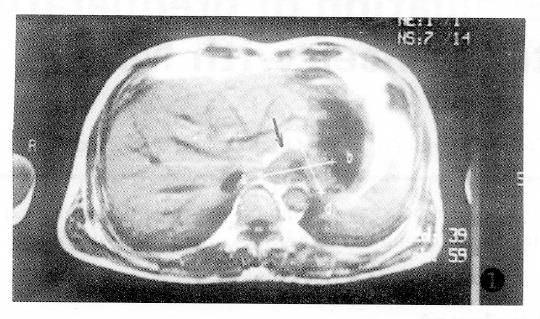
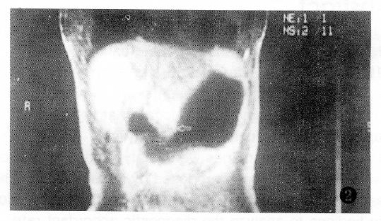
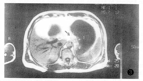
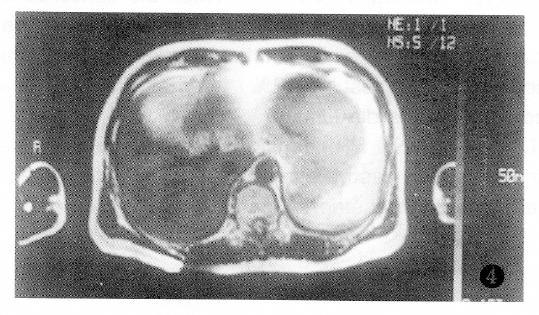
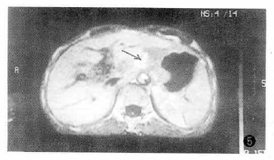
The MR images were analyzed by two radiologists before patients underwent surgery. The locations, appearances, sizes as well as the relation of the tumors to adjacent organs were evaluated. Location was classified according to four areas: Cardia fundus, corpus, antrum and entire stomach. A gastric wall > 1 cm thick was considered abnormal[1,5,6]. According to the criteria[5] proposed by Matsushita M, the degree of serosal invasion observed by MR imaging was classified as MRT-1 (no abnormal findings), MRT-2 (presence of a clear hypointense band or clear fat plane surrounding the lesion) (Figures 1 and 2), MRT-3 (presence of irregular margin or fat signal blurring around the lesion) (Figures 3 and 4), or MRT-4 (presence of contiguous extention of lesion signal to adjacent organs or definite involvement of adjacent structures), (Figure 5). The evaluation of both the extent and depth of gastric cancer relied primarily on the observation of images on the T-1WI[6]. This was due to the excellent contrast between the hypointensity of water signal in the stomach lumen, the isointensity of the gastric wall, and the hyperintensity of the fat plane outside the stomach.
The findings of a thickened wall that was characterized as a lesion with MRI corresponded to the gastric cancer lesion observed during surgery. On the T-1WI, thickened gastric wall ranged from 1.3-3.9 cm (mean 2.29 cm). The stomach was sometimes deformed and stenosed. In patients with advanced gastric cancer, the moderate low signal of the tumor extended to contiguous organs through hyperintense fat intervals. On the T-2WI, moderate high tumor signal did not contrast sharply with the high signal of inside water and outside fat, so the tumor margin could not be easily identified. MRI results were compared with findings from pathologic examination (Table 1).
| Magnetic Resonance Imaging diagnosis | Histologic diagnosis | ||
| T2 | T3 | T4 | |
| MRT2 | 7 | 2 | |
| MRT3 | 2 | 17 | 3 |
| MRT4 | 3 | ||
From the Table, we can see that the MR diagnostic accuracy was 77.8% (seven out of nine) for T2, 77.3% (17 out of 22) for T3, and 100% (three out of three) for T4 tumors, with the overall accuracy equaling 79.4%. Moreover, the accuracy in determining the degree of serosal invasion was 88.2% (27 out of 34) when T3 and T4 lesions were regarded as a single group. Statistically, MRI staging showed a significant correlation with histopathologic staging using the Spearman correlation test (rs′ = 0.743, P < 0.1). However, seven of 34 cases were staged incorrectly using MR imaging. Two of the stage T3 tumors were understaged as stage MRT-2 because there were no hyperintense signals of fat plane in emaciated patients. Two of the stage T2 tumors were overstaged as stage MRT-3 due to motion artifacts. Three of the stage T4 tumors were understaged as stage MRT-3 because peritoneal and omentum invasion were not detected. Analysis of the concordance between MRI and histologic staging results based on tumor location (Table 2), revealed that the staging accuracy was highest in gastric cardia fundus tumors.
| Location | Cases | Concordant number | Concordant rate |
| Cardia fundus | 11 | 10 | 90.9 |
| Corpus | 14 | 11 | 78.6 |
| Antrum | 7 | 5 | 71.4 |
| Entire stomach | 2 | 1 |
Preoperative staging in advanced gastric cancer is very important for surgeons to judge the probability of gastrectomy, select the appropriate operative approach and evaluate prognosis[7]. It is also helpful for patients who have to undergo radiotherapy[8]. There have been many reports about preoperative staging in advanced gastric cancer with CT[1,7,9] and endoscopic US[2,3]. The staging accuracy by CT and US is 42%-69% and 81%-92%, respectively. Anatomically, the stomach is obliquely in contact with the left lobe of the liver, which interferes with the accuracy of determining hepatic invasion by CT due to its partial volume effect. Furthermore, the limited spatial resolution often misses tiny metastases[1]. Cho JS suggested that dynamic CT be used to improve the diagnostic accuracy in the preoperative staging of gastric cancer[9]. As endoscopic US is invasive and its performance requires special skills[1], it is difficult to perform on patients with advanced gastric cancer. For MRI, however, no special preparation is required. MRI not only provides axial images, but also coronal, sagittal, and oblique images. The shape, size, tumor location and its relation with adjacent structures can be found easily by concurrently analyzing various images. Oral water administration can both distend the gastric wall as well as wash out the gastric juice attached to the gastric wall so that the advanced cancer wall area cannot be distended. As a result, is appears in an MRI as a thickened area and can be easily differentiated from the normal wall. Water also acts as a negative contrast agent. On T1 WI, the gastric wall appears very well due to the sharp contrast between the inside hypointense water and the outside hyperintense fat plane. Slight extraserosal invasion, which is difficult to visualize with CT, causes blurring of the fat layer on an MRI and so it is easily identified. It is thus very helpful for staging gastric cancer. Our study shows that there is a significant correlation between preoperative MRIs and pathologic staging of gastric cancer. The staging accuracy was somewhat superior to the previously published results that used CT. The 88% accuracy in determining extraserosal invasion indicated that MRI staging was more valuable for advanced gastric cancer. The higher staging accuracy found for gastric cardia fundus tumors was likely due to the relative stability of the cardia region, resulting in minimal motion artifacts and enhanced image clarity[6].
In our study, preoperative staging with MRI was incorrect in seven cases. Most of them were due to obvious motion artifacts or the emaciation of patients. For extremely emaciated patient, there is a minimal fat interval between the stomach and nearby organs, which might result in inaccurate staging[8]. In two cases, the metastases to the peritoneal and omentum were undetected, possibly due to the limited MR resolution of low field strength and the relatively small size of the lesions.
In conclusion, preoperative staging in advanced gastric cancer with MRI is particularly valuable for cardia fundus lesions. The evaluation of both the extent and depth of tumors relies primarily on the observation of images on the T1WI.
Original title:
S- Editor: Filipodia L- Editor: Jennifer E- Editor: Liu WX
| 1. | Minami M, Kawauchi N, Itai Y, Niki T, Sasaki Y. Gastric tumors: radiologic-pathologic correlation and accuracy of T staging with dynamic CT. Radiology. 1992;185:173-178. [RCA] [PubMed] [DOI] [Full Text] [Cited by in Crossref: 126] [Cited by in RCA: 118] [Article Influence: 3.6] [Reference Citation Analysis (0)] |
| 2. | Botet JF, Lightdale CJ, Zauber AG, Gerdes H, Winawer SJ, Urmacher C, Brennan MF. Preoperative staging of gastric cancer: comparison of endoscopic US and dynamic CT. Radiology. 1991;181:426-432. [RCA] [PubMed] [DOI] [Full Text] [Cited by in Crossref: 212] [Cited by in RCA: 222] [Article Influence: 6.5] [Reference Citation Analysis (0)] |
| 3. | Akahoshi K, Misawa T, Fujishima H, Chijiiwa Y, Maruoka A, Ohkubo A, Nawata H. Preoperative evaluation of gastric cancer by endoscopic ultrasound. Gut. 1991;32:479-482. [PubMed] |
| 4. | China National Coroperative Group of Gastric Cancer. [Surgical treatment of gastric cancer in China: an analysis of 11, 734 cases]. Zhonghua WaiKe Zazhi. 1982;20:577-580. [PubMed] |
| 5. | Matsushita M, Oi H, Murakami T, Takata N, Kim T, Kishimoto H, Nakamura H, Okamoto S, Okamura J. Extraserosal invasion in advanced gastric cancer: evaluation with MR imaging. Radiology. 1994;192:87-91. [RCA] [PubMed] [DOI] [Full Text] [Cited by in Crossref: 34] [Cited by in RCA: 39] [Article Influence: 1.3] [Reference Citation Analysis (0)] |
| 6. | Gao YG, Chai YQ, Chai ZL (compile). Diagnosis of MRI. Beijing: The People's Military Doctor Publication 1993; 528-529. |
| 7. | Zhang WS, Xu JX, Ming SL, Zhang J. The evaluation of CT examination on gastric and esophageal tumor. Zhonghua Shiyong WaiKe Zazhi. 1989;9:527-528. |
| 8. | Lee KR, Levine E, Moffat RE, Bigongiari LR, Hermreck AS. Computed tomographic staging of malignant gastric neoplasms. Radiology. 1979;133:151-155. [RCA] [PubMed] [DOI] [Full Text] [Cited by in Crossref: 62] [Cited by in RCA: 43] [Article Influence: 0.9] [Reference Citation Analysis (0)] |
| 9. | Cho JS, Kim JK, Rho SM, Lee HY, Jeong HY, Lee CS. Preoperative assessment of gastric carcinoma: value of two-phase dynamic CT with mechanical iv. injection of contrast material. AJR Am J Roentgenol. 1994;163:69-75. [RCA] [PubMed] [DOI] [Full Text] [Cited by in Crossref: 87] [Cited by in RCA: 91] [Article Influence: 2.9] [Reference Citation Analysis (0)] |


