Published online Jun 15, 1997. doi: 10.3748/wjg.v3.i2.72
Revised: January 31, 1997
Accepted: March 1, 1997
Published online: June 15, 1997
AIM: To determine whether medullary catecholaminergic neurons expressing Fos induced by chemical stimulation of the stomach project to the paraventricular nucleus of hypothalamus (PVH) in rats.
METHODS: Horseradish peroxidase (HRP) was introduced stereotaxically into the PVH of rats. Histochemical analysis of coronal sections through the medulla were analyzed using triple-label immunohistochemistry to identify cells that were retrogradely labeled with HRP, Fos (ABC method), and tyrosin hydroxylase (TH) (PAP method).
RESULTS: Seven kinds of labeled neurons were found in the nucleus tractus solitarii (NTS), the ventrolateral medulla (VLM) and the reticular formation (RF) of the medulla: Fos-like immunoreactive (FosLI) neurons, TH-like immunoreactive (TH-LI) neurons and HRP retrogradely single-labeled neurons, FosLI/HRP, FosLI/TH-LI and HRP/TH-LI double-labeled neurons, and FosLI/HRP/TH-LI triple-labeled neurons.
CONCLUSION: Ascending projections from the NTS, VLM and RF to the PVH might be involved in the transmitting process of visceral noxious stimulation.
- Citation: Dong YX, Xiong KH, Rao ZR, Shi JW. Fos expression in catecholaminergic medullary neurons induced by chemical stimulation of stomach projecting to the paraventricular nucleus of the hypothalamus in rats. World J Gastroenterol 1997; 3(2): 72-74
- URL: https://www.wjgnet.com/1007-9327/full/v3/i2/72.htm
- DOI: https://dx.doi.org/10.3748/wjg.v3.i2.72
It is well known that catecholaminergic medullary neurons are mainly located in the A1 and A2 regions of the brain stem, while a few of them are distributed in the reticular formation (RF) between A1 and A2 regions[1,2]. It has been previously reported that only a few neurons in nucleus tractus solitarii (NTS) sent projective fibers to the paraventricular nucleus of hypothalamus (PVH)[3-5]; however, recent studies have demonstrated that many neurons in the NTS and ventrolateral medulla (VLM) send their axons directly to the PVH[6], with some of them originating from A1 and A2 regions[2]. The paraventricular nucleus of hypothalamus (PVH) is an important part of the hypothalamus, and hypothalamus is known to play an important role in regulating visceral activities. Additionally, after injection of formalin into the stomach, numerous Fos-like immunoreactive (FosLI) neurons were detected within the central nervous system of the rats, including the NTS, VLM and RF[7]. Therefore, in this study, we investigated whether the catecholaminergic neurons in rat medulla projecting to the PVH responded to the gastrointestinal noxious stimulation.
Twelve male Sprague-Dawley rats, weighing 180-230 g, were used. The rats were kept in a dark, warm and quiet facility for 36 h before experiment. Then the rats were randomly divided into one experimental group (n = 6) and three control groups (n = 2 for each group).
The rats were anesthetized with sodium pentobarbital (40 mg/kg, i.p.) and stereotaxically injected unilaterally with 0.1 μL of a 30% solution of horseradish peroxidase (HRP) dissolved in 0.9% saline into the PVH. Forty-eight hours later, the rats were anesthetized again by placing the animals into a transparent glass box containing methoxyflurane gas and then administered with 2 mL of 1% formalin directly into the stomach through a plastic tube. Within 3-5 min after the injection, the animals awoke. Two hours later, the rats were re-anesthetized with an overdose of sodium pentobarbital, perfused through the ascending aorta with 150 mL of 0.9% saline, followed by 500 mL of a fixative containing 4% paraformaldehyde in 0.1 mol/L phosphate buffer (PB, pH7.4), and 200 mL of 10% sucrose in PB. The brains were removed and stored in 30% sucrose in 0.1 mol/L PB overnight at 4 °C. The brains were cut through the medulla into coronal sections 40 μm thick on a freezing microtome.
The sections were processed with a triple labeling technique[8]. For the histochemical demonstration of injected and transported HRP, tetramethylbenzidine (TMB) was used as a chromogen and sodium tungstate as a stabilizer. The HRP reaction products were intensified with diaminobenzidine (DAB) and CoCl2. Then, the sections were used for immunohistochemical staining for Fos according to the avidin biotin peroxidase complex (ABC) method. The sections were successively incubated with sheep anti Fos protein antibody (1:4000; Cambridge Biochemical Research, United Kingdom) for 48 h at 4 °C, followed by biotinylated rabbit anti sheep IgG (1:300; Vector) for 4 h at room temperature (RT) and finally streptavidin HRP complex (1:300; Vector) for 1 h at RT. Then, the sections were treated using the glucose oxidase DAB nickel method, the reactions usually lasted between 30-50 min at RT.
Finally, the sections were washed in 0.01 M phosphate buffered saline (PBS), and were then used for immunocytochemical staining for tyrosine hydroxylase (TH) by the peroxidase anti peroxidase (PAP) method. The sections were successively incubated with a monoclonal antibody against TH (1:300; Biochringer Mannbein Biochemical) for 48 h at 4 °C, followed by goat anti mouse IgG (1:100; Sigma) and finally a mouse PAP complex (1:300; Sigma), each for 24 h at 4 °C. The sections were then treated with 0.05% DAB and 0.03% H2O2 in 0.05 mol/L Tris HCl buffer (pH7.6) for 10-20 min at RT.
The control experiments (groups 1 to 3) were performed in six rats as follows: Two rats were reared as described above without any treatment; two rats were only anesthetized by methoxyflurane gas without any formalin injected into the stomach and two received HRP injection into the PVH without any additional treatment. After survival for 36 h, 2 h and 48 h, respectively, the rats were anesthetized with sodium pentobarbital (40 mg/kg, i.p.) and transcardially perfused as described above. The sections obtained from these six control rats were processed for immunohistochemical staining for Fos only.
Some of the sections obtained from the experimental rats were selected for the primary antibody substitution test (control group 4), in which the anti-Fos sera and monoclonal antibody against TH were replaced by 0.01 mol/L PBS (1:50-100).
Under light microscope, seven kinds of labeled cells (i.e. HRP, TH-LI and FosLI single-labeled neurons; hRP/TH-LI, FosLI/TH-LI and FosLI/HRP double-labeled neurons; hRP/FosLI/TH-LI triple-labeled neurons) were observed in the medulla.
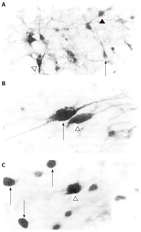
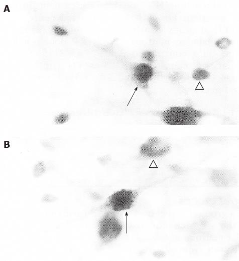
HRP reaction products were seen as black punctate granules in the cytoplasm (Figure 1A), while FosLI neurons appeared with a dark, round or ovoid nuclei and exhibited no staining in the cytoplasm (Figure 1C and Figure 2A). The cytoplasm of TH-LI neurons were homogeneously brown (Figure 1A, 1B and Figure 2B). Thus, HRP/TH-LI double-labeled neurons were identified by the presence of black punctate granules on a homogeneous brown background (Figure 1B); FosLI/TH-LI double-labeled neurons were characterized by the presence of a dark nucleus with a homogeneous brown cytoplasm (Figure 1A and 2A). Numerous black punctate granules surrounding a dark nucleus were seen in FosLI/HRP double-labeled neurons (Figure 1C). HRP/FosLI/TH-LI triple-labeled neurons contained a dark nucleus and black punctate granules on homogeneous brown background of the cytoplasm (Figure 2B). As the three different reaction products were clear and definite, the differently labeled neurons could easily be distinguished.
Retrogradely labeled HRP neurons were predominately observed in the medulla ipsilateral to the HRP injection site. Most of them were distributed in the NTS and VLM at the middle and caudal levels of the medulla. Most HRP labeled cell bodies in the NTS and VLM were fusiform or oval in shape, and medium or small in size with a diameter ranging from 15 to 30 μm. A few HRP labeled neurons were scattered in the medulla reticular formation (RF) between the NTS and VLM.
TH-LI neurons were mainly distributed in the A1 and A2 regions at the middle and caudal levels of the medulla. TH-LI neurons in NTS and VLM were fusiform or oval in shape and small or medium in size with diameters ranging from 20 to 30 μm. A few TH-LI neurons were also found in the RF.
FosLI neurons were bilaterally distributed and were frequently observed in the medial subnucleus (SolM) and commissural subnucleus (SolC) of the NTS at the middle and caudal levels of the medulla, although they were distributed throughout the entire rostrocaudal extent of the medulla, and additionally in the VLM and RF.
The number of labeled neurons was counted in one rat (Table 1). The total number of HRP labeled neurons was 162, while TH-LI neurons was 263, and FosLI neurons was 1275 in the medulla. FosLI/TH-LI double-labeled neurons (314) were most numerous among all double-labeled neurons and constituted 14.4% (314/2185) of the total population of labeled neurons. On the other hand, HRP/TH-LI and FosLI/HRP double-labeled neurons were much less frequent than FosLI/TH-LI double-labeled neurons, with 77 HRP/TH-LI neurons constituting 3.5% (77/2185) of the total labeled neurons. FosLI/HRP double-labeled neurons only constituted 2.0% (43/2185) of the total labeled neurons. HRP/FosLI/TH-LI triple-labeled neurons only constituted 2.3% (51/2185) of all labeled neurons.
| HRP | TH-LI | FosLI | HRP/ TH-LI | FosLI/ TH-LI | FosLI/ HRP | HRP/FosLI/ TH-LI | Total | |
| NTS | 119 | 153 | 937 | 41 | 77 | 28 | 33 | 1388 |
| RF | 11 | 32 | 204 | 7 | 19 | 6 | 2 | 281 |
| VLM | 32 | 78 | 134 | 29 | 218 | 9 | 16 | 516 |
| Total (%) | 162 (7.4) | 263 (12.0) | 1275 (58.4) | 77 (3.5) | 314 (14.4) | 43 (2.0) | 51 (2.3) | 2185 (100) |
Rats that did not receive either the formalin injection into the stomach or the HRP injection (group 1) did not express any FosLI neurons in the medulla.
Rats that did not receive a formalin injection into their stomachs, but did inhale the methoxyflurane gas anesthesia (group 2) did express a minimal amount of FosLI neurons in the medial septal nucleus and the PVH at 2 h after inhalation.
Rats subjected to HRP injection into the PVH and sacrificed 48 h later (group 3) did not express FosLI neurons in the medulla.
The sections that were used to verify the antibodies using the substitution test (group 4) were negative, with neither FosLI nor TH-LI detected in the medulla of these sections.
A previous study had demonstrated that after a tritiated amino acid tracer was injected into the NTS of rats, the longest distance of the labeled ascending fibers and terminals was observed at the level of the pons[4]. A different study found that the injection of HRP into the PVH led to a significant number of HRP labeled neurons in the VLM[3]. Other more recent studies have confirmed that neurons in the NTS and VLM send axons to the PVH[3,5,6,9]. Our results have proved that the projection fibers to the PVH originated mainly from the SolM and SolC and NTS and the VLM, with fewer projections from RF at the middle and caudal segments of medulla. However, the transmitters, modulaters and function of these projecting neurons were not known. Phaseolus vulgaris leucoagglutinin (PHA-L) anterograde tracing study has demonstrated that the neurons in the A1 and A2 regions project to the PVH[2]. In this study, we found that FosLI neurons were observed within the NTS, VLM and RF of the rat following gastrointestinal (visceral) noxious stimulation[7].
Our results show that 77% (1683/2185) of the total population of labeled neurons in the NTS, VLM and RF responded to visceral noxious stimulation, and were Fos positive. About 15% (333/2185) of all seven kinds of labeled neurons in the NTS, VLM and RF sent their axons to the PVH, about 5.9% of the neurons projected to the PVH exhibited TH-LI, and some of these TH-LI neurons were Fos positive. These results suggest that TH-LI neurons in the NTS, VLM and RF might be involved in the transmission of nociceptive information from the stomach to the PVH.
The authors are grateful to Profoess Li YQ for his critical reading of the manuscript and helpful suggestions.
Original title:
S- Editor: Filipodia L- Editor: Jennifer E- Editor: Liu WX
| 1. | Mantyh PW, Hunt SP. Neuropeptides are present in projection neurones at all levels in visceral and taste pathways: from periphery to sensory cortex. Brain Res. 1984;299:297-312. [RCA] [PubMed] [DOI] [Full Text] [Cited by in Crossref: 133] [Cited by in RCA: 129] [Article Influence: 3.1] [Reference Citation Analysis (0)] |
| 2. | Cunningham ET, Sawchenko PE. Anatomical specificity of noradrenergic inputs to the paraventricular and supraoptic nuclei of the rat hypothalamus. J Comp Neurol. 1988;274:60-76. [RCA] [PubMed] [DOI] [Full Text] [Cited by in Crossref: 569] [Cited by in RCA: 574] [Article Influence: 15.5] [Reference Citation Analysis (0)] |
| 3. | Ciriello J, Caverson MM. Ventrolateral medullary neurons relay cardiovascular inputs to the paraventricular nucleus. Am J Physiol. 1984;246:R968-R978. [PubMed] |
| 4. | Norgren R. Projections from the nucleus of the solitary tract in the rat. Neuroscience. 1978;3:207-218. [RCA] [PubMed] [DOI] [Full Text] [Cited by in Crossref: 578] [Cited by in RCA: 547] [Article Influence: 11.6] [Reference Citation Analysis (0)] |
| 5. | Ciriello J, Caverson MM, Polosa C. Function of the ventrolateral medulla in the control of the circulation. Brain Res. 1986;396:359-391. [RCA] [PubMed] [DOI] [Full Text] [Cited by in Crossref: 290] [Cited by in RCA: 282] [Article Influence: 7.2] [Reference Citation Analysis (0)] |
| 6. | Ricardo JA, Koh ET. Anatomical evidence of direct projections from the nucleus of the solitary tract to the hypothalamus, amygdala, and other forebrain structures in the rat. Brain Res. 1978;153:1-26. [RCA] [PubMed] [DOI] [Full Text] [Cited by in Crossref: 1105] [Cited by in RCA: 1045] [Article Influence: 22.2] [Reference Citation Analysis (0)] |
| 7. | Chen LW, Jin GR, Rao ZR, Shi JW. C-fos expression within central nervous system of the rat following gastrointestinal visceral oxious stimulation. Shenjing Jiepoxue Zazhi. 1993;9:185-191. |
| 8. | Rao ZR, Chen LW, Jin GR, Han ZA, Dong YX, Xiong KH. Simultaneous demonstration of Fos protein, tyrosine hydroxylase (TH) and HRP retrograde labeling in same neurons-A triple labeling technique. Acta Anatomica Sinca. 1994;25:219-223. |
| 9. | Kannan H, Yamashita H. Connections of neurons in the region of the nucleus tractus solitarius with the hypothalamic paraventricular nucleus: their possible involvement in neural control of the cardiovascular system in rats. Brain Res. 1985;329:205-212. [RCA] [PubMed] [DOI] [Full Text] [Cited by in Crossref: 114] [Cited by in RCA: 117] [Article Influence: 2.9] [Reference Citation Analysis (0)] |


