Copyright
©The Author(s) 2023.
World J Gastroenterol. Sep 28, 2023; 29(36): 5180-5197
Published online Sep 28, 2023. doi: 10.3748/wjg.v29.i36.5180
Published online Sep 28, 2023. doi: 10.3748/wjg.v29.i36.5180
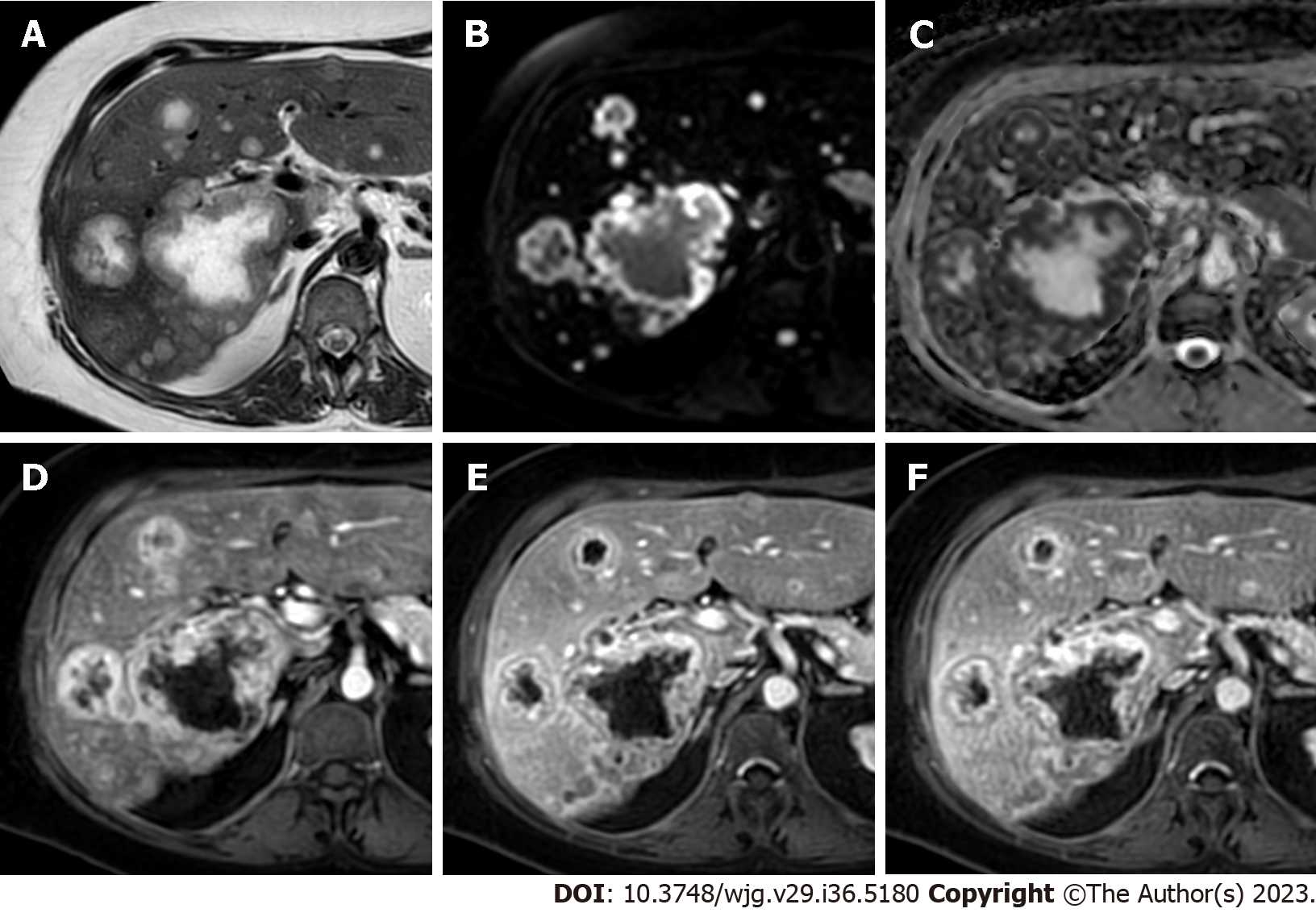
Figure 5 Multiple liver metastases from breast cancer.
A: ECA-magnetic resonance imaging of a 51-year-old woman shows multiple liver lesions with central marker hyperintensity on T2-weighted images; B: Diffusion weighted imaging sequence demonstrates lesions’ peripheral restricted diffusion; C: Apparent diffusion coefficient map shows a corresponding peripheral hypointensity of the lesions; D: On post-contrast arterial phase the lesions demonstrate a peripheral hyperenhancement rim with a central hypo-perfused area; E: On portal venous a corresponding peripheral washout is observed; F: On delayed post-contrast phase the peripheral hypoenhancement persists.
- Citation: Maino C, Vernuccio F, Cannella R, Cortese F, Franco PN, Gaetani C, Giannini V, Inchingolo R, Ippolito D, Defeudis A, Pilato G, Tore D, Faletti R, Gatti M. Liver metastases: The role of magnetic resonance imaging. World J Gastroenterol 2023; 29(36): 5180-5197
- URL: https://www.wjgnet.com/1007-9327/full/v29/i36/5180.htm
- DOI: https://dx.doi.org/10.3748/wjg.v29.i36.5180


