Basic Study Open Access
Copyright ©The Author(s) 2023. Published by Baishideng Publishing Group Inc. All rights reserved.
World J Gastroenterol. Aug 14, 2023; 29(30): 4657-4670
Published online Aug 14, 2023. doi: 10.3748/wjg.v29.i30.4657
Fecal microbiota transplantation alleviates experimental colitis through the Toll-like receptor 4 signaling pathway
Xin Wen, Rui Xie, Hong-Gang Wang, Min-Na Zhang, Le He, Meng-Hui Zhang, Xiao-Zhong Yang, Department of Gastroenterology, The Affiliated Huaian No. 1 People’s Hospital of Nanjing Medical University, Huai’an 223300, Jiangsu Province, China
ORCID number: Xin Wen (0000-0001-8904-1340); Hong-Gang Wang (0000-0003-4761-0407); Min-Na Zhang (0000-0003-1567-3788); Meng-Hui Zhang (0000-0002-8679-386X); Xiao-Zhong Yang (0000-0003-2036-5878).
Author contributions: Wen X, Xie R, and Wang HG contributed equally to this work; Wen X, Xie R, and Wang HG conceived and designed this work, and drafted and revised the manuscript; Wen X, Zhang MN, He L, and Zhang MH performed the experiments, collected samples, and analyzed the data; Yang XZ and Wang HG worked on the concept and guidance of this study; Yang XZ and Wang HG provided the funding support and project administration; All authors have read and approved the final manuscript.
Supported by the Scientific Research Project of Jiangsu Provincial Health Commission, No. H2018082; Huai’an Natural Science Research Project Project, No. HAB201926; and Scientific Research Project of Translational Medicine Innovation Team of Huai’an First People’s Hospital, No. YZHT201905.
Institutional animal care and use committee statement: The animal experimental protocol was approved by experimental animal ethics committee of the Affiliated Huaian No. 1 People’s Hospital of Nanjing Medical University (Approval No. DW-P-2018-008-01).
Conflict-of-interest statement: The authors have no conflicts interest to declare.
Data sharing statement: The data presented in the study are available in article. The datasets analysed during the current study are available in the NCBI Sequence Read Archive (SRA) database, submission number: SUB11829874.
ARRIVE guidelines statement: The authors have read the ARRIVE guidelines, and the manuscript was prepared and revised according to the ARRIVE guidelines.
Open-Access: This article is an open-access article that was selected by an in-house editor and fully peer-reviewed by external reviewers. It is distributed in accordance with the Creative Commons Attribution NonCommercial (CC BY-NC 4.0) license, which permits others to distribute, remix, adapt, build upon this work non-commercially, and license their derivative works on different terms, provided the original work is properly cited and the use is non-commercial. See: https://creativecommons.org/Licenses/by-nc/4.0/
Corresponding author: Xiao-Zhong Yang, MD, PhD, Chief Doctor, Doctor, Professor, Department of Gastroenterology, The Affiliated Huaian No. 1 People’s Hospital of Nanjing Medical University, No. 1 Huanghe Road, Huai’an 223300, Jiangsu Province, China. hayyyxzh@njmu.edu.cn
Received: May 22, 2023
Peer-review started: May 22, 2023
First decision: June 20, 2023
Revised: July 3, 2023
Accepted: July 11, 2023
Article in press: July 11, 2023
Published online: August 14, 2023
Processing time: 79 Days and 23.3 Hours

Abstract
BACKGROUND

Fecal microbiota transplantation (FMT) has shown promising therapeutic effects on mice with experimental colitis and patients with ulcerative colitis (UC). FMT modulates the Toll-like receptor 4 (TLR4) signaling pathway to treat some other diseases. However, it remains unknown whether this modulation is also involved in the treatment of UC.

AIM

To clarify the necessity of TLR4 signaling pathway in FMT on dextran sodium sulphate (DSS)-induced mice and explain the mechanism of FMT on UC, through association analysis of gut microbiota with colon transcriptome in mice.

METHODS

A mouse colitis model was constructed with wild-type (WT) and TLR4-knockout (KO) mice. Fecal microbiota was transplanted by gavage. Colon inflammation severity was measured by disease activity index (DAI) scoring and hematoxylin and eosin staining. Gut microbiota structure was analyzed through 16S ribosomal RNA sequencing. Gene expression in the mouse colon was obtained by transcriptome sequencing.

RESULTS

The KO (DSS + Water) and KO (DSS + FMT) groups displayed indistinguishable body weight loss, colon length, DAI score, and histology score, which showed that FMT could not inhibit the disease in KO mice. In mice treated with FMT, the relative abundance of Akkermansia decreased, and Lactobacillus became dominant. In particular, compared with those in WT mice, the scores of DAI and colon histology were clearly decreased in the KO-DSS group. Microbiota structure showed a significant difference between KO and WT mice. Akkermansia were the dominant genus in healthy KO mice. The ineffectiveness of FMT in KO mice was related to the decreased abundance of Akkermansia. Gene Ontology enrichment analysis showed that differentially expressed genes between each group were mainly involved in cytoplasmic translation and cellular response to DNA damage stimulus. The top nine genes correlating with Akkermansia included Aqp4, Clca4a, Dpm3, Fau, Mcrip1, Meis3, Nupr1 L, Pank3, and Rps13 (|R| > 0.9, P < 0.01).

CONCLUSION

FMT may ameliorate DSS-induced colitis by regulating the TLR4 signaling pathway. TLR4 modulates the composition of gut microbiota and the expression of related genes to ameliorate colitis and maintain the stability of the intestinal environment. Akkermansia bear great therapeutic potential for colitis.

Key Words: Toll-like receptor 4; Fecal microbiota transplantation; Colitis; Akkermansia; Lactobacillus; Aquaporin 4; Transcriptome sequencing

Core Tip: Recent studies have shown that fecal microbiota transplantation (FMT) has a therapeutic role in patients with inflammatory bowel disease. The Toll-like receptor 4 (TLR4) signaling pathway may play a critical role in intestinal injury and repair. Here, we conducted animal experiments to explore the role of TLR4 in dextran sodium sulphate-induced colitis in mice and the treatment of FMT.


INTRODUCTION

Recent studies support that inflammatory bowel disease (IBD) can be categorized as a “microbial dysbiosis disease,” because of its progression synchronizing the dysbacteriosis of gut microbiota[1]. Host physiology such as barrier function, metabolism, immune responses, and homeostasis involves microbiome-induced cell signaling, proliferation, and neurotransmitter biosynthesis[2]. In IBD patients, intestinal bacterial diversity decreases and the bacterial community structure changes[3]. In dextran sodium sulphate (DSS)-induced colitis in mice, some probiotics, including Lactbacillus and Bifidobacterium, are significantly reduced[4]. New evidence indicates that IBD is not merely a consequence of chronic inflammation, but also of disruption of the gut microbiome and destruction of the intestinal epithelial barrier[5].

Gut microbiota play a role in inflammation-related activities. Fecal microbiota transplantation (FMT) has shown high efficacy and safety in treating ulcerative colitis (UC)[6,7] due to its immunomodulatory and anti-inflammatory functions[8]. Our previous study showed that FMT can counter DSS-induced colitis in mice by increasing the relative abundance of Lactobacillus[9]. FMT has also shown therapeutic potential for a range of other diseases, such as hepatic disorders and metabolic syndrome[10]. Recent studies have demonstrated that Toll-like receptor 4 (TLR4) is exploited by FMT in treating many diseases such as spleen deficiency diarrhea[11], Parkinson’s disease[12,13], developmental arsenic neurotoxicity[14], fluorosis[15], and acute lung injury[16]. Previous studies have indicated that FMT intervention can inhibit activation of the nuclear factor kappa B (NF-κB) signaling pathway[17], which is downstream of TLR4. However, there have been limited studies investigating the role of TLR4 in FMT for UC.

As a class of transmembrane proteins that recognize invading microbes and activate immune cells, Toll-like receptors (TLRs) regulate gene transcription and the acquired intestinal immune response[18]. In the etiology of IBD, microbes in the intestinal lumen induce abnormal immune responses, along with excessive leakage of bacterial antigens into the mucosa[19]. TLR4, an important immune activator, is highly expressed in the intestinal epithelial cells and lamina propria cells of UC patients[20]. It binds to ligands to activate cytokine signaling, recruit inflammatory cells, and damage intestinal mucosal barrier, all of which aggravate intestinal inflammatory lesions. More importantly, substantial evidence supports a pro-inflammatory role of the TLR4 signaling pathway in UC. Expression levels of TLR4 are positively correlated with disease activity indices (DAIs), endoscopy scores, and histopathological scores[21]. DSS-induced colitis deteriorates in mice with TLR4 overexpression[22,23], but is stably maintained in TLR4-deficient mice[24,25]. Multiple experiments have shown that inhibiting the TLR4 signaling pathway can prevent DSS-induced colitis[26,27]. While TLR4 plays a crucial role in intestinal injury and repair, its role in shaping colonic bacterial homeostasis and microbiota-related immunity remains poorly understood.

Our previous studies confirmed the efficacy FMT on IBD, but the mechanism has not been reported[9]. Therefore, we explored the role of TLR4 in the mechanism by which FMT treats DSS-induced colitis in the mice.

MATERIALS AND METHODS
Animals

Wild-type (WT) C57BL/10J mice and TLR4-knockout (KO) mice on the C57BL/10J background (female; 6 to 8 wk of age; weighing 18-20 g; specific pathogen-free (SPF) grade) were purchased from the Model Animal Research Center of Nanjing University (Nanjing, China). All mice were reared in an SPF condition at the experimental animal center of the Affiliated Huaian No. 1 People’s Hospital of Nanjing Medical University. Throughout the acclimatization and study periods, all mice were maintained in a 12 h-light/12 h-dark cycle (21 °C ± 2 °C with a relatively constant humidity of 45% ± 10%) and had access to food and water ad libitum. All mice were group-housed and reared in a standard cage, with TLR4 KO mice kept separately from C57BL/10J mice in different cages.

DSS-induced colitis

DSS (36-50 kDa) was purchased from MP Biomedicals LLC (Irvine, CA, United States) and dissolved in distilled water. Experimental colitis was induced as previously described with minor changes[9]. For different groups, the mice were administered 2.5% (w/v) DSS in drinking water for 7 d. Mice in the KO (DSS + FMT) group were fed fecal microbiota from healthy WT mice from day 8 (once every 2 d) until the end of the experiment, while mice in the KO (DSS + water) group were fed normal saline at the same time. The mice were evaluated daily by scoring via the disease activity index (DAI)[28]. The DAI score was calculated on a 0-4 scale as previously described[29].

Fecal preparation and transplantation

The process of FMT was performed as previously described[9]. Briefly, feces from donor mice (healthy WT mice) were collected and resuspended in sterile normal saline at 0.125 g/mL. Then 0.2 mL of this suspension was administered to mice once every 2 d by oral gavage. This process lasted 7 d.

Histopathology

Mice were euthanized by cervical dislocation, and their abdominal cavity was opened immediately. The colon tissue was dissected; colons were measured for colon length, and tissues were examined for gross macroscopic appearance and stool consistency. The distal colon segment was placed in 10% neutral buffered formalin for 24 h, embedded in paraffin, and cut into sections 4 μm in thickness. Then the sections were stained with hematoxylin and eosin (H&E). H&E-stained sections were examined for inflammation and tissue damage by an experienced pathologist in a blinded manner. Tissue histology was scored by summing the scores of the following parameters according to a previous study[30]: Extent of inflammation, aberrant crypt foci, lymphocyte infiltration, and aberrant colon wall.

Fecal DNA extraction and 16S ribosomal RNA sequencing

Fecal DNA extraction and 16S ribosomal RNA (rRNA) sequencing were performed as previously reported[9]. The V3-V4 hypervariable region of the bacterial 16S-rRNA gene was amplified with primers 338F (5’-ACTCCTACGGGAGGCAGCAG-3’) and 806R (5’-GGACTACHVGGGTWTCTAA T-3’) with the ABI GeneAmp® 9700 PCR thermocycler (Applied Biosystems, Foster City, CA, United States)[9]. All PCR products were extracted from a 2% agarose gel and purified using the AxyPrep DNA Gel Extraction Kit (Axygen Biosciences, Union City, CA, United States)[31]. Purified amplicons were sequenced on the Illumina MiSeq PE300 platform (Illumina, San Diego, CA, United States). The raw 16S rRNA gene sequencing reads were demultiplexed, quality-filtered by fastp version 0.20.0, and merged by FLASH version 1.2.7. Operational taxonomic units (OTUs) with 97% similarity cutoff[32] were clustered using UPARSE version 7.1[33], and chimeric sequences were identified and removed. Bacterial alpha-diversity was determined by sampling-based OTU analysis. Analysis of species accumulation curves was performed to assess the rationality and efficiency of the sequencing depth. Principal component analysis (PCA) was implemented in R programming. Using the Wilcoxon rank-sum test, the bacterial taxonomic analysis was performed for comparison at the bacterial phylum, class, order, family, genus levels between two groups. Based on the matrix of normalized relative abundance, bacteria with significantly different abundances between assigned taxa were determined by linear discriminant analysis effect size (LEfSe) with the Kruskal-Wallis rank-sum test (P < 0.05). LDA was used to assess the effect size of each feature (LDA score [log10] = 3 as the cut-off value).

Transcriptome analysis

Total RNA was extracted from inflammatory colonic tissue. For sequencing, a 1 cm colon tissue was sampled from the site about 2 cm from the anus, regardless of whether there was visible inflammation. The tissue samples with minimum and maximum histological scores were removed. Then the colon samples from four randomly chosen animals in each group was used for sequencing. Methods for amplifying and sequencing followed those previously published[9,29]. Briefly, 2 μg RNA per sample was used to sequence on the Illumina Hiseq 4000 platform. Differential expression analysis was performed using the DESeq R package (1.10.1) according to the manufacturer’s protocol. Then, to explore the potential function of the differentially expressed genes (DEGs), GOseq R package[34] and KOBAS software[35] were used to test the enrichment of DEGs in Gene Ontology (GO) functional annotations[36] and Kyoto Encyclopedia of Genes and Genomes (KEGG) pathways[37].

Correlation analysis for gut microbiota and transcriptome

We used Metastats software to confirm the difference in the relative abundance of microbiota among the samples (P ≤ 0.05). We used DEG sequencing to carry out transcriptome difference analysis (the threshold was Padj < 0.05 & |log2FC| > 1). Finally, R psych software package was used to analyze the Spearman association between the transcriptome and intestinal microflora. Those with |R| > 0.8 and P < 0.05 (strong correlation) were screened for mapping.

Statistical analysis

Differences were analyzed using the t-test with Graphpad Prism 8.0 software (GraphPad Software Inc., La Jolla, CA, United States). Results are shown as the mean ± standard error of the mean. P < 0.05 was considered statistically significant.

RESULTS
FMT does not improve acute colitis induced by DSS in TLR4-KO mice

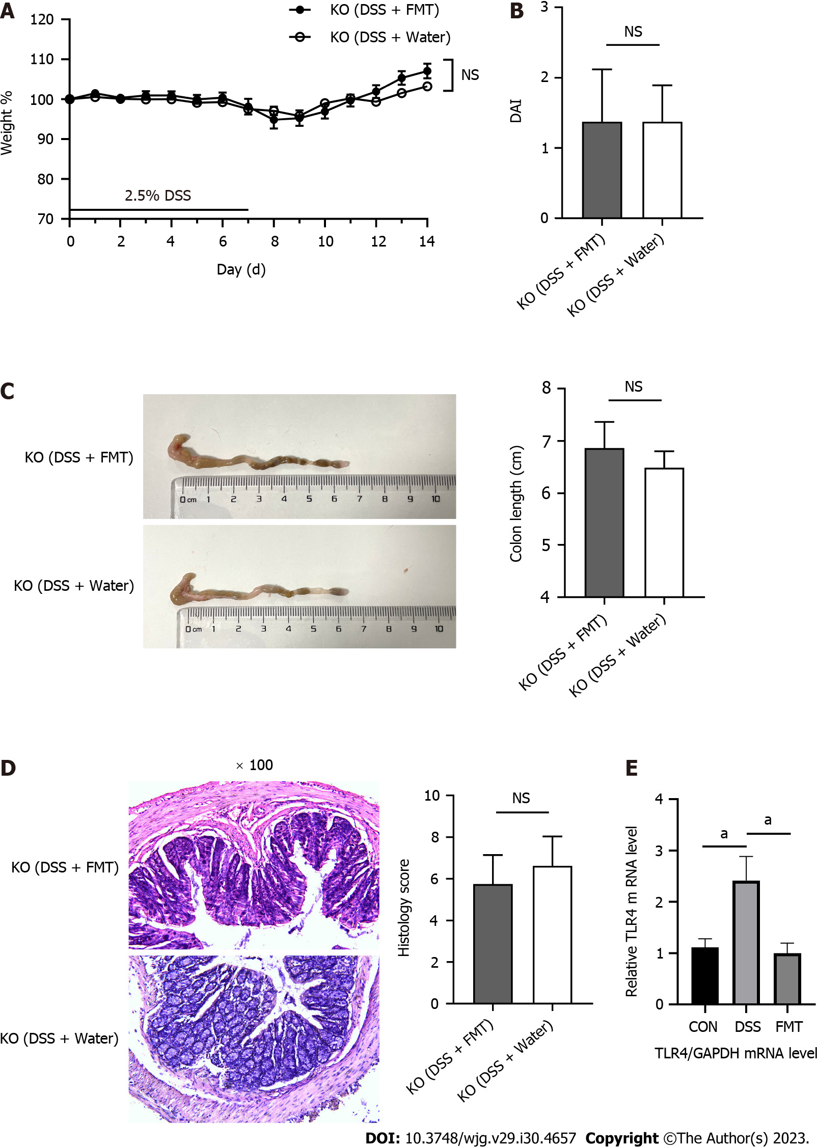
In our previous experiment, we found that FMT is effective to treat colitis[9]. We further explored whether this efficacy is related to the TLR4 pathway. Acute DSS-induced colitis was induced in eight animals per group using 2.5% DSS in the drinking water. After gavage with fecal microbiota from the WT mice, mice in the KO (DSS + water) and KO (DSS + FMT) groups displayed indistinguishable body weight loss, colon length, DAI score, and histology score (Figure 1A-D). Beyond our expectation, FMT had no effect on colonic inflammation in TLR4-KO mice. We compared the expression of TLR4 gene in the intestine of WT mice before and after FMT. The transcriptome sequence data indicated that DSS increased, but FMT effectively decreased the expression of TLR4 (Figure 1E).

Figure 1
Figure 1 Fecal microbiota transplantation did not alleviate acute colitis induced by dextran sodium sulphate in Toll-like receptor 4 knockout mice. A: Body weight of mice during the course of colitis; B: The bar chart represents the disease activity index (DAI) score of mice on day 14; C: Representative images of colons from mice (left) and statistical analysis of colon length (right); D: Representative hematoxylin and eosin staining of colon tissues, original magnification 100 ×, and histological scores (right); E: Relative quantification of the transcription level of Toll-like receptor 4 (TLR4) among groups. aP < 0.05. DSS: Dextran sodium sulphate; FMT: Fecal microbiota transplantation; KO: Knockout.
FMT changes the intestinal flora of TLR4-KO mice

We investigated whether FMT changed the composition of gut microbiota in the KO (DSS + water) and KO (DSS + FMT) groups. We employed LEfSe to evaluate the bacterial taxa (at genus level) in the two groups (Figure 2A). The dominating taxa in KO (DSS + FMT) group were enriched in Lactobacillus, which indicated that we had successfully transplanted the gut microbiota of healthy WT mice. Meanwhile, the KO (DSS + FMT) group had a lower abundance of Akkermansia, indicating that FMT could alter the relative abundance of Akkermansia in KO mice (Figure 2B).

Figure 2
Figure 2 Fecal microbiota transplantation changed the gut microbiota of Toll-like receptor 4 knockout mice. A: Linear discriminant analysis (LDA) effect size (LEfSe) analysis in two groups with an LDA score > 3.0; B: The relative abundance of Akkermansia in Toll-like receptor 4 knockout mice. aP < 0.05, bP < 0.01. DSS: Dextran sodium sulphate; FMT: Fecal microbiota transplantation.
TLR4 KO alleviates DSS-induced colitis

We used TLR4-deficient mice and WT mice to determine whether TLR4 may protect mice from DSS-induced colitis. Mice in the KO-DSS (n = 8) and WT-DSS (n = 7) groups were given distilled drinking water containing 2.5% DSS for 7 d (Figure 3A). Compared with WT mice, KO mice showed lower susceptibility to DSS, as manifested by their much smaller body weight loss (Figure 3B), lower DAI (Figure 3C), and longer colons (Figure 3D). Compared to the WT-DSS group, mice in the KO-DSS group exhibited a more intact colon structure, less severe crypt damage, and reduced inflammatory infiltration (Figure 3E). In summary, KO mice showed increased tolerance to DSS-induced colitis.

Figure 3
Figure 3 Toll-like receptor 4 knockout alleviated dextran sodium sulphate-induced inflammation in the colon. A: Scheme of the animal experimental design; B: The change in body weight of mice from days 0 to 7 during the disease course (knockout-dextran sodium sulphate [KO-DSS]: n = 8; wild type [WT]-DSS: n = 7); C: The bar chart represents the disease activity index (DAI) score on day 7; D: Representative colons (left) and statistical analysis (right) of colonic length; E: Representative hematoxylin and eosin staining of colon tissues (left), original magnification 100 ×, and histological scores (right). aP < 0.05, bP < 0.01, and cP < 0.001 were considered statistically significant. FMT: Fecal microbiota transplantation.
TLR4 deficiency influences the diversity and composition of gut microbiota

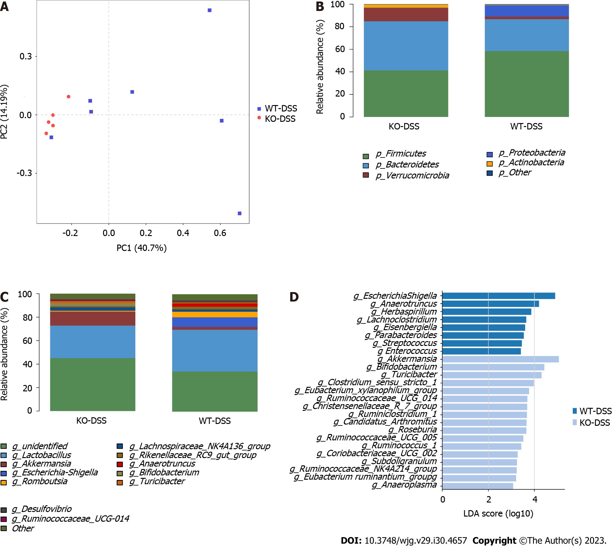
We further investigated whether the protection against DSS-induced colitis was due to TLR4 KO or microbiota re-composition. We detected the gut microbiota of WT and KO mice in the basal and DSS-treated states. We analyzed the beta-diversity of microbiota based on PCA. An evident clustering separation between OTUs revealed the different community structures between each two groups, suggesting that these communities are distinct in terms of their compositional structure (Figure 4A and 5A).

Figure 4
Figure 4 Diversities and compositions of gut microbiota in knockdown-control and wild type-control groups. A: β-diversity evaluated using the weighted UniFrac-based PCA (knockdown-control [KO-CON]: n = 7; wild type [WT]-CON: n = 7); B and C: Bar graphs showing the relative abundances of different bacteria at the phylum and genus levels; D: Linear discriminant analysis (LDA) effect size analysis in groups with an LDA score > 3.0 between two groups.
Figure 5
Figure 5 Diversities and compositions of gut microbiota in knockout-dextran sodium sulphate and wild type-dextran sodium sulphate groups. A: Multiple sample principal component analysis (knockout-dextran sodium sulphate [KO-DSS]: n = 5; wild type [WT]-DSS: n = 7); B and C: Bar graphs showing the relative abundances of different bacteria at the phylum and genus levels; D: Linear discriminant analysis (LDA) effect size (LEfSe) analysis in groups with an LDA score > 3.0.

At the phylum level, TLR4 deficiency decreased the abundance of Bacteroidetes and increased the abundances of Actinobacteria and Verrucomicrobia (P < 0.05; Figure 4B), compared to those in WT mice. After DSS induction, a significant increase of phylum Proteobacteria was observed in the WT-DSS group compared to the KO-DSS group (P < 0.05; Figure 5B). Verrucomicrobia was the most abundant phylum among those with significant differences (P < 0.05). At the genus level, Akkermansia abundance was significantly higher in KO mice than in WT mice either healthy or diseased (P < 0.05; Figure 4C and 5C). To further investigate the potential effect of microbiota composition on DSS-induced colitis, we used the LEfSe to detect the marked differences in the dominant bacterial communities between the two groups (Figure 4D and 5D). Specifically, Lactobacillus and Peptococcus were enriched in the WT-CON group (Figure 4D), while Escherichia Shigella and Anaerotruncus were enriched in the WT-DSS group (Figure 5D). Interestingly, Akkermansia and Bifidobacterium were enriched either in healthy and diseased KO mice (Figure 4D and 5D). The collective results of our study indicated clear differences in the intestinal microbiome between WT mice and KO mice, both in healthy conditions and during illness. These findings highlight the important role of TLR4 in shaping the composition and diversity of the intestinal microbiota.

TLR4-KO-shaped microbiota affect the transcriptome in the colon of mice

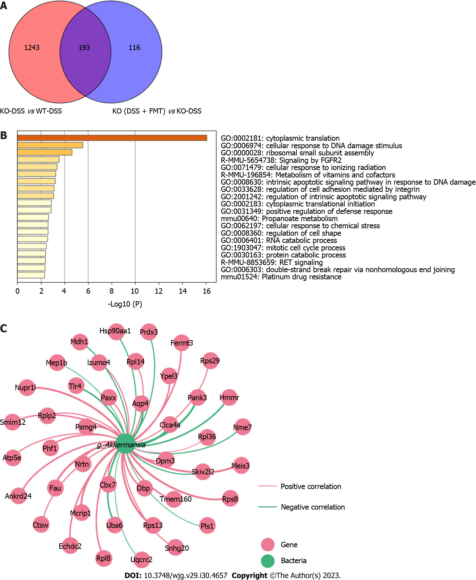
To further explore whether FMT can change the gene expression related to TLR4, we investigated the DEGs between groups. Compared to those in the WT-DSS group, 1436 genes were differentially expressed in the KO-DSS group, and 309 genes in the KO (DSS + FMT) group. Furthermore, 193 DEGs were found among the KO-DSS group, WT-DSS group, and KO (DSS + FMT) group (Figure 6A). GO enrichment analysis showed that these DEGs were mainly involved in cytoplasmic translation and cellular response to DNA damage stimulus (Figure 6B). According to 16S rRNA sequencing analysis, we found that Akkermansia was dominant in the KO group. To characterize potential gene-microbe interactions, we computed gene-microbe correlations with Spearman correlation efficients (Figure 6C). The top nine genes correlating with Akkermansia included Aqp4, Clca4a, Dpm3, Fau, Mcrip1, Meis3, Nupr1 L, Pank3, and Rps13 (|R| > 0.9, P < 0.01).

Figure 6
Figure 6 Colonic transcriptome profile and gene-microbe correlation. A: Venn diagram illustrates genes regulated by fecal microbiota transplantation (FMT) and Toll-like receptor 4 knockout (KO); B: The top 20 Gene Ontology terms enriched in these 193 differentially expressed genes (DEGs); C: Network visualizing 193 DEGs associated with Akkermansia (|R| > 0.8, P < 0.05). CON: Control; DSS: Dextran Sodium Sulphate; WT: Wild type.
DISCUSSION

Researchers have found that patients with active UC can benefit from FMT[38]. Moreover, our previous study also verified that FMT can treat colitis in mice. In the present study, the expression of TLR4 was upregulated by DSS, and downregulated after FMT. It therefore stands to reason that, by inhibiting TLR4, a protective effect from intestinal inflammation will be induced. Considering the ubiquitous involvement of the TLR4 signaling pathway in the activities of the mucosa, we designed this animal study to elucidate its interaction with FMT in UC. In this study, TLR4 KO significantly alleviated the clinical and histological manifestations of DSS-induced colitis. Notably, the increased relative abundance of the predominant Akkermansia species contributed to the heightened resistance against colon inflammation. Through further investigation, we discovered that genetic KO of TLR4 significantly impacted the structure and composition of the gut microbiota, resulting in a shift towards an anti-inflammatory configuration. This shift plays a crucial role in promoting enhanced resistance and tolerance to colitis.

At the phylum level, DSS changed the relative abundances of Bacteroidetes, Actinobacteria, and Verrucomicrobia in TLR4-KO mice compared to WT mice. Possibly, the high abundance of anti-inflammatory Akkermansia in the gut microbiota curbs the aggravation of colitis, despite the absence of TLR4 signaling. Akkermansia was the dominant genus in healthy KO mice, while after the treatment of FMT, their level decreased. Compared with that in the WT group, the status of colitis in the KO group was not significantly attenuated by FMT, suggesting that the therapeutic effect of FMT on colitis is closely related to the TLR4 signaling pathway and Akkermansia.

In the gut, the expression of TLRs changes with the composition of microbiota[39], as well as the activity of the intestinal epithelium such as inflammation[40]. In the present study, we observed the difference in microbial composition between WT-DSS and KO-DSS groups. At the phylum level, the KO-DSS group had a higher relative abundance of Actinobacteria and Verrucomicrobia, while WT-DSS had a higher relative abundance of Proteobacteria. In addition, Verrucomicrobia demonstrated the most significant difference at the phylum level. Lo Sasso et al[41] analyzed the composition of gut microbiota in UC patients via fecal microbiota whole-genome sequencing, finding increased abundance of Proteobacteria and decreased abundance of Verrucomicrobia. In addition, one study characterized the mucosal microbiome of pediatric UC patients, noting a significant decrease in the phylum Verrucomicrobia at the phylum level[42]. It has been reported that the abundance of Proteobacteria increases in UC mice[43]. Moreover, the relative abundance of Proteobacteria in DSS-induced mice rises remarkably, compared with that in WT mice, which can be restored to normal after Lizhong therapy[44]. Consistently, this study proves that DSS can raise the abundance of Proteobacteria in WT mice, rather than KO mice.

In particular, we found that the abundance of Akkermansia increased in the KO-DSS group, but then dropped notably after FMT, indicating its role in the effect of FMT on UC. As previously reported, the abundance of Akkermansia decreases in UC patients[45], but it is unclear whether this is a cause or consequence of UC. Akkermansia can protect intestinal barrier function and reduce the production of inflammatory cytokines[46]. On the other hand, Akkermansia can increase the production of short-chain fatty acids and antioxidant enzymes, indicating that Akkermansia may proliferate to alleviate colitis[47]. According to our experiment, the relative abundance of Akkermansia was negatively correlated with the severity of colitis in our animal models. Akkermansia bear great therapeutic potential for colitis. Studies on human and mice have revealed that the injection of beneficial bacteria such as Lactobacillus, Akkermansia, and Bifidobacterium can alleviate the inflammation in UC patients[48-50]. In a systematic review of three studies, the abundance of Akkermansia decreased in all UC patients[51]. A high abundance of Akkermansia can modulate host metabolism to prevent seizures[52]. Several Akkermansia species have demonstrated the ability to modulate immune responses and protect barrier function[53].

Despite the widely recognized beneficial properties of Akkermansia as a potential probiotic, it is crucial to take into account the potential occurrence of adverse effects. Patients with colorectal cancer have a higher abundance of A. muciniphila[3]. A prior study demonstrated that the genetic deletion of TLR4 exacerbates the severity of colon inflammation, resulting in the decreased abundance of Akkermansia[54]. This conflicting conclusion may be explained by various factors, such as the different mouse species and different experimental models used. When the equilibrium of the gut microbiota is disturbed, beneficial microbes have the potential to shift towards virulent species, leading to adverse effects on the host. Studies have suggested a potential link between Akkermansia and TLR4 signaling. A study demonstrated that the administration of anthocyanins extracted from Lycium ruthenicum (ACs) increases the abundance of Akkermansia, thus inhibiting the lipopolysaccharide/NF-κB/TLR4 pathway to improve intestinal function[55]. It has also been observed that inhibition of the TLR4 signaling pathway can increase the abundance of Akkermansia[56]. Akkermansia promotes the integrity of the intestinal barrier and regulates immune homeostasis, potentially by interacting with TLR4[57,58]. In this study, the composition and structure of gut microbiota presented a significant difference between KO-DSS mice and WT-DSS mice. Based on the above results, we advocate that Akkermansia can increase resistance to acute colitis in TLR4-KO mice. However, more in-depth investigations are needed to determine if Akkermansia negatively associated with TLR4 are a potential target of FMT in treating UC.

TLR4 is differentially expressed in patients with early and advanced UC, indicating a close correlation between TLR4 and UC[59]. Inhibition of TLR4 significantly decreases the expression of cell cycle regulatory genes. Furthermore, TLR4 signaling in colonic epithelial cells promotes the recruitment of inflammatory cells through microRNA 155-mediated posttranscriptional regulation[60]. In the current study, our results showed that FMT downregulated the expression of genes related to the TLR4/myosin light chain kinase signaling pathway in WT mice, highlighting the importance of TLR4 in the effectiveness of FMT. Functional analysis revealed that most DEGs were enriched in cytoplasmic translation and cellular response to DNA damage stimulus. The top nine DEGs strongly related to Akkermansia were primarily associated with cell cycle regulation, transcriptional control, apoptosis, stress responses, and inflammatory responses. Their functions aligned with the main processes identified in GO analysis, indicating their involvement in crucial biological pathways. These functions highlight its potential role in modulating various cellular activities. Aquaporin 4 (AQP4), a water channel protein that facilitates transmembrane water movement, has the strongest correlation[61]. AQPs are widely distributed in mammals’ secretory and absorptive epithelial cells and are responsible for transport and trafficking processes. In colonic inflammation, AQP4 is abundantly expressed in the basolateral membrane of colonic epithelial cells in humans and mice. The permeability of cell membranes is positively correlated with AQP4 expression[62]. AQP4 overexpression facilitates the entry of water into cytes, thereby contributing to cytotoxic edema[63-65]. AQP4 deficiency alleviates experimental colitis in the mice[66]. Although we did not use the same mouse KO model in the present study, the effect of AQP4 on colonic inflammation is consistent with that of TLR4. Activating the high mobility group box 1 protein/TLR4/NF-κB pathway can increase the expression of AQP4[67,68]. Furthermore, lipopolysaccharide, a potent TLR4 agonist, significantly increases the mRNA level of AQP4 expression through TLR4 signaling in the cortex and astrocytes[62]. We speculate that TLR4 deficiency can protect against colitis by increasing the abundance of Akkermansia and reducing the expression of AQP4. As shown by previous results, FMT can relieve colitis in WT mice[9]. However, in this study, FMT did not exert effects on colonic inflammation in TLR4-KO mice. It was intriguing to determine that the abundance of Akkermansia, which was dominant in TLR4-KO mice, was significantly decreased after FMT. This may be related to the decreased relative abundance of Akkermansia. While the DEGs mentioned above may have roles in immune regulation, inflammation, or cellular processes that can intersect with TLR4 signaling, their specific relationships with TLR4 are not extensively characterized. Notwithstanding, further studies are needed to determine whether FMT also targets Akkermansia to regulate the expression of related DEGs in countering colon inflammation.

In this study, we assessed the microbial diversity and composition in DSS-induced mice. The bacteria inhabited in the mucosa may play major roles in the development of IBD. So it is necessary to explore the function of microbiota in mucosal tissues in future study. However, animal studies have certain limitations in evaluating the mechanism of TLR4. Therefore, clinical studies should be designed to unveil the interplay among TLR4, gut microbiota, and UC.

CONCLUSION

TLR4 modulates the composition of gut microbiota and regulates the expression of microbiome-related genes to ameliorate colitis and maintain the stability of the intestinal environment. For the first time, we find that FMT may ameliorate DSS-induced colitis by regulating the TLR4 signaling pathway. Our findings will make the treatment of patients more targeted and is worthy of clinical trials in the future.

ARTICLE HIGHLIGHTS
Research background

It is well known that microbiota dysbiosis contributes to the occurrence of inflammatory bowel disease (IBD). Fecal microbiota transplantation (FMT) has shown promising therapeutic effects on both clinical and basic studies of ulcerative colitis (UC). Substantial evidence supports a negative pro-inflammatory role of Toll-like receptor 4 (TLR4) signaling pathway in IBD. However, it remains unknown whether this modulation is also involved in the treatment of FMT on UC.

Research motivation

FMT treats other diseases by regulating the TLR4 signaling pathway. Previous studies have shown that the expression of TLR4 is higher in the intestinal mucosa of patients with effective FMT and lower in patients with poor FMT. We speculate that the TLR4 signaling pathway may be involved in the therapeutic mechanism of FMT on IBD.

Research objectives

To clarify the necessity of TLR4 signaling pathway in FMT on regulating gut microbiota in dextran sodium sulphate (DSS)-induced colitis.

Research methods

Experimental colitis was constructed in wild-type (WT) and TLR4-knockout (KO) mice and fecal microbiota was transplanted by gavage. Colon inflammation severity in mouse model was measured by disease activity index (DAI) score and hematoxylin and eosin (H&E) staining. Gut microbiota alteration was analyzed through 16S ribosomal RNA sequencing. The difference of gene expression in mouse colon was obtained by transcriptome sequencing of colon tissue.

Research results

In KO mice treated with FMT or water, these two groups displayed indistinguishable body weight loss, colon length, DAI score, and histology score, which showed that FMT could hardly alter the disease progress in KO mice. Next, compared with WT mice, the scores of DAI and colon histology clearly decreased in the KO-DSS group. KO mice experienced enhanced resistibility to DSS-induced colitis. There was a significant difference in the microbiota structure between KO and WT mice. Akkermansia was the dominant genus in healthy KO mice. But unexpectedly, after treatment with FMT, the relative abundance of Akkermansia decreased, while the level of Lactobacillus in the intestine of mice was maintained. The ineffectiveness in KO mice after FMT was related to the decrease of Akkermansia. GO enrichment analysis showed that DEGs between each group were mainly involved in cytoplasmic translation and cellular response to DNA damage stimulus. Finally, we listed the top nine genes related to Akkermansia.

Research conclusions

FMT may ameliorate DSS-induced colitis by regulating the TLR4 signaling pathway.

Research perspectives

This study provides new insights into the underlying mechanisms of FMT as a treatment for UC, which greatly helps to optimize FMT treatment in the future.

Footnotes

Provenance and peer review: Unsolicited article; Externally peer reviewed.

Peer-review model: Single blind

Specialty type: Gastroenterology and hepatology

Country/Territory of origin: China

Peer-review report’s scientific quality classification

Grade A (Excellent): 0

Grade B (Very good): B, B

Grade C (Good): 0

Grade D (Fair): 0

Grade E (Poor): 0

P-Reviewer: El-Nakeep S, Egypt; Xu QB, China S-Editor: Chen YL L-Editor: Filipodia P-Editor: Yuan YY

References
1.  Ni J, Wu GD, Albenberg L, Tomov VT. Gut microbiota and IBD: causation or correlation? Nat Rev Gastroenterol Hepatol. 2017;14:573-584.  [PubMed]  [DOI]  [Cited in This Article: ]  [Cited by in Crossref: 1021]  [Cited by in F6Publishing: 1024]  [Article Influence: 146.3]  [Reference Citation Analysis (0)]
2.  Schirmer M, Garner A, Vlamakis H, Xavier RJ. Microbial genes and pathways in inflammatory bowel disease. Nat Rev Microbiol. 2019;17:497-511.  [PubMed]  [DOI]  [Cited in This Article: ]  [Cited by in Crossref: 249]  [Cited by in F6Publishing: 480]  [Article Influence: 120.0]  [Reference Citation Analysis (0)]
3.  Ma Y, Zhang Y, Xiang J, Xiang S, Zhao Y, Xiao M, Du F, Ji H, Kaboli PJ, Wu X, Li M, Wen Q, Shen J, Yang Z, Li J, Xiao Z. Metagenome Analysis of Intestinal Bacteria in Healthy People, Patients With Inflammatory Bowel Disease and Colorectal Cancer. Front Cell Infect Microbiol. 2021;11:599734.  [PubMed]  [DOI]  [Cited in This Article: ]  [Cited by in Crossref: 8]  [Cited by in F6Publishing: 27]  [Article Influence: 9.0]  [Reference Citation Analysis (0)]
4.  Liu Y, Luo L, Luo Y, Zhang J, Wang X, Sun K, Zeng L. Prebiotic Properties of Green and Dark Tea Contribute to Protective Effects in Chemical-Induced Colitis in Mice: A Fecal Microbiota Transplantation Study. J Agric Food Chem. 2020;68:6368-6380.  [PubMed]  [DOI]  [Cited in This Article: ]  [Cited by in Crossref: 54]  [Cited by in F6Publishing: 59]  [Article Influence: 14.8]  [Reference Citation Analysis (0)]
5.  Mehandru S, Colombel JF. The intestinal barrier, an arbitrator turned provocateur in IBD. Nat Rev Gastroenterol Hepatol. 2021;18:83-84.  [PubMed]  [DOI]  [Cited in This Article: ]  [Cited by in Crossref: 60]  [Cited by in F6Publishing: 156]  [Article Influence: 52.0]  [Reference Citation Analysis (0)]
6.  Thomas H. IBD: FMT induces clinical remission in ulcerative colitis. Nat Rev Gastroenterol Hepatol. 2017;14:196.  [PubMed]  [DOI]  [Cited in This Article: ]  [Cited by in Crossref: 5]  [Cited by in F6Publishing: 5]  [Article Influence: 0.7]  [Reference Citation Analysis (0)]
7.  Moayyedi P, Surette MG, Kim PT, Libertucci J, Wolfe M, Onischi C, Armstrong D, Marshall JK, Kassam Z, Reinisch W, Lee CH. Fecal Microbiota Transplantation Induces Remission in Patients With Active Ulcerative Colitis in a Randomized Controlled Trial. Gastroenterology. 2015;149:102-109.e6.  [PubMed]  [DOI]  [Cited in This Article: ]  [Cited by in Crossref: 930]  [Cited by in F6Publishing: 1008]  [Article Influence: 112.0]  [Reference Citation Analysis (0)]
8.  Lima SF, Gogokhia L, Viladomiu M, Chou L, Putzel G, Jin WB, Pires S, Guo CJ, Gerardin Y, Crawford CV, Jacob V, Scherl E, Brown SE, Hambor J, Longman RS. Transferable Immunoglobulin A-Coated Odoribacter splanchnicus in Responders to Fecal Microbiota Transplantation for Ulcerative Colitis Limits Colonic Inflammation. Gastroenterology. 2022;162:166-178.  [PubMed]  [DOI]  [Cited in This Article: ]  [Cited by in Crossref: 58]  [Cited by in F6Publishing: 55]  [Article Influence: 27.5]  [Reference Citation Analysis (0)]
9.  Wen X, Wang HG, Zhang MN, Zhang MH, Wang H, Yang XZ. Fecal microbiota transplantation ameliorates experimental colitis via gut microbiota and T-cell modulation. World J Gastroenterol. 2021;27:2834-2849.  [PubMed]  [DOI]  [Cited in This Article: ]  [Cited by in CrossRef: 30]  [Cited by in F6Publishing: 47]  [Article Influence: 15.7]  [Reference Citation Analysis (3)]
10.  Green JE, Davis JA, Berk M, Hair C, Loughman A, Castle D, Athan E, Nierenberg AA, Cryan JF, Jacka F, Marx W. Efficacy and safety of fecal microbiota transplantation for the treatment of diseases other than Clostridium difficile infection: a systematic review and meta-analysis. Gut Microbes. 2020;12:1-25.  [PubMed]  [DOI]  [Cited in This Article: ]  [Cited by in Crossref: 43]  [Cited by in F6Publishing: 85]  [Article Influence: 21.3]  [Reference Citation Analysis (0)]
11.  Xu J, Liu C, Shi K, Sun X, Song C, Xu K, Liu Y. Atractyloside-A ameliorates spleen deficiency diarrhea by interfering with TLR4/MyD88/NF-κB signaling activation and regulating intestinal flora homeostasis. Int Immunopharmacol. 2022;107:108679.  [PubMed]  [DOI]  [Cited in This Article: ]  [Cited by in Crossref: 1]  [Reference Citation Analysis (0)]
12.  Zhao Z, Ning J, Bao XQ, Shang M, Ma J, Li G, Zhang D. Fecal microbiota transplantation protects rotenone-induced Parkinson's disease mice via suppressing inflammation mediated by the lipopolysaccharide-TLR4 signaling pathway through the microbiota-gut-brain axis. Microbiome. 2021;9:226.  [PubMed]  [DOI]  [Cited in This Article: ]  [Cited by in Crossref: 64]  [Cited by in F6Publishing: 219]  [Article Influence: 73.0]  [Reference Citation Analysis (0)]
13.  Zhong Z, Chen W, Gao H, Che N, Xu M, Yang L, Zhang Y, Ye M. Fecal Microbiota Transplantation Exerts a Protective Role in MPTP-Induced Parkinson's Disease via the TLR4/PI3K/AKT/NF-κB Pathway Stimulated by α-Synuclein. Neurochem Res. 2021;46:3050-3058.  [PubMed]  [DOI]  [Cited in This Article: ]  [Cited by in Crossref: 4]  [Cited by in F6Publishing: 37]  [Article Influence: 12.3]  [Reference Citation Analysis (0)]
14.  Zhao Q, Hao Y, Yang X, Mao J, Tian F, Gao Y, Tian X, Yan X, Qiu Y. Mitigation of maternal fecal microbiota transplantation on neurobehavioral deficits of offspring rats prenatally exposed to arsenic: Role of microbiota-gut-brain axis. J Hazard Mater. 2023;457:131816.  [PubMed]  [DOI]  [Cited in This Article: ]  [Cited by in F6Publishing: 7]  [Reference Citation Analysis (0)]
15.  Zhang S, Zhao T, Wang Y, Mi J, Liu J, Fan X, Niu R, Sun Z. Intestinal microbiota regulates colonic inflammation in fluorosis mice by TLR/NF-κB pathway through short-chain fatty acids. Food Chem Toxicol. 2023;178:113866.  [PubMed]  [DOI]  [Cited in This Article: ]  [Cited by in F6Publishing: 7]  [Reference Citation Analysis (0)]
16.  Tang J, Xu L, Zeng Y, Gong F. Effect of gut microbiota on LPS-induced acute lung injury by regulating the TLR4/NF-κB signaling pathway. Int Immunopharmacol. 2021;91:107272.  [PubMed]  [DOI]  [Cited in This Article: ]  [Cited by in Crossref: 181]  [Cited by in F6Publishing: 215]  [Article Influence: 71.7]  [Reference Citation Analysis (0)]
17.  Zhang W, Zou G, Li B, Du X, Sun Z, Sun Y, Jiang X. Fecal Microbiota Transplantation (FMT) Alleviates Experimental Colitis in Mice by Gut Microbiota Regulation. J Microbiol Biotechnol. 2020;30:1132-1141.  [PubMed]  [DOI]  [Cited in This Article: ]  [Cited by in Crossref: 28]  [Cited by in F6Publishing: 109]  [Article Influence: 27.3]  [Reference Citation Analysis (0)]
18.  Ley RE, Peterson DA, Gordon JI. Ecological and evolutionary forces shaping microbial diversity in the human intestine. Cell. 2006;124:837-848.  [PubMed]  [DOI]  [Cited in This Article: ]  [Cited by in Crossref: 2162]  [Cited by in F6Publishing: 2190]  [Article Influence: 121.7]  [Reference Citation Analysis (0)]
19.  Park JH, Peyrin-Biroulet L, Eisenhut M, Shin JI. IBD immunopathogenesis: A comprehensive review of inflammatory molecules. Autoimmun Rev. 2017;16:416-426.  [PubMed]  [DOI]  [Cited in This Article: ]  [Cited by in Crossref: 166]  [Cited by in F6Publishing: 199]  [Article Influence: 28.4]  [Reference Citation Analysis (0)]
20.  Ocansey DKW, Wang L, Wang J, Yan Y, Qian H, Zhang X, Xu W, Mao F. Mesenchymal stem cell-gut microbiota interaction in the repair of inflammatory bowel disease: an enhanced therapeutic effect. Clin Transl Med. 2019;8:31.  [PubMed]  [DOI]  [Cited in This Article: ]  [Cited by in Crossref: 50]  [Cited by in F6Publishing: 46]  [Article Influence: 9.2]  [Reference Citation Analysis (0)]
21.  Tan Y, Zou KF, Qian W, Chen S, Hou XH. Expression and implication of toll-like receptors TLR2, TLR4 and TLR9 in colonic mucosa of patients with ulcerative colitis. J Huazhong Univ Sci Technolog Med Sci. 2014;34:785-790.  [PubMed]  [DOI]  [Cited in This Article: ]  [Cited by in Crossref: 21]  [Cited by in F6Publishing: 23]  [Article Influence: 2.3]  [Reference Citation Analysis (0)]
22.  Shi D, Das J, Das G. Inflammatory bowel disease requires the interplay between innate and adaptive immune signals. Cell Res. 2006;16:70-74.  [PubMed]  [DOI]  [Cited in This Article: ]  [Cited by in Crossref: 33]  [Cited by in F6Publishing: 35]  [Article Influence: 1.9]  [Reference Citation Analysis (0)]
23.  Tun X, Yasukawa K, Yamada K. Involvement of nitric oxide with activation of Toll-like receptor 4 signaling in mice with dextran sodium sulfate-induced colitis. Free Radic Biol Med. 2014;74:108-117.  [PubMed]  [DOI]  [Cited in This Article: ]  [Cited by in Crossref: 28]  [Cited by in F6Publishing: 28]  [Article Influence: 2.8]  [Reference Citation Analysis (0)]
24.  Shi YJ, Gong HF, Zhao QQ, Liu XS, Liu C, Wang H. Critical role of toll-like receptor 4 (TLR4) in dextran sulfate sodium (DSS)-Induced intestinal injury and repair. Toxicol Lett. 2019;315:23-30.  [PubMed]  [DOI]  [Cited in This Article: ]  [Cited by in Crossref: 21]  [Cited by in F6Publishing: 32]  [Article Influence: 6.4]  [Reference Citation Analysis (0)]
25.  Tam JSY, Coller JK, Hughes PA, Prestidge CA, Bowen JM. Toll-like receptor 4 (TLR4) antagonists as potential therapeutics for intestinal inflammation. Indian J Gastroenterol. 2021;40:5-21.  [PubMed]  [DOI]  [Cited in This Article: ]  [Cited by in Crossref: 20]  [Cited by in F6Publishing: 33]  [Article Influence: 11.0]  [Reference Citation Analysis (0)]
26.  Ge X, Wen H, Fei Y, Xue R, Cheng Z, Li Y, Cai K, Li L, Li M, Luo Z. Structurally dynamic self-healable hydrogel cooperatively inhibits intestinal inflammation and promotes mucosal repair for enhanced ulcerative colitis treatment. Biomaterials. 2023;299:122184.  [PubMed]  [DOI]  [Cited in This Article: ]  [Cited by in Crossref: 1]  [Cited by in F6Publishing: 4]  [Article Influence: 4.0]  [Reference Citation Analysis (0)]
27.  Elkholy SE, Maher SA, Abd El-Hamid NR, Elsayed HA, Hassan WA, Abdelmaogood AKK, Hussein SM, Jaremko M, Alshawwa SZ, Alharbi HM, Imbaby S. The immunomodulatory effects of probiotics and azithromycin in dextran sodium sulfate-induced ulcerative colitis in rats via TLR4-NF-κB and p38-MAPK pathway. Biomed Pharmacother. 2023;165:115005.  [PubMed]  [DOI]  [Cited in This Article: ]  [Cited by in Crossref: 2]  [Cited by in F6Publishing: 12]  [Article Influence: 12.0]  [Reference Citation Analysis (0)]
28.  Wang H, Liu N, Yang Z, Zhao K, Pang H, Shao K, Zhou Z, Li S, He N. Preventive effect of pectic oligosaccharides on acute colitis model mice: modulating epithelial barrier, gut microbiota and Treg/Th17 balance. Food Funct. 2022;13:9999-10012.  [PubMed]  [DOI]  [Cited in This Article: ]  [Reference Citation Analysis (0)]
29.  Wang HG, Zhang MN, Wen X, He L, Zhang MH, Zhang JL, Yang XZ. Cepharanthine ameliorates dextran sulphate sodium-induced colitis through modulating gut microbiota. Microb Biotechnol. 2022;15:2208-2222.  [PubMed]  [DOI]  [Cited in This Article: ]  [Cited by in Crossref: 39]  [Cited by in F6Publishing: 27]  [Article Influence: 13.5]  [Reference Citation Analysis (0)]
30.  Li C, Ai G, Wang Y, Lu Q, Luo C, Tan L, Lin G, Liu Y, Li Y, Zeng H, Chen J, Lin Z, Xian Y, Huang X, Xie J, Su Z. Oxyberberine, a novel gut microbiota-mediated metabolite of berberine, possesses superior anti-colitis effect: Impact on intestinal epithelial barrier, gut microbiota profile and TLR4-MyD88-NF-κB pathway. Pharmacol Res. 2020;152:104603.  [PubMed]  [DOI]  [Cited in This Article: ]  [Cited by in Crossref: 76]  [Cited by in F6Publishing: 83]  [Article Influence: 20.8]  [Reference Citation Analysis (0)]
31.  Yu J, Zhang W, Dao Y, Yang M, Pang X. Characterization of the Fungal Community in Fritillariae Cirrhosae Bulbus through DNA Metabarcoding. J Fungi (Basel). 2022;8.  [PubMed]  [DOI]  [Cited in This Article: ]  [Reference Citation Analysis (0)]
32.  Edgar RC. UPARSE: highly accurate OTU sequences from microbial amplicon reads. Nat Methods. 2013;10:996-998.  [PubMed]  [DOI]  [Cited in This Article: ]  [Cited by in Crossref: 8951]  [Cited by in F6Publishing: 9397]  [Article Influence: 854.3]  [Reference Citation Analysis (0)]
33.  Stackebrandt E, Goebel BM. Taxonomic note: A place for DNA:DNA reassociation and 16s rRNA sequence analysis in the present species definition in bacteriology. Physics. 1994;44:846-849.  [PubMed]  [DOI]  [Cited in This Article: ]  [Cited by in Crossref: 4703]  [Cited by in F6Publishing: 4736]  [Article Influence: 157.9]  [Reference Citation Analysis (0)]
34.  Yan A, Ban Y, Gao Z, Chen X, Wang L. PathwaySplice: an R package for unbiased pathway analysis of alternative splicing in RNA-Seq data. Bioinformatics. 2018;34:3220-3222.  [PubMed]  [DOI]  [Cited in This Article: ]  [Cited by in Crossref: 2]  [Cited by in F6Publishing: 2]  [Article Influence: 0.4]  [Reference Citation Analysis (0)]
35.  Mao X, Cai T, Olyarchuk JG, Wei L. Automated genome annotation and pathway identification using the KEGG Orthology (KO) as a controlled vocabulary. Bioinformatics. 2005;21:3787-3793.  [PubMed]  [DOI]  [Cited in This Article: ]  [Cited by in Crossref: 2085]  [Cited by in F6Publishing: 2339]  [Article Influence: 123.1]  [Reference Citation Analysis (0)]
36.  Young MD, Wakefield MJ, Smyth GK, Oshlack A. Gene ontology analysis for RNA-seq: accounting for selection bias. Genome Biol. 2010;11:R14.  [PubMed]  [DOI]  [Cited in This Article: ]  [Cited by in Crossref: 5094]  [Cited by in F6Publishing: 4472]  [Article Influence: 319.4]  [Reference Citation Analysis (0)]
37.  Kanehisa M, Araki M, Goto S, Hattori M, Hirakawa M, Itoh M, Katayama T, Kawashima S, Okuda S, Tokimatsu T, Yamanishi Y. KEGG for linking genomes to life and the environment. Nucleic Acids Res. 2008;36:D480-D484.  [PubMed]  [DOI]  [Cited in This Article: ]  [Cited by in Crossref: 3661]  [Cited by in F6Publishing: 4273]  [Article Influence: 251.4]  [Reference Citation Analysis (0)]
38.  Zhang WH, Jin ZY, Yang ZH, Zhang JY, Ma XH, Guan J, Sun BL, Chen X. Fecal Microbiota Transplantation Ameliorates Active Ulcerative Colitis by Downregulating Pro-inflammatory Cytokines in Mucosa and Serum. Front Microbiol. 2022;13:818111.  [PubMed]  [DOI]  [Cited in This Article: ]  [Cited by in Crossref: 1]  [Cited by in F6Publishing: 1]  [Article Influence: 0.5]  [Reference Citation Analysis (0)]
39.  Yang H, Wang W, Romano KA, Gu M, Sanidad KZ, Kim D, Yang J, Schmidt B, Panigrahy D, Pei R, Martin DA, Ozay EI, Wang Y, Song M, Bolling BW, Xiao H, Minter LM, Yang GY, Liu Z, Rey FE, Zhang G. A common antimicrobial additive increases colonic inflammation and colitis-associated colon tumorigenesis in mice. Sci Transl Med. 2018;10.  [PubMed]  [DOI]  [Cited in This Article: ]  [Cited by in Crossref: 84]  [Cited by in F6Publishing: 105]  [Article Influence: 21.0]  [Reference Citation Analysis (0)]
40.  Dheer R, Santaolalla R, Davies JM, Lang JK, Phillips MC, Pastorini C, Vazquez-Pertejo MT, Abreu MT. Intestinal Epithelial Toll-Like Receptor 4 Signaling Affects Epithelial Function and Colonic Microbiota and Promotes a Risk for Transmissible Colitis. Infect Immun. 2016;84:798-810.  [PubMed]  [DOI]  [Cited in This Article: ]  [Cited by in Crossref: 79]  [Cited by in F6Publishing: 106]  [Article Influence: 13.3]  [Reference Citation Analysis (0)]
41.  Lo Sasso G, Khachatryan L, Kondylis A, Battey JND, Sierro N, Danilova NA, Grigoryeva TV, Markelova MI, Khusnutdinova DR, Laikov AV, Salafutdinov II, Romanova YD, Siniagina MN, Vasiliev IY, Boulygina EA, Solovyeva VV, Garanina EE, Kitaeva KV, Ivanov KY, Chulpanova DS, Kletenkov KS, Valeeva AR, Odintsova AK, Ardatskaya MD, Abdulkhakov RA, Ivanov NV, Peitsch MC, Hoeng J, Abdulkhakov SR. Inflammatory Bowel Disease-Associated Changes in the Gut: Focus on Kazan Patients. Inflamm Bowel Dis. 2021;27:418-433.  [PubMed]  [DOI]  [Cited in This Article: ]  [Cited by in Crossref: 23]  [Cited by in F6Publishing: 35]  [Article Influence: 11.7]  [Reference Citation Analysis (0)]
42.  Shah R, Cope JL, Nagy-Szakal D, Dowd S, Versalovic J, Hollister EB, Kellermayer R. Composition and function of the pediatric colonic mucosal microbiome in untreated patients with ulcerative colitis. Gut Microbes. 2016;7:384-396.  [PubMed]  [DOI]  [Cited in This Article: ]  [Cited by in Crossref: 69]  [Cited by in F6Publishing: 75]  [Article Influence: 9.4]  [Reference Citation Analysis (0)]
43.  Zhang XJ, Yuan ZW, Qu C, Yu XT, Huang T, Chen PV, Su ZR, Dou YX, Wu JZ, Zeng HF, Xie Y, Chen JN. Palmatine ameliorated murine colitis by suppressing tryptophan metabolism and regulating gut microbiota. Pharmacol Res. 2018;137:34-46.  [PubMed]  [DOI]  [Cited in This Article: ]  [Cited by in Crossref: 68]  [Cited by in F6Publishing: 119]  [Article Influence: 19.8]  [Reference Citation Analysis (0)]
44.  Zou J, Shen Y, Chen M, Zhang Z, Xiao S, Liu C, Wan Y, Yang L, Jiang S, Shang E, Qian D, Duan J. Lizhong decoction ameliorates ulcerative colitis in mice via modulating gut microbiota and its metabolites. Appl Microbiol Biotechnol. 2020;104:5999-6012.  [PubMed]  [DOI]  [Cited in This Article: ]  [Cited by in Crossref: 27]  [Cited by in F6Publishing: 57]  [Article Influence: 14.3]  [Reference Citation Analysis (0)]
45.  Zhang T, Ji X, Lu G, Zhang F. The potential of Akkermansia muciniphila in inflammatory bowel disease. Appl Microbiol Biotechnol. 2021;105:5785-5794.  [PubMed]  [DOI]  [Cited in This Article: ]  [Cited by in Crossref: 25]  [Cited by in F6Publishing: 90]  [Article Influence: 30.0]  [Reference Citation Analysis (0)]
46.  Kong C, Yan X, Liu Y, Huang L, Zhu Y, He J, Gao R, Kalady MF, Goel A, Qin H, Ma Y. Ketogenic diet alleviates colitis by reduction of colonic group 3 innate lymphoid cells through altering gut microbiome. Signal Transduct Target Ther. 2021;6:154.  [PubMed]  [DOI]  [Cited in This Article: ]  [Cited by in Crossref: 27]  [Cited by in F6Publishing: 91]  [Article Influence: 30.3]  [Reference Citation Analysis (0)]
47.  Wu Z, Huang S, Li T, Li N, Han D, Zhang B, Xu ZZ, Zhang S, Pang J, Wang S, Zhang G, Zhao J, Wang J. Gut microbiota from green tea polyphenol-dosed mice improves intestinal epithelial homeostasis and ameliorates experimental colitis. Microbiome. 2021;9:184.  [PubMed]  [DOI]  [Cited in This Article: ]  [Cited by in Crossref: 117]  [Cited by in F6Publishing: 319]  [Article Influence: 106.3]  [Reference Citation Analysis (0)]
48.  Wang L, Tang L, Feng Y, Zhao S, Han M, Zhang C, Yuan G, Zhu J, Cao S, Wu Q, Li L, Zhang Z. A purified membrane protein from Akkermansia muciniphila or the pasteurised bacterium blunts colitis associated tumourigenesis by modulation of CD8(+) T cells in mice. Gut. 2020;69:1988-1997.  [PubMed]  [DOI]  [Cited in This Article: ]  [Cited by in Crossref: 184]  [Cited by in F6Publishing: 296]  [Article Influence: 74.0]  [Reference Citation Analysis (0)]
49.  von Schillde MA, Hörmannsperger G, Weiher M, Alpert CA, Hahne H, Bäuerl C, van Huynegem K, Steidler L, Hrncir T, Pérez-Martínez G, Kuster B, Haller D. Lactocepin secreted by Lactobacillus exerts anti-inflammatory effects by selectively degrading proinflammatory chemokines. Cell Host Microbe. 2012;11:387-396.  [PubMed]  [DOI]  [Cited in This Article: ]  [Cited by in Crossref: 152]  [Cited by in F6Publishing: 161]  [Article Influence: 13.4]  [Reference Citation Analysis (0)]
50.  Bian X, Wu W, Yang L, Lv L, Wang Q, Li Y, Ye J, Fang D, Wu J, Jiang X, Shi D, Li L. Administration of Akkermansia muciniphila Ameliorates Dextran Sulfate Sodium-Induced Ulcerative Colitis in Mice. Front Microbiol. 2019;10:2259.  [PubMed]  [DOI]  [Cited in This Article: ]  [Cited by in Crossref: 184]  [Cited by in F6Publishing: 332]  [Article Influence: 66.4]  [Reference Citation Analysis (0)]
51.  Pittayanon R, Lau JT, Leontiadis GI, Tse F, Yuan Y, Surette M, Moayyedi P. Differences in Gut Microbiota in Patients With vs Without Inflammatory Bowel Diseases: A Systematic Review. Gastroenterology. 2020;158:930-946.e1.  [PubMed]  [DOI]  [Cited in This Article: ]  [Cited by in Crossref: 203]  [Cited by in F6Publishing: 340]  [Article Influence: 85.0]  [Reference Citation Analysis (0)]
52.  Olson CA, Vuong HE, Yano JM, Liang QY, Nusbaum DJ, Hsiao EY. The Gut Microbiota Mediates the Anti-Seizure Effects of the Ketogenic Diet. Cell. 2018;173:1728-1741.e13.  [PubMed]  [DOI]  [Cited in This Article: ]  [Cited by in Crossref: 579]  [Cited by in F6Publishing: 568]  [Article Influence: 94.7]  [Reference Citation Analysis (0)]
53.  Chang ZP, Deng GF, Shao YY, Xu D, Zhao YN, Sun YF, Zhang SQ, Hou RG, Liu JJ. Shaoyao-Gancao Decoction Ameliorates the Inflammation State in Polycystic Ovary Syndrome Rats via Remodeling Gut Microbiota and Suppressing the TLR4/NF-κB Pathway. Front Pharmacol. 2021;12:670054.  [PubMed]  [DOI]  [Cited in This Article: ]  [Cited by in Crossref: 8]  [Cited by in F6Publishing: 26]  [Article Influence: 8.7]  [Reference Citation Analysis (0)]
54.  Liu Y, Yang M, Tang L, Wang F, Huang S, Liu S, Lei Y, Wang S, Xie Z, Wang W, Zhao X, Tang B, Yang S. TLR4 regulates RORγt(+) regulatory T-cell responses and susceptibility to colon inflammation through interaction with Akkermansia muciniphila. Microbiome. 2022;10:98.  [PubMed]  [DOI]  [Cited in This Article: ]  [Cited by in Crossref: 36]  [Cited by in F6Publishing: 80]  [Article Influence: 40.0]  [Reference Citation Analysis (0)]
55.  Tian B, Zhao J, Zhang M, Chen Z, Ma Q, Liu H, Nie C, Zhang Z, An W, Li J. Lycium ruthenicum Anthocyanins Attenuate High-Fat Diet-Induced Colonic Barrier Dysfunction and Inflammation in Mice by Modulating the Gut Microbiota. Mol Nutr Food Res. 2021;65:e2000745.  [PubMed]  [DOI]  [Cited in This Article: ]  [Cited by in Crossref: 81]  [Cited by in F6Publishing: 86]  [Article Influence: 28.7]  [Reference Citation Analysis (0)]
56.  Liu Y, Zhang H, Xie A, Sun J, Yang H, Li J, Li Y, Chen F, Mei Y, Liang Y. Lactobacillus rhamnosus and L. plantarum Combination Treatment Ameliorated Colitis Symptoms in a Mouse Model by Altering Intestinal Microbial Composition and Suppressing Inflammatory Response. Mol Nutr Food Res. 2023;67:e2200340.  [PubMed]  [DOI]  [Cited in This Article: ]  [Cited by in F6Publishing: 12]  [Reference Citation Analysis (0)]
57.  Zhou S, Yang L, Hu L, Qin W, Cao Y, Tang Z, Li H, Hu X, Fang Z, Li S, Huang Z, Chen H. Blueberry extract alleviated lipopolysaccharide-induced inflammation responses in mice through activating the FXR/TGR5 signaling pathway and regulating gut microbiota. J Sci Food Agric. 2023;103:4638-4648.  [PubMed]  [DOI]  [Cited in This Article: ]  [Reference Citation Analysis (0)]
58.  Shi J, Wang F, Tang L, Li Z, Yu M, Bai Y, Weng Z, Sheng M, He W, Chen Y. Akkermansia muciniphila attenuates LPS-induced acute kidney injury by inhibiting TLR4/NF-κB pathway. FEMS Microbiol Lett. 2022;369.  [PubMed]  [DOI]  [Cited in This Article: ]  [Cited by in Crossref: 1]  [Cited by in F6Publishing: 5]  [Article Influence: 2.5]  [Reference Citation Analysis (0)]
59.  Song R, Li Y, Hao W, Wang B, Yang L, Xu F. Identification and analysis of key genes associated with ulcerative colitis based on DNA microarray data. Medicine (Baltimore). 2018;97:e10658.  [PubMed]  [DOI]  [Cited in This Article: ]  [Cited by in Crossref: 7]  [Cited by in F6Publishing: 7]  [Article Influence: 1.2]  [Reference Citation Analysis (0)]
60.  Guo J, Liao M, Wang J. TLR4 signaling in the development of colitis-associated cancer and its possible interplay with microRNA-155. Cell Commun Signal. 2021;19:90.  [PubMed]  [DOI]  [Cited in This Article: ]  [Cited by in Crossref: 5]  [Cited by in F6Publishing: 21]  [Article Influence: 7.0]  [Reference Citation Analysis (0)]
61.  Verkman AS, Anderson MO, Papadopoulos MC. Aquaporins: important but elusive drug targets. Nat Rev Drug Discov. 2014;13:259-277.  [PubMed]  [DOI]  [Cited in This Article: ]  [Cited by in Crossref: 374]  [Cited by in F6Publishing: 413]  [Article Influence: 41.3]  [Reference Citation Analysis (0)]
62.  Song TT, Bi YH, Gao YQ, Huang R, Hao K, Xu G, Tang JW, Ma ZQ, Kong FP, Coote JH, Chen XQ, Du JZ. Systemic pro-inflammatory response facilitates the development of cerebral edema during short hypoxia. J Neuroinflammation. 2016;13:63.  [PubMed]  [DOI]  [Cited in This Article: ]  [Cited by in Crossref: 75]  [Cited by in F6Publishing: 88]  [Article Influence: 11.0]  [Reference Citation Analysis (0)]
63.  Saadoun S, Papadopoulos MC. Aquaporin-4 in brain and spinal cord oedema. Neuroscience. 2010;168:1036-1046.  [PubMed]  [DOI]  [Cited in This Article: ]  [Cited by in Crossref: 133]  [Cited by in F6Publishing: 140]  [Article Influence: 9.3]  [Reference Citation Analysis (0)]
64.  Hemley SJ, Bilston LE, Cheng S, Chan JN, Stoodley MA. Aquaporin-4 expression in post-traumatic syringomyelia. J Neurotrauma. 2013;30:1457-1467.  [PubMed]  [DOI]  [Cited in This Article: ]  [Cited by in Crossref: 26]  [Cited by in F6Publishing: 31]  [Article Influence: 2.8]  [Reference Citation Analysis (0)]
65.  Zu J, Wang Y, Xu G, Zhuang J, Gong H, Yan J. Curcumin improves the recovery of motor function and reduces spinal cord edema in a rat acute spinal cord injury model by inhibiting the JAK/STAT signaling pathway. Acta Histochem. 2014;116:1331-1336.  [PubMed]  [DOI]  [Cited in This Article: ]  [Cited by in Crossref: 40]  [Cited by in F6Publishing: 42]  [Article Influence: 4.2]  [Reference Citation Analysis (0)]
66.  Wang L, Tang H, Wang C, Hu Y, Wang S, Shen L. Aquaporin 4 deficiency alleviates experimental colitis in mice. FASEB J. 2019;33:8935-8944.  [PubMed]  [DOI]  [Cited in This Article: ]  [Cited by in Crossref: 10]  [Cited by in F6Publishing: 16]  [Article Influence: 3.2]  [Reference Citation Analysis (0)]
67.  Deng S, Qiu K, Tu R, Zheng H, Lu W. Relationship Between Pregnancy and Acute Disseminated Encephalomyelitis: A Single-Case Study. Front Immunol. 2020;11:609476.  [PubMed]  [DOI]  [Cited in This Article: ]  [Cited by in Crossref: 2]  [Cited by in F6Publishing: 2]  [Article Influence: 0.7]  [Reference Citation Analysis (0)]
68.  Sun L, Li M, Ma X, Feng H, Song J, Lv C, He Y. Inhibition of HMGB1 reduces rat spinal cord astrocytic swelling and AQP4 expression after oxygen-glucose deprivation and reoxygenation via TLR4 and NF-κB signaling in an IL-6-dependent manner. J Neuroinflammation. 2017;14:231.  [PubMed]  [DOI]  [Cited in This Article: ]  [Cited by in Crossref: 49]  [Cited by in F6Publishing: 51]  [Article Influence: 7.3]  [Reference Citation Analysis (0)]