Copyright
©The Author(s) 2023.
World J Gastroenterol. Jul 21, 2023; 29(27): 4344-4355
Published online Jul 21, 2023. doi: 10.3748/wjg.v29.i27.4344
Published online Jul 21, 2023. doi: 10.3748/wjg.v29.i27.4344
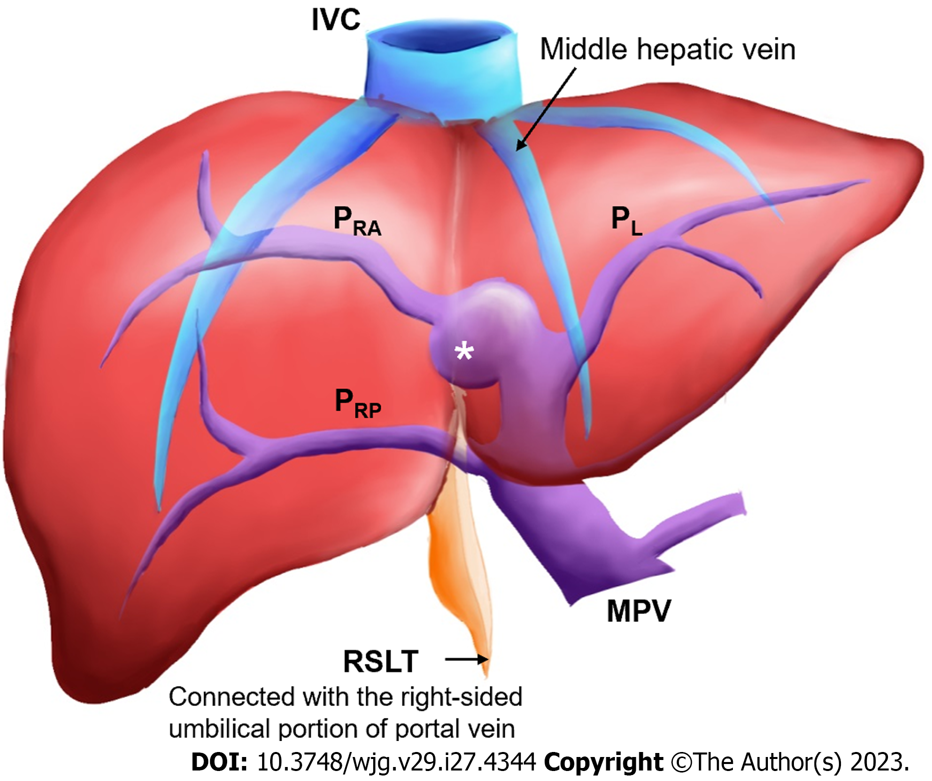
Figure 1 Coronal schematic of the most common anatomical variation in a liver with right-sided ligamentum teres.
The white asterisk indicates the umbilical portion of the portal vein. IVC: Inferior vena cava; MHV: Middle hepatic vein; PRA: Right anterior portal vein; PL: Left portal vein; PRP: Right posterior portal vein; RSLT: Right-sided ligamentum teres; MPV: Main portal vein.
- Citation: Lin HY, Lee RC, Chai JW, Hsu CY, Chou Y, Hwang HE, Liu CA, Chiu NC, Yen HH. Predicting portal venous anomalies by left-sided gallbladder or right-sided ligamentum teres hepatis: A large scale, propensity score-matched study. World J Gastroenterol 2023; 29(27): 4344-4355
- URL: https://www.wjgnet.com/1007-9327/full/v29/i27/4344.htm
- DOI: https://dx.doi.org/10.3748/wjg.v29.i27.4344


