Copyright
©The Author(s) 2022.
World J Gastroenterol. Feb 28, 2022; 28(8): 868-877
Published online Feb 28, 2022. doi: 10.3748/wjg.v28.i8.868
Published online Feb 28, 2022. doi: 10.3748/wjg.v28.i8.868
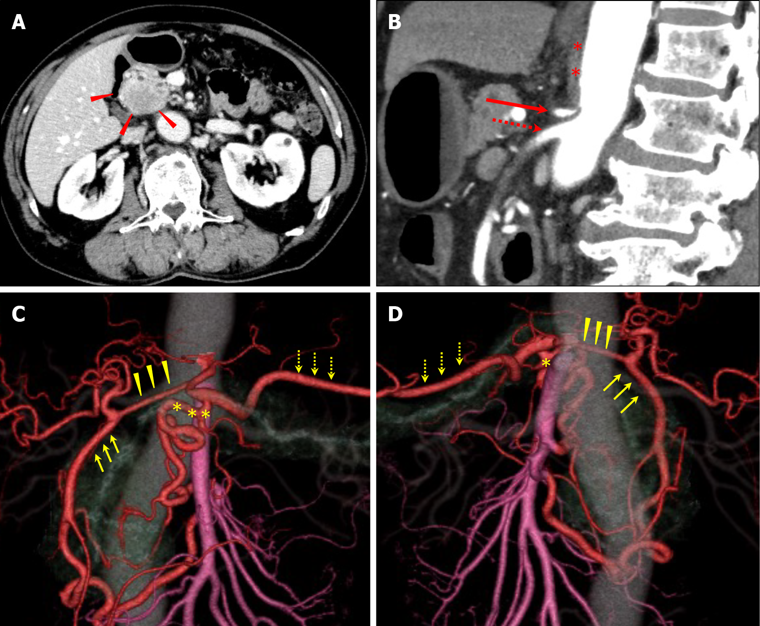
Figure 1 Pre-treatment imaging findings.
A: There was a tumor, with poor contrast, in the pancreatic head on computed tomography (CT) imaging. Red arrowheads: the tumor; B and C: Three-dimensional reconstruction imaging showed developed collateral pathways around the pancreatic head. One connected the superior mesenteric artery (SMA) and common hepatic artery (CHA) via the gastroduodenal artery (GDA) and another connected the SMA and splenic artery (SPA) via the dorsal pancreatic artery (DPA); D: The sagittal view of the CT showed celiac axis (CA) stenosis due to compression by MAL which developed caudally. Yellow arrows: GDA; yellow arrowheads: CHA; yellow asterisks: DPA; yellow dotted arrows: SPA; red asterisks: MAL; red arrow: CA; red dotted arrow: SMA.
- Citation: Yoshida E, Kimura Y, Kyuno T, Kawagishi R, Sato K, Kono T, Chiba T, Kimura T, Yonezawa H, Funato O, Kobayashi M, Murakami K, Takagane A, Takemasa I. Treatment strategy for pancreatic head cancer with celiac axis stenosis in pancreaticoduodenectomy: A case report and review of literature. World J Gastroenterol 2022; 28(8): 868-877
- URL: https://www.wjgnet.com/1007-9327/full/v28/i8/868.htm
- DOI: https://dx.doi.org/10.3748/wjg.v28.i8.868


