Copyright
©The Author(s) 2022.
World J Gastroenterol. Aug 28, 2022; 28(32): 4540-4556
Published online Aug 28, 2022. doi: 10.3748/wjg.v28.i32.4540
Published online Aug 28, 2022. doi: 10.3748/wjg.v28.i32.4540
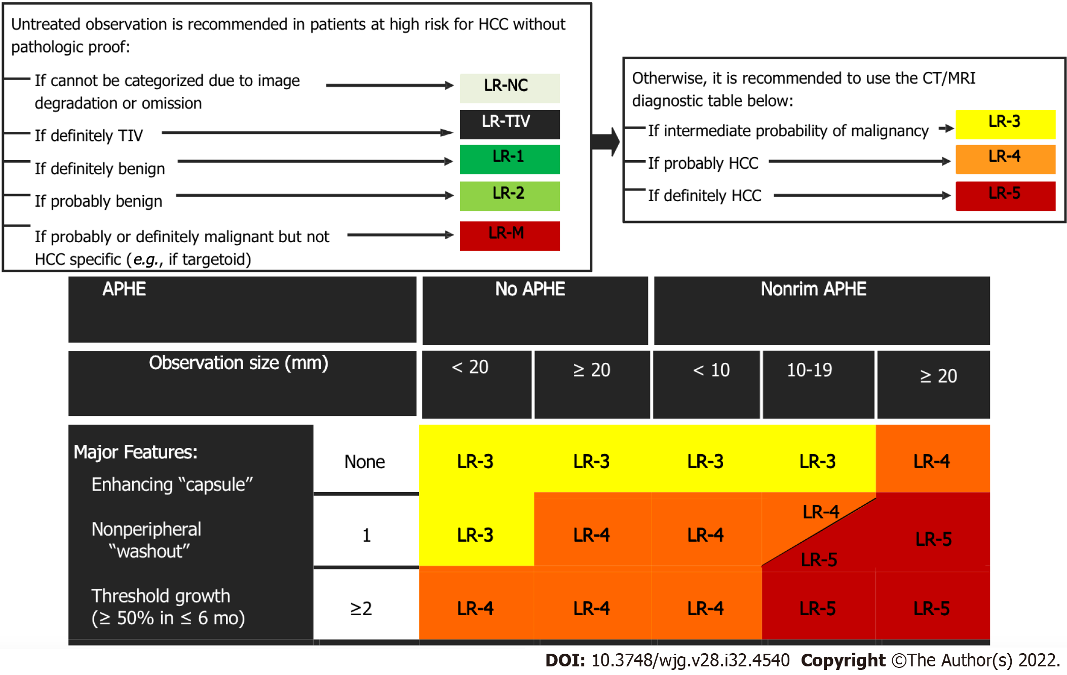
Figure 4 Diagnostic algorithm of liver lesions in computed tomography/magnetic resonance imaging (Liver Imaging Reporting and Data System v2018 CORE).
Observations in the cell LR-4 and LR-5 are categorized based on one additional major feature: LR-4 if enhancing “capsule” and LR-5 if nonperipheral “washout” or threshold growth. APHE: Arterial phase hyperenhancement; CT: Computed tomography; HCC: Hepatocellular carcinoma; MRI: Magnetic resonance imaging; TIV: Tumor in vein.
- Citation: Liava C, Sinakos E, Papadopoulou E, Giannakopoulou L, Potsi S, Moumtzouoglou A, Chatziioannou A, Stergioulas L, Kalogeropoulou L, Dedes I, Akriviadis E, Chourmouzi D. Liver Imaging Reporting and Data System criteria for the diagnosis of hepatocellular carcinoma in clinical practice: A pictorial minireview. World J Gastroenterol 2022; 28(32): 4540-4556
- URL: https://www.wjgnet.com/1007-9327/full/v28/i32/4540.htm
- DOI: https://dx.doi.org/10.3748/wjg.v28.i32.4540


