Copyright
©The Author(s) 2021.
World J Gastroenterol. Jun 28, 2021; 27(24): 3581-3594
Published online Jun 28, 2021. doi: 10.3748/wjg.v27.i24.3581
Published online Jun 28, 2021. doi: 10.3748/wjg.v27.i24.3581
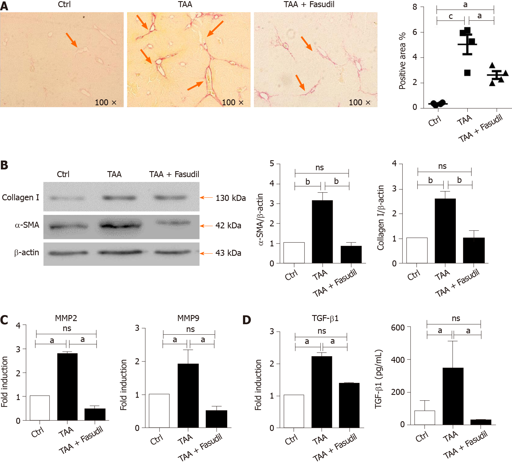
Figure 2 Fasudil prevents thioacetamide-induced liver fibrosis.
A: Histological observation of collagen deposition by sirius red staining under a light-field microscope with × 100 magnification (left). Quantitative analysis of sirius red staining of each group (right); B: Expression of alpha smooth muscle actin (a-SMA) and collagen I in liver tissues was assayed by western blotting. Right panel shows the quantification of α-SMA and collagen I protein levels; C: Levels of matrix metalloproteinase 2 (MMP2) and MMP9 mRNA in liver tissues were detected by real time-reverse transcription polymerase chain reaction (RT-PCR); D: Hepatic expression of transforming growth factor beta 1 (TGF-β1) mRNA was detected by real time-RT-PCR, the secretion level of TGF-β1 in serum was assayed by enzyme-linked immunoassay. Data represent the mean ± SD from at least three independent experiments. aP < 0.05, bP < 0.01.
- Citation: Han QJ, Mu YL, Zhao HJ, Zhao RR, Guo QJ, Su YH, Zhang J. Fasudil prevents liver fibrosis via activating natural killer cells and suppressing hepatic stellate cells. World J Gastroenterol 2021; 27(24): 3581-3594
- URL: https://www.wjgnet.com/1007-9327/full/v27/i24/3581.htm
- DOI: https://dx.doi.org/10.3748/wjg.v27.i24.3581


