Copyright
©The Author(s) 2020.
World J Gastroenterol. Jul 21, 2020; 26(27): 3865-3888
Published online Jul 21, 2020. doi: 10.3748/wjg.v26.i27.3865
Published online Jul 21, 2020. doi: 10.3748/wjg.v26.i27.3865
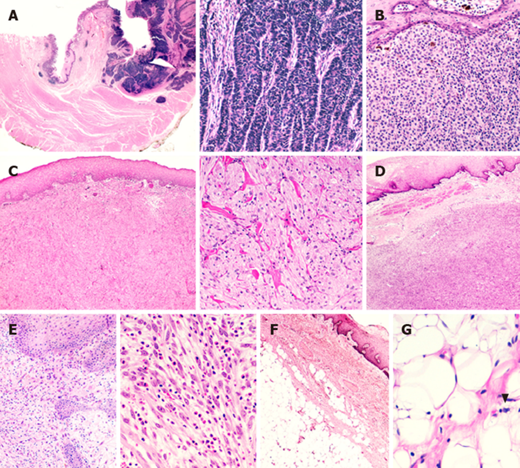
Figure 1 Representative rare histotypes of oesophageal neoplastic lesions.
A: Neuroendocrine carcinoma with full-thickness oesophageal wall involvement; at higher magnification, note the organoid growth pattern of the lesion; B: A case of primitive oesophageal melanoma with melanin deposits; C: Granular cell tumour of the oesophageal wall; note the sheets or nests of plump, round or polygonal cells with eosinophilic granular cytoplasm at higher magnification; D: Oesophageal gastrointestinal stromal tumour. E: Inflammatory myofibroblastic tumour characterized by spindled myofibroblasts and ganglion-like cells dispersed in a myxoid background with a lymphocytic, plasma cellular and eosinophilic infiltrate. F: Oesophageal lipoma; G: High-power field of an atypical oesophageal lipomatous tumour (i.e., ”well-differentiated liposarcoma”) (arrowhead showing a lipoblast).
- Citation: Businello G, Dal Pozzo CA, Sbaraglia M, Mastracci L, Milione M, Saragoni L, Grillo F, Parente P, Remo A, Bellan E, Cappellesso R, Pennelli G, Michelotto M, Fassan M. Histopathological landscape of rare oesophageal neoplasms. World J Gastroenterol 2020; 26(27): 3865-3888
- URL: https://www.wjgnet.com/1007-9327/full/v26/i27/3865.htm
- DOI: https://dx.doi.org/10.3748/wjg.v26.i27.3865


