Published online Apr 7, 2020. doi: 10.3748/wjg.v26.i13.1439
Peer-review started: December 21, 2019
First decision: January 3, 2020
Revised: January 20, 2020
Accepted: March 9, 2020
Article in press: March 9, 2020
Published online: April 7, 2020
Processing time: 108 Days and 14.2 Hours
Lanthanum carbonate is used for treatment of hyperphosphatemia mostly in patients with chronic renal failure. Although lanthanum carbonate is safe, recently, lanthanum deposition in the gastrointestinal mucosa of patients has been reported in the literature. This review provides an overview of gastroduodenal lanthanum deposition and focuses on disease’s endoscopic, radiological, and histological features, prevalence, and outcome, by reviewing relevant clinical studies, case reports, and basic research findings, to better understand the endoscopic manifestation of gastrointestinal lanthanum deposition. The possible relationship between gastric lanthanum deposition pattern and gastric mucosal atrophy is also illustrated; in patients without gastric mucosal atrophy, gastric lanthanum deposition appears as diffuse white lesions in the posterior wall and lesser curvature of the gastric body. In the gastric mucosa with atrophy, lanthanum-related lesions likely appear as annular or granular whitish lesions. Moreover, these white lesions are probably more frequently observed in the lower part of the stomach, where intestinal metaplasia begins.
Core tip: This review provides an overview of the endoscopic and pathological diagnosis of gastroduodenal lanthanum deposition. Previously reported case reports, case series, and retrospective studies are reviewed, focusing on disease’s endoscopic, histological, and computed tomography features, prevalence, and outcome. Although gastroduodenal deposition presents with white appearance at esophagogastroduodenoscopy, macroscopic features and locations of gastric lesions possibly vary depending on the presence or absence of mucosal atrophy. Our hypotheses are also related to the pattern of lanthanum deposition in the gastric mucosa with/without atrophy, which will aid endoscopists to understand this disease entity.
- Citation: Iwamuro M, Urata H, Tanaka T, Okada H. Review of the diagnosis of gastrointestinal lanthanum deposition. World J Gastroenterol 2020; 26(13): 1439-1449
- URL: https://www.wjgnet.com/1007-9327/full/v26/i13/1439.htm
- DOI: https://dx.doi.org/10.3748/wjg.v26.i13.1439
Lanthanum carbonate (Fosrenol®) is used for treatment of hyperphosphatemia and in the management of chronic renal failure, especially in dialysis patients[1-3]. Since patients with chronic renal failure generally have a reduced ability to excrete phosphorus from the bloodstream, the levels of serum phosphorus increase as their kidney function deteriorates, resulting in hyperphosphatemia. Persistent hyperphosphatemia causes osteoporosis and deposition of calcium phosphate on the blood vessel walls, leading to arteriosclerosis[4,5]. Moreover, hyperphosphatemia is associated with an increased risk of cardiovascular events and mortality[6]. Therefore, serum phosphorus levels should be controlled within their normal ranges during the management of dialysis patients.
For the treatment of hyperphosphatemia in dialysis patients, aluminum-containing agents have long been used as phosphate binders since the 1970s. However, chronic administration of aluminum salts results in accumulation of aluminum in the central nervous system, bone and hematopoietic cells, leading to encephalopathy, osteomalacia, myopathy, and microcytic anemia[7]. Due to these severe toxic effects, long-term use of aluminum salts is not allowed for dialysis patients. Alternatively, calcium carbonate, calcium acetate, polymers such as sevelamer hydrochloride, and lanthanum carbonate have been developed and used in clinical practice. Among these, calcium carbonate occasionally increases serum calcium levels and causes extraskeletal calcification. Sevelamer hydrochloride also exhibits adverse events such as digestive symptoms mainly due to constipation. In contrast, lanthanum carbonate has been widely used for patients with chronic renal failure because it is safe and well-tolerated by the patients.
After ingestion of lanthanum carbonate, the lanthanum ion (La3+) is released in the stomach and binds to dietary phosphate in the intestinal tract. Lanthanum phosphate is an insoluble complex in the gut that is not absorbed by the digestive tract, thereby it is excreted from the body together with feces. The absorption rate of lanthanum carbonate from the gastrointestinal tract into the blood is less than 0.002%[8]. Because the slightly absorbed lanthanum is reportedly excreted from the body via bile[9], lanthanum carbonate can be safely used even in patients with impaired renal function. Although absorbed lanthanum is known to deposit in the bones and liver, the amount of deposition is quite small and does not cause any damage to the organs[10,11]. However, more recently, patients with lanthanum deposition in the gastrointestinal mucosa have been reported in the literature. Here, we review relevant clinical studies, case reports, and basic research findings, including our articles, to better understand the endoscopic manifestation of gastrointestinal lanthanum deposition.
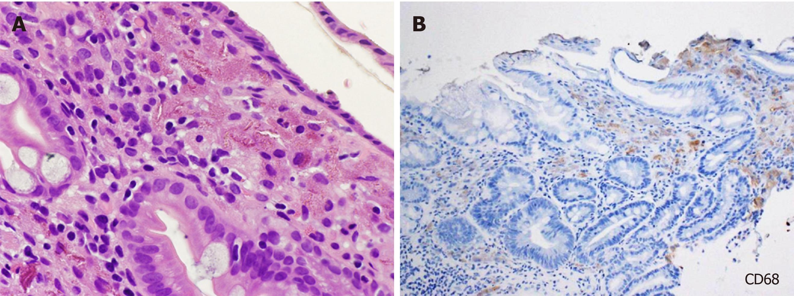
Hematoxylin and eosin staining of biopsy specimens containing lanthanum showed deposition of fine, amorphous, eosinophilic material (Figure 1A). Deposited materials have been variably described as inclusion-like materials with irregularly branching or coiled configurations[12], granular, brown material, sometimes needle-shaped or crystalloid and irregular eosinophilic material with slit-like clefts[13,14], variably dense and granular deposits[15], many colorless and coarse granular materials[16], gray or brown pigments or crystal-like structures[17], amphophilic and/or yellowish-brown fine granules, and amphophilic and/or brownish rods or curly strings[18]. Deposited materials are generally phagocytosed by macrophages in the lamina propria in the stomach and duodenum, which are more clearly visualized with immuno-histochemical staining, such as anti-CD68 staining (Figure 1B). Nakamura et al[19] revealed that the macrophages are positive for both CD68 and CD206 staining and speculated that M2 macrophages potentially play a role in the clearance of lanthanum from the gastroduodenal mucosa[19].
Diagnosis of lanthanum deposition can be made with (1) conventional light microscopy observation of the fine, amorphous, eosinophilic material; and (2) medication information of patient’s current or past use of lanthanum carbonate. Analysis of elements by energy-dispersive X-ray spectrometry (EDX) is not always required, unless the amount of lanthanum deposition is subtle or pathological features are atypical[20-24].
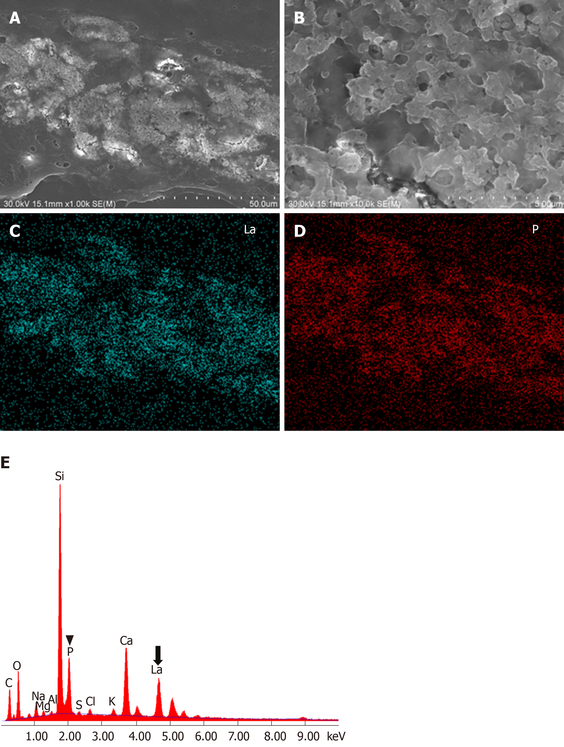
In scanning electron microscopy (SEM), deposited lanthanum is visible as bright areas (Figure 2A). Higher magnification shows that the deposition is composed of aggregates of particles, measuring 0.5–3 m in diameter (Figure 2B). Elemental mapping by energy dispersive X-ray spectroscopy is useful to visualize distribution of lanthanum (Figure 2C) and phosphate (Figure 2D), which is identical to that of the bright areas. EDX also shows presence of lanthanum and phosphate elements (Figure 2E). Since both lanthanum and phosphate are generally detected simultaneously, deposited material is considered as lanthanum phosphate. SEM observation and EDX analysis enable analysis of the elemental composition and distribution of elements, leading to direct proof of lanthanum deposition. In contrast to light microscopy, deposited lanthanum is easily identified as bright areas with SEM[22].
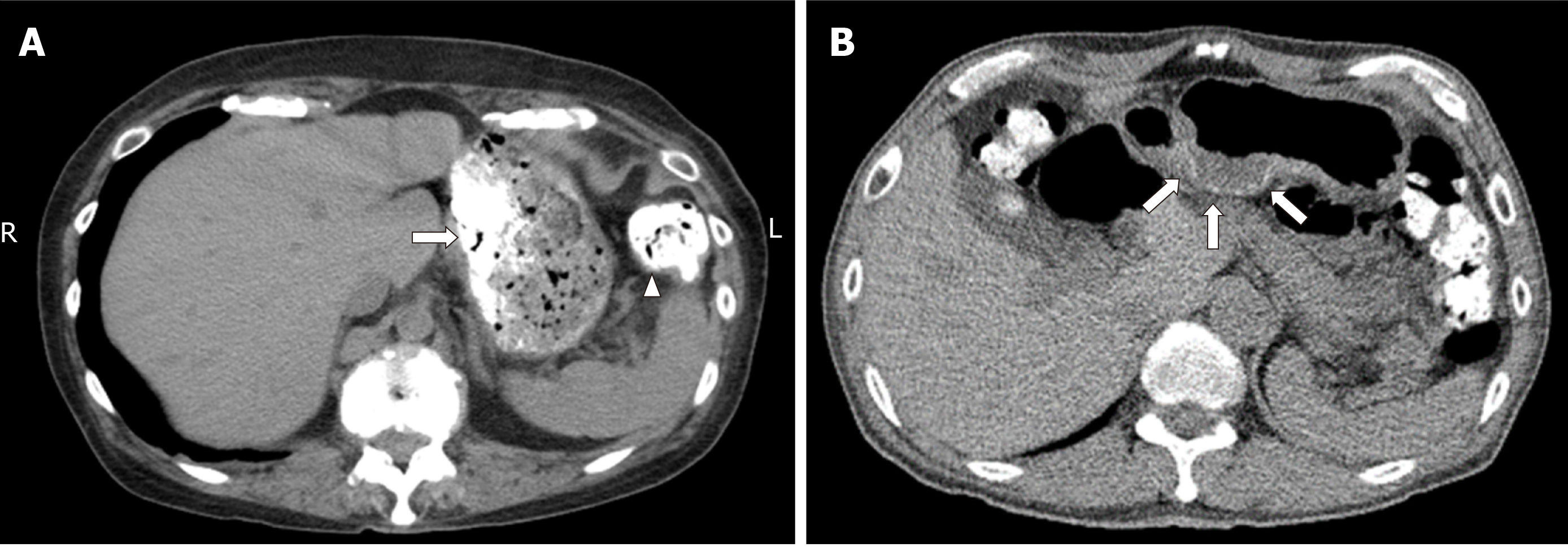
Because lanthanum carbonate is not transparent to X-rays, it has a radiopaque appearance in plain abdominal radiography and computed tomography (CT) scanning images[25]. Figure 3A shows ingested lanthanum carbonate in the stomach (arrow) and colon (arrowhead) that is displayed as high-density substance. In this patient, lanthanum carbonate and food contents are separated in the stomach, while lanthanum carbonate in the colon is combined with digested materials and forms a lump of feces. In another patient, deposited lanthanum in the stomach is observed as a high-density layer within the gastric mucosa (Figure 3B). Namie et al[26] reported the identification of a high-density layer in the stomach in 42 out of 70 (60%) patients treated with lanthanum carbonate.
Lanthanum carbonate has been marketed as a phosphate binder since 2005 in United States and was released in Japan in 2009. Despite its general tolerance and safety profile, lanthanum deposition in the gastroduodenal mucosa of lanthanum carbonate users was first reported in 2015. The endoscopic features of the gastric lesions were initially portrayed as numerous small, irregular white spots[13], scattered ulcerations[15], polyp, erosions, ulcer[12,14], white thickenings of annular shape or those on the gastric folds[26], slightly granular, white mucosa[20], granular white depositions in reddish mucosa[21], white spots[27], mucosal irregularity with reddish and/or whitish color, and even nonremarkable mucosa[18]. Based on the accumulation of cases reported in the literature and clinical practice, typical endoscopic findings of gastric lanthanum deposition have been currently recognized as white lesions.
In our earlier work, we reviewed four patients showing gastric white lesions (Bw) and peripheral mucosa where the white substance was not endoscopically observed (Bp) during biopsy[28]. We performed SEM analysis and EDX spectrometry to quantify the lanthanum elements (wt%) in the biopsy specimens. We showed that the amount of lanthanum was significantly higher in Bw than in Bp (0.15–0.31 wt% vs 0.00–0.13 wt%) (P < 0.05), revealing that pathological lanthanum deposition coincides with endoscopically observed white lesions in the gastric mucosa. On the other hand, although its deposited amount was small, lanthanum was detectable in EDX analysis of the gastric mucosa where the white substance was not endoscopically visible[22,28]. Consequently, a subtle amount of lanthanum deposition is not detected under endoscopic observation, while it can be detected with optical microscopy or electron microscopy.
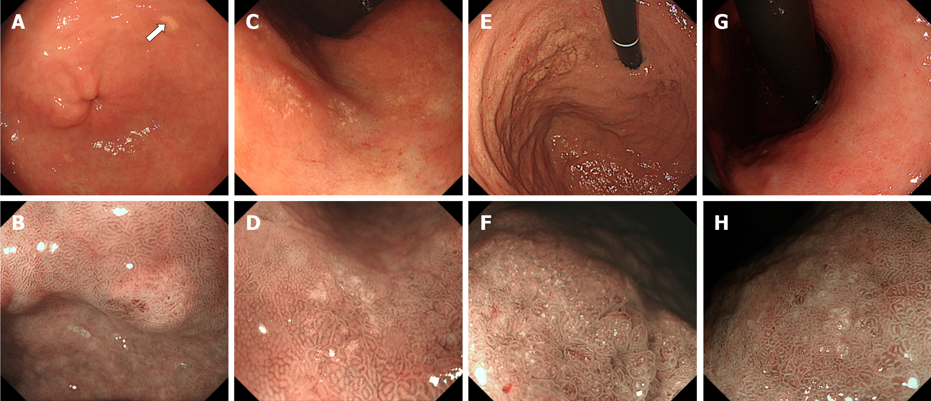
We also reviewed gastric lesions of lanthanum deposition and subclassified endoscopic features into “whitish spots” (Figure 4A and B), “annular whitish mucosa” (Figure 4C and D), and “diffuse whitish mucosa” (Figure 4E and F)[29]. Subsequently we added “fine granular whitish deposition” as a new subtype of endoscopic features (Figure 4G and H), because we noticed that whitish spots are relatively infrequent compared with the other three subtypes, while small amount of lanthanum deposition often appears as fine, granular white lesions[23]. We defined annular whitish mucosa as a lesion (s) ≤ 20 mm in diameter with a white color in the periphery. A diffuse whitish mucosa presents a white area > 20 mm in diameter, where reddish areas may be intermixed. Fine granular whitish deposition is a tiny or faint whitish lesion (s) ≤ 1 mm in diameter. Whitish spot is a whitish lesion ≤ 20 mm in diameter with a uniform white color, resembling gastric xanthoma. Generally, diffuse whitish mucosa is observed in the non-atrophic mucosa of the gastric body, whereas annular whitish mucosa and fine granular whitish deposition are observed in the atrophic mucosa. In the following sections, we discuss endoscopic features of lanthanum deposition focusing on gastric mucosal atrophy.
Possible association with gastric lanthanum deposition and underlying regenerative changes, intestinal metaplasia, and/or foveolar hyperplasia has been reported in 2015. Makino et al[13] reported that macrophages containing lanthanum existed in the gastric mucosa with atrophic pyloric glands and foveolar epithelium fully replaced by intestinal metaplasia. Tonooka et al[30] also found atrophic mucosa with metaplastic epithelia in their patient. They speculated that altered mucosal structure resulted in increased epithelial permeability, finally leading to lanthanum deposition in the stomach. Ban et al[18] found a significant correlation between deposition grade of lanthanum and mucosal alterations such as regenerative changes, intestinal metaplasia, or foveolar hyperplasia, reinforcing the tendency of lanthanum deposition in higher degree in the microscopically altered gastric mucosa. Ji et al[31] investigated the mucosal barrier defects in patients with intestinal metaplasia using laser confocal endomicroscopy. In vivo functional imaging revealed that lanthanum nitrate did not permeated normal gastric epithelium, whereas it permeated gastric mucosa with intestinal metaplasia. These results indicated that gastric mucosa with regenerative changes, intestinal metaplasia, and/or foveolar hyperplasia allows permeation of lanthanum.
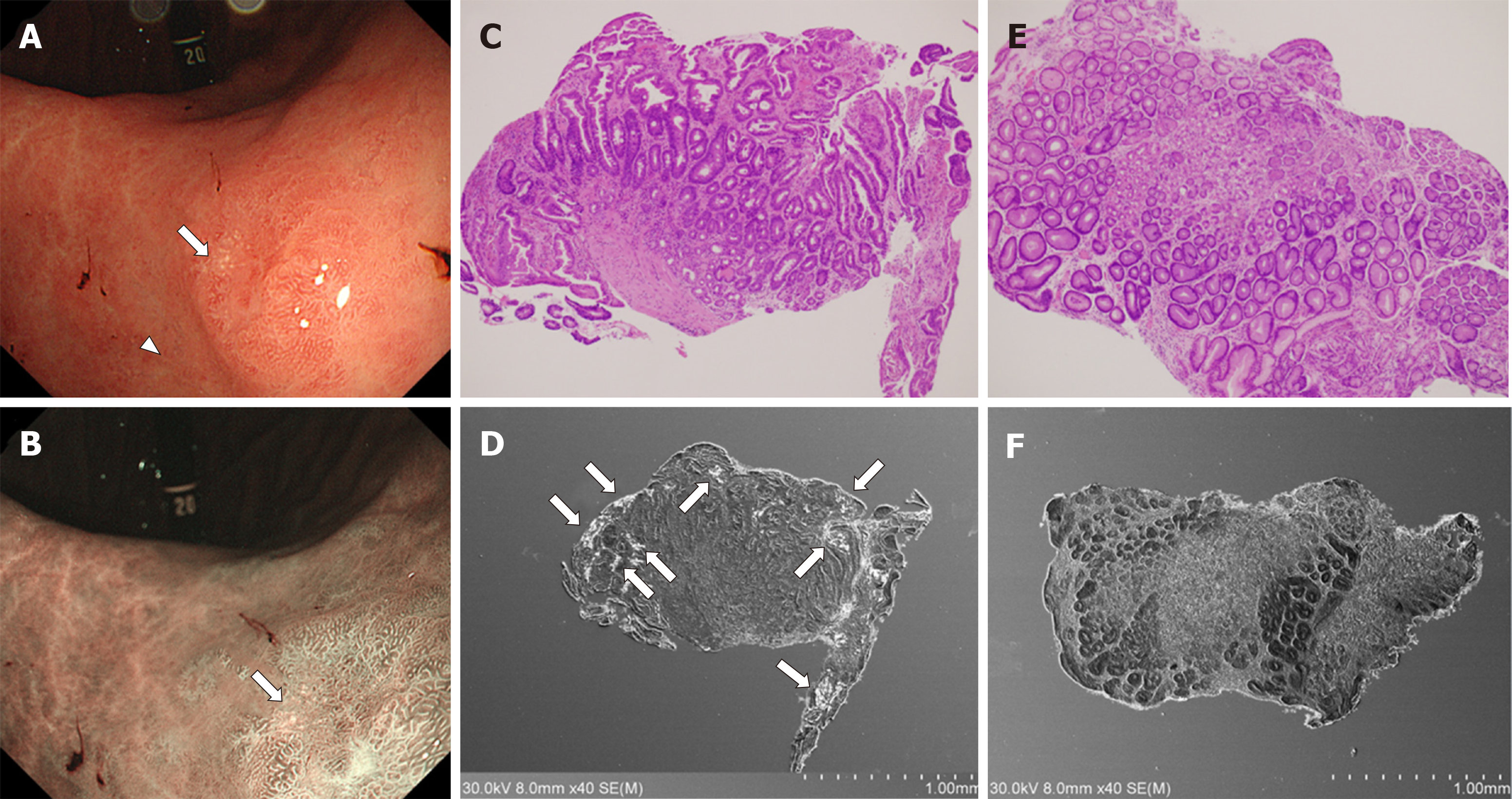
We recently reviewed endoscopic features of gastric lanthanum deposition in 10 patients with gastric atrophy (under review). Although gastric lanthanum deposition appears as whitish lesions, this presentation was not observed in 1 out of 10 patients. In the gastric mucosa with atrophy, the antrum (n = 5) and angle (n = 5) were most frequently involved and lanthanum deposition presented with annular and/or granular whitish lesions. Whitish lesions were also found in the gastric body with mucosal atrophy that appeared as annular (n = 1), granular (n = 1), and diffuse whitish lesions (n = 1). Consequently, we speculate that, in the atrophic gastric mucosa, lanthanum-related lesions typically present with annular or granular whitish lesions. In our earlier study, we investigated pathological features of four patients with annular whitish lesions[28]. We took one biopsy sample from white lesions and the other sample from the surrounding mucosa approximately 5 mm away from the white lesions. Intestinal metaplasia was identified in 3 out of 4 samples acquired from the annular whitish lesion, whereas the surrounding mucosa contained no intestinal metaplasia (Figure 5). Thus, we speculate that lanthanum deposition appears as “annular” or “granular” lesions in the atrophic mucosa, because intestinal metaplasia unevenly exists in the background gastric mucosa and is susceptible to lanthanum deposition. In addition, lanthanum-related lesions are probably more frequently observed in the gastric antrum and angle, because intestinal metaplasia generally appears at the lower part of the stomach.
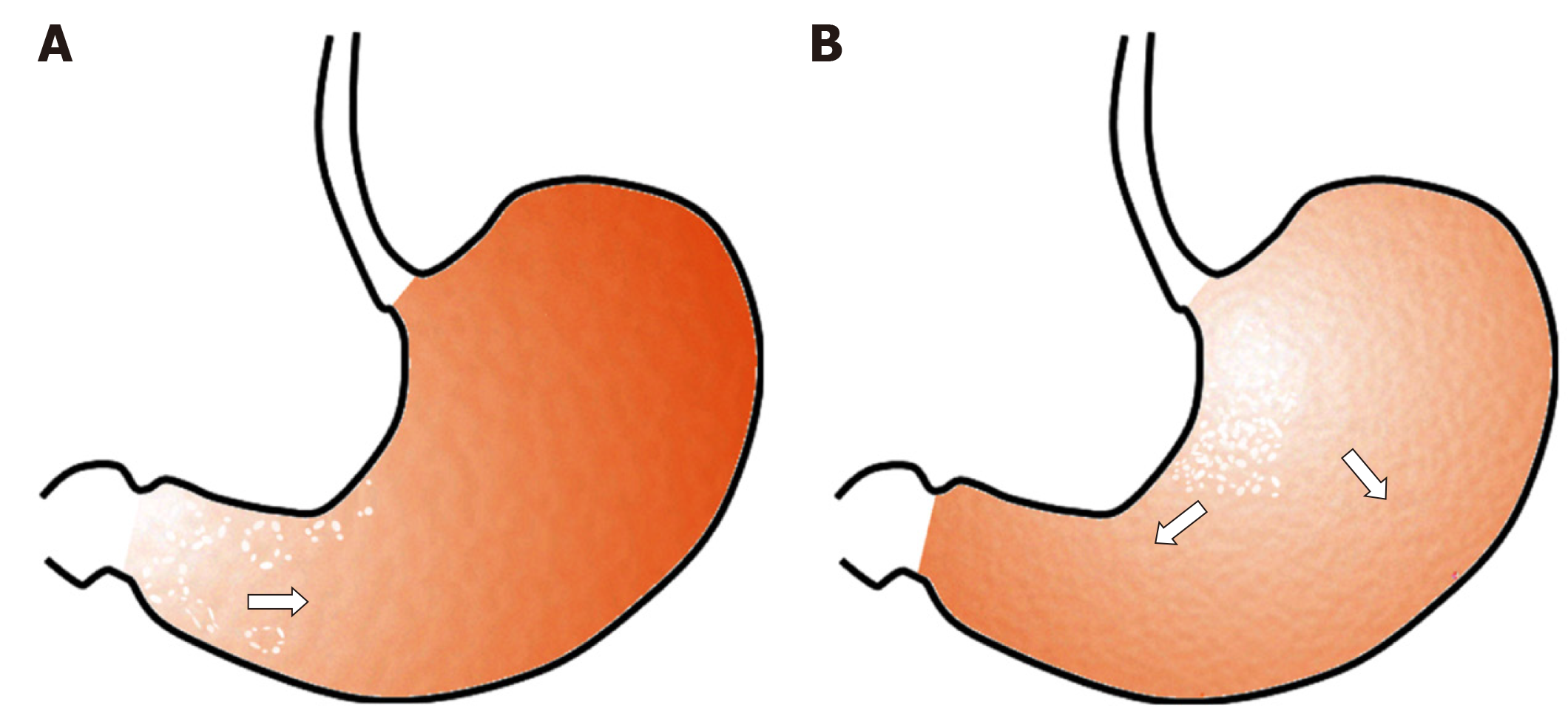
We postulate that in the atrophic mucosa, particularly in areas with intestinal metaplasia, lanthanum deposition presents with annular and/or granular whitish lesions, predominantly in the gastric antrum and angle. As the intestinal metaplasia expands, the size of the areas with lanthanum deposition may increase (Figure 6A).
As described above, several reports have revealed that lanthanum deposition develops within the gastric mucosa showing regenerative changes, intestinal metaplasia, and/or foveolar hyperplasia. Because all these histopathological features generally arise as Helicobacter pylori (H. pylori)-induced mucosal alterations, lanthanum deposition had been considered to occur in the stomach in close association with H. pylori-infection. In contrast, we previously reported two patients with lanthanum deposition in the stomach who were serologically and histopathologically negative for H. pylori[32]. In our patients, lanthanum deposition was identified as diffuse whitish lesions, which were predominantly observed in the lesser curvature and posterior wall of the gastric body, rather than in the antrum or angle. Based on this observation, we hypothesized that in the gastric mucosa without atrophy, lanthanum primarily deposits in the lesser curvature and posterior wall of the gastric body and presents diffuse whitish lesions (Figure 6B). The area of lanthanum deposition probably expands as time elapses unless the patient stops lanthanum carbonate.
We speculate that gastric body-predominant lanthanum deposition occurs because of the direct physical contact between ingested lanthanum carbonate and the gastric body mucosa[32]. Figure 3A shows a CT image of a patient, who had been administered lanthanum carbonate. Even after abstinence from food and medicine for 10 h, a substantial amount of lanthanum was observed as a high-density substance (Figure 3A, arrow), predominantly in the gastric body. Thus, the gastric body is expected to be involved in lanthanum deposition due to prolonged contact with ingested lanthanum.
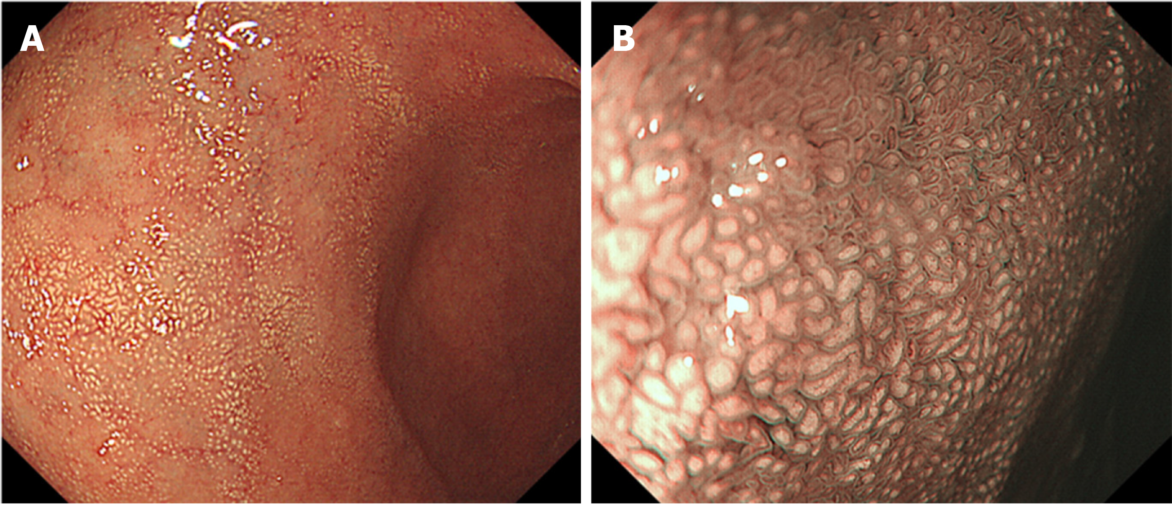
Although several authors have described various endoscopic features of lanthanum-related duodenal lesions as duodenal ulcer[12], granular and micronodular mucosa[33], granular mucosa[34], and duodenitis[27], we first reported white villi as a characteristic appearance of duodenal lanthanum deposition[35]. Subsequently, we retrospectively reviewed endoscopic and pathological features in patients with pathologically proven lanthanum deposition in the gastrointestinal tract[36]. We revealed that, among 19 patients who underwent biopsy from the duodenum, lanthanum deposition was detected in 17 patients (89.5%). Moreover, white villi were observed in 15 patients (88.2%). These results indicate that the duodenum is often involved in lanthanum deposition, which generally presents with white villi (Figure 7). However, these deposits may not be detected during esophagogastroduodenoscopy in some cases due to the subtle degree of deposition. We consider that endoscopic biopsy should be performed in the duodenum as well as in the stomach, regardless of the presence or absence of white villi, for accurate determination of lanthanum deposition in the gastrointestinal tract[36].
Deposition of lanthanum in organs other than the stomach and duodenum has scarcely been reported. In 2009, Davis et al[37]reported lanthanum deposition in the mesenteric lymph nodes in postmortem examination of a dialysis patient. Yabuki et al[17] and Tonooka et al[30] also described involvement of the gastric regional lymph nodes in patients who underwent gastrectomy for gastric cancer. Goto et al[14] reported lanthanum deposition in a tubular adenoma of the transverse colon.
In animal models, Lacour et al[38] investigated lanthanum concentrations in various rat tissues after oral administration for 28 d. Although lanthanum concentration did not increase in the brain and heart of rats, significantly elevated levels of lanthanum concentration were observed in the liver, lungs, femur, muscles, and kidneys of lanthanum-treated rats. Of note, compared with lanthanum-treated rats with normal kidney function, rats with impaired kidney function showed significantly higher tissue lanthanum concentration in the brain, liver, heart, lungs, femur, and muscles. Therefore, in patients with chronic kidney disease, a long period of careful observation may be required to reveal organ-specific properties of lanthanum accumulation.
Coexistence of gastric cancer and lanthanum deposition in the gastric mucosa has been reported by several authors[13,17,30,39]. Makino et al[13] reported lanthanum-related gastric lesions as numerous small, irregular white spots, which exist in the periphery of the area with gastric cancer. Yabuki et al[17] also described lanthanum deposition in non-neoplastic area, while deposition was not significant in the gastric adenocarcinoma lesion. The authors speculated that different permeability between adenocarcinoma and background mucosa resulted in the uneven distribution of lanthanum deposition. Tonooka et al[30] and Takatsuna et al[39] also described quite small amounts of lanthanum deposition in gastric cancer lesions. Based on these observations, the area with gastric cancer is probably spared from lanthanum deposition. Although understanding this phenomenon will help endoscopists to easily identify gastric cancer lesions in the lanthanum-deposited stomach, this concept requires further investigation.
The actual prevalence of gastroduodenal lanthanum deposition among end-stage renal disease patients treated with lanthanum carbonate has not yet been clarified. Murakami et al[29] performed endoscopic biopsy in 9 out of 90 patients with lanthanum carbonate prescription. Gastric lanthanum deposition was histologically diagnosed in seven patients (7/9, 77.8%). Goto et al[14] reported that lanthanum was detected in the stomach of 12 out of 14 patients who received lanthanum carbonate and underwent endoscopic biopsy (12/14, 85.7%). Namie et al[26] found high-density lesions within the gastric mucosa on CT scanning in 42 out of 70 patients who were administered lanthanum carbonate (42/70, 60.0%). Therefore, prevalence of lanthanum deposition in the stomach is estimated to be 60%–85%. However, because only retrospective studies have been performed, sampling biases are inevitable and prospective studies are required to address this issue.
Our retrospective study revealed that, during continuous lanthanum carbonate use, lanthanum-related lesions in the stomach were endoscopically unchanged in two patients, whereas whitish lesions became apparent and spread in three patients[29], indicating that gastric lanthanum deposition progresses in several patients in a time-dependent manner. In contrast, Rothenberg et al[15] reported resolution of histiocytosis in the stomach and significant decrease in the duodenum three months after cessation of lanthanum carbonate in a patient with gastroduodenal lanthanum deposition. However, Namie et al[26] reported that CT and pathological findings of lanthanum-related gastric lesions were unchanged eight months after discontinuation of lanthanum carbonate. Awad et al[40] also noted that lanthanum-related lesions remained unchanged six months after drug cessation. Moreover, Hoda et al[33] detected lanthanum deposition in a biopsy specimen from the stomach acquired seven years after stopping lanthanum carbonate intake. It has not been clarified to date whether lanthanum deposition in tissues is reversible or not.
The pathological significance of lanthanum deposition in the gastrointestinal mucosa has not been clarified, and there is no consensus on whether to stop administration of lanthanum carbonate in such cases. Yabuki et al[17] investigated histological changes in the gastric mucosa of lanthanum carbonate-consuming rats. The authors found a variety of alterations including glandular atrophy, stromal fibrosis, proliferation of mucous neck cells, intestinal metaplasia, squamous cell papilloma, erosion, and ulcer in the stomach of rats. They speculated that deposited lanthanum was able to cause mucosal injury and abnormal cell proliferation, leading to structural changes in the mucosa and neoplastic lesions in the stomach. In this context, we consider that endoscopists should accurately diagnose lanthanum deposition in the gastroduodenal tract in lanthanum-carbonate users and recommend them to undergo regular endoscopy examinations in order to track progression of white lesions and elucidate whether lanthanum deposition is related to health problems.
Lanthanum deposition in the stomach and duodenum is recognized as whitish lesions during endoscopy examination. Accurate diagnosis and keeping track of the health of patients with gastroduodenal lanthanum deposition are essential to elucidate pathogenicity of lanthanum deposition in the gastrointestinal tract.
Manuscript source: Invited manuscript
Specialty type: Gastroenterology and hepatology
Country of origin: Japan
Peer-review report classification
Grade A (Excellent): A
Grade B (Very good): B
Grade C (Good): 0
Grade D (Fair): 0
Grade E (Poor): 0
P-Reviewer: Casadesus D, Chow WK S-Editor: Zhang L L-Editor: A E-Editor: Ma YJ
| 1. | Shinoda T, Yamasaki M, Chida Y, Takagi M, Tanaka Y, Ando R, Suzuki T, Tagawa H. Improvement of MBD parameters in dialysis patients by a switch to, and combined use of lanthanum carbonate: Josai Dialysis Forum collaborative study. Ther Apher Dial. 2013;17 Suppl 1:29-34. [RCA] [PubMed] [DOI] [Full Text] [Cited by in Crossref: 1] [Cited by in RCA: 1] [Article Influence: 0.1] [Reference Citation Analysis (0)] |
| 2. | Kasai S, Sato K, Murata Y, Kinoshita Y. Randomized crossover study of the efficacy and safety of sevelamer hydrochloride and lanthanum carbonate in Japanese patients undergoing hemodialysis. Ther Apher Dial. 2012;16:341-349. [RCA] [PubMed] [DOI] [Full Text] [Cited by in Crossref: 22] [Cited by in RCA: 23] [Article Influence: 1.8] [Reference Citation Analysis (0)] |
| 3. | Shigematsu T; Lanthanum Carbonate Research Group. One year efficacy and safety of lanthanum carbonate for hyperphosphatemia in Japanese chronic kidney disease patients undergoing hemodialysis. Ther Apher Dial. 2010;14:12-19. [RCA] [PubMed] [DOI] [Full Text] [Cited by in Crossref: 6] [Cited by in RCA: 7] [Article Influence: 0.5] [Reference Citation Analysis (0)] |
| 4. | Lau WL, Festing MH, Giachelli CM. Phosphate and vascular calcification: Emerging role of the sodium-dependent phosphate co-transporter PiT-1. Thromb Haemost. 2010;104:464-470. [RCA] [PubMed] [DOI] [Full Text] [Full Text (PDF)] [Cited by in Crossref: 92] [Cited by in RCA: 89] [Article Influence: 5.9] [Reference Citation Analysis (0)] |
| 5. | Giachelli CM. The emerging role of phosphate in vascular calcification. Kidney Int. 2009;75:890-897. [RCA] [PubMed] [DOI] [Full Text] [Full Text (PDF)] [Cited by in Crossref: 385] [Cited by in RCA: 349] [Article Influence: 21.8] [Reference Citation Analysis (0)] |
| 6. | Kendrick J, Kestenbaum B, Chonchol M. Phosphate and cardiovascular disease. Adv Chronic Kidney Dis. 2011;18:113-119. [RCA] [PubMed] [DOI] [Full Text] [Cited by in Crossref: 43] [Cited by in RCA: 53] [Article Influence: 3.8] [Reference Citation Analysis (0)] |
| 7. | Salusky IB. A new era in phosphate binder therapy: what are the options? Kidney Int Suppl. 2006;S10-S15. [RCA] [PubMed] [DOI] [Full Text] [Cited by in Crossref: 31] [Cited by in RCA: 26] [Article Influence: 1.4] [Reference Citation Analysis (0)] |
| 8. | Giotta N, Marino AM. Pharmacoeconomic analysis: Analysis of cost-effectiveness of lanthanum-carbonate (Lc) in uncontrolled hyperphosphatemia in dialysis. Value Health. 2015;18:A511. [DOI] [Full Text] |
| 9. | Pennick M, Dennis K, Damment SJ. Absolute bioavailability and disposition of lanthanum in healthy human subjects administered lanthanum carbonate. J Clin Pharmacol. 2006;46:738-746. [RCA] [PubMed] [DOI] [Full Text] [Cited by in Crossref: 88] [Cited by in RCA: 88] [Article Influence: 4.6] [Reference Citation Analysis (0)] |
| 10. | Spasovski GB, Sikole A, Gelev S, Masin-Spasovska J, Freemont T, Webster I, Gill M, Jones C, De Broe ME, D'Haese PC. Evolution of bone and plasma concentration of lanthanum in dialysis patients before, during 1 year of treatment with lanthanum carbonate and after 2 years of follow-up. Nephrol Dial Transplant. 2006;21:2217-2224. [RCA] [PubMed] [DOI] [Full Text] [Cited by in Crossref: 124] [Cited by in RCA: 115] [Article Influence: 6.1] [Reference Citation Analysis (0)] |
| 11. | Damment SJ, Pennick M. Clinical pharmacokinetics of the phosphate binder lanthanum carbonate. Clin Pharmacokinet. 2008;47:553-563. [RCA] [PubMed] [DOI] [Full Text] [Cited by in Crossref: 67] [Cited by in RCA: 68] [Article Influence: 4.0] [Reference Citation Analysis (0)] |
| 12. | Haratake J, Yasunaga C, Ootani A, Shimajiri S, Matsuyama A, Hisaoka M. Peculiar histiocytic lesions with massive lanthanum deposition in dialysis patients treated with lanthanum carbonate. Am J Surg Pathol. 2015;39:767-771. [RCA] [PubMed] [DOI] [Full Text] [Cited by in Crossref: 41] [Cited by in RCA: 42] [Article Influence: 4.2] [Reference Citation Analysis (0)] |
| 13. | Makino M, Kawaguchi K, Shimojo H, Nakamura H, Nagasawa M, Kodama R. Extensive lanthanum deposition in the gastric mucosa: the first histopathological report. Pathol Int. 2015;65:33-37. [RCA] [PubMed] [DOI] [Full Text] [Cited by in Crossref: 40] [Cited by in RCA: 50] [Article Influence: 4.5] [Reference Citation Analysis (0)] |
| 14. | Goto K, Ogawa K. Lanthanum Deposition Is Frequently Observed in the Gastric Mucosa of Dialysis Patients with Lanthanum Carbonate Therapy: A Clinicopathologic Study of 13 Cases, Including 1 Case of Lanthanum Granuloma in the Colon and 2 Nongranulomatous Gastric Cases. Int J Surg Pathol. 2016;24:89-92. [RCA] [PubMed] [DOI] [Full Text] [Cited by in Crossref: 26] [Cited by in RCA: 56] [Article Influence: 5.6] [Reference Citation Analysis (0)] |
| 15. | Rothenberg ME, Araya H, Longacre TA, Pasricha PJ. Lanthanum-Induced Gastrointestinal Histiocytosis. ACG Case Rep J. 2015;2:187-189. [RCA] [PubMed] [DOI] [Full Text] [Full Text (PDF)] [Cited by in Crossref: 25] [Cited by in RCA: 23] [Article Influence: 2.3] [Reference Citation Analysis (0)] |
| 16. | Yasunaga C, Haratake J, Ohtani A. Specific Accumulation of Lanthanum Carbonate in the Gastric Mucosal Histiocytes in a Dialysis Patient. Ther Apher Dial. 2015;19:622-624. [RCA] [PubMed] [DOI] [Full Text] [Cited by in Crossref: 13] [Cited by in RCA: 13] [Article Influence: 1.3] [Reference Citation Analysis (0)] |
| 17. | Yabuki K, Shiba E, Harada H, Uchihashi K, Matsuyama A, Haratake J, Hisaoka M. Lanthanum deposition in the gastrointestinal mucosa and regional lymph nodes in dialysis patients: Analysis of surgically excised specimens and review of the literature. Pathol Res Pract. 2016;212:919-926. [RCA] [PubMed] [DOI] [Full Text] [Cited by in Crossref: 19] [Cited by in RCA: 18] [Article Influence: 2.0] [Reference Citation Analysis (0)] |
| 18. | Ban S, Suzuki S, Kubota K, Ohshima S, Satoh H, Imada H, Ueda Y. Gastric mucosal status susceptible to lanthanum deposition in patients treated with dialysis and lanthanum carbonate. Ann Diagn Pathol. 2017;26:6-9. [RCA] [PubMed] [DOI] [Full Text] [Cited by in Crossref: 28] [Cited by in RCA: 32] [Article Influence: 3.6] [Reference Citation Analysis (0)] |
| 19. | Nakamura T, Tsuchiya A, Kobayashi M, Naito M, Terai S. M2-polarized macrophages relate the clearance of gastric lanthanum deposition. Clin Case Rep. 2019;7:570-572. [RCA] [PubMed] [DOI] [Full Text] [Full Text (PDF)] [Cited by in Crossref: 3] [Cited by in RCA: 4] [Article Influence: 0.7] [Reference Citation Analysis (1)] |
| 20. | Iwamuro M, Sakae H, Okada H. White Gastric Mucosa in a Dialysis Patient. Gastroenterology. 2016;150:322-323. [RCA] [PubMed] [DOI] [Full Text] [Cited by in Crossref: 13] [Cited by in RCA: 13] [Article Influence: 1.4] [Reference Citation Analysis (0)] |
| 21. | Iwamuro M, Kanzaki H, Tanaka T, Kawano S, Kawahara Y, Okada H. Lanthanum phosphate deposition in the gastric mucosa of patients with chronic renal failure. Nihon Shokakibyo Gakkai Zasshi. 2016;113:1216-1222. [RCA] [PubMed] [DOI] [Full Text] [Cited by in RCA: 6] [Reference Citation Analysis (0)] |
| 22. | Iwamuro M, Urata H, Tanaka T, Ando A, Nada T, Kimura K, Yamauchi K, Kusumoto C, Otsuka F, Okada H. Lanthanum Deposition in the Stomach: Usefulness of Scanning Electron Microscopy for Its Detection. Acta Med Okayama. 2017;71:73-78. [RCA] [PubMed] [DOI] [Full Text] [Cited by in RCA: 8] [Reference Citation Analysis (0)] |
| 23. | Iwamuro M, Kanzaki H, Kawano S, Kawahara Y, Tanaka T, Okada H. Endoscopic features of lanthanum deposition in the gastroduodenal mucosa. Gastroenterol Endosc. 2017;59:1428-1434. [DOI] [Full Text] |
| 24. | Iwamuro M, Urata H, Tanaka T, Okada H. Gastric lanthanum phosphate deposition masquerading as white globe appearance. Dig Liver Dis. 2019;51:168. [RCA] [PubMed] [DOI] [Full Text] [Cited by in Crossref: 5] [Cited by in RCA: 5] [Article Influence: 0.8] [Reference Citation Analysis (0)] |
| 25. | Cerny S, Kunzendorf U. Images in clinical medicine. Radiographic appearance of lanthanum. N Engl J Med. 2006;355:1160. [RCA] [PubMed] [DOI] [Full Text] [Cited by in Crossref: 26] [Cited by in RCA: 27] [Article Influence: 1.4] [Reference Citation Analysis (0)] |
| 26. | Namie S, Hamabe S, Kawatomi M, Kawatomi M, Oda H, Nakazawa M, Nishino T. Investigation of deposition of lanthanum on gastric mucosa in hemodialysis patients with lanthanum therapy. J Jpn Soc Dial Ther. 2015;48:169-177. [RCA] [DOI] [Full Text] [Cited by in Crossref: 9] [Cited by in RCA: 9] [Article Influence: 0.9] [Reference Citation Analysis (0)] |
| 27. | Hattori K, Maeda T, Nishida S, Imanishi M, Sakaguchi M, Amari Y, Moriya T, Hirose Y. Correlation of lanthanum dosage with lanthanum deposition in the gastroduodenal mucosa of dialysis patients. Pathol Int. 2017;67:447-452. [RCA] [PubMed] [DOI] [Full Text] [Cited by in Crossref: 17] [Cited by in RCA: 19] [Article Influence: 2.4] [Reference Citation Analysis (0)] |
| 28. | Iwamuro M, Urata H, Tanaka T, Kawano S, Kawahara Y, Kimoto K, Okada H. Lanthanum deposition corresponds to white lesions in the stomach. Pathol Res Pract. 2018;214:934-939. [RCA] [PubMed] [DOI] [Full Text] [Cited by in Crossref: 9] [Cited by in RCA: 9] [Article Influence: 1.3] [Reference Citation Analysis (0)] |
| 29. | Murakami N, Yoshioka M, Iwamuro M, Nasu J, Nose S, Shiode J, Okada H, Yamamoto K. Clinical Characteristics of Seven Patients with Lanthanum Phosphate Deposition in the Stomach. Intern Med. 2017;56:2089-2095. [RCA] [PubMed] [DOI] [Full Text] [Full Text (PDF)] [Cited by in Crossref: 21] [Cited by in RCA: 20] [Article Influence: 2.5] [Reference Citation Analysis (0)] |
| 30. | Tonooka A, Uda S, Tanaka H, Yao A, Uekusa T. Possibility of lanthanum absorption in the stomach. Clin Kidney J. 2015;8:572-575. [RCA] [PubMed] [DOI] [Full Text] [Full Text (PDF)] [Cited by in Crossref: 22] [Cited by in RCA: 27] [Article Influence: 2.7] [Reference Citation Analysis (0)] |
| 31. | Ji R, Zuo XL, Yu T, Gu XM, Li Z, Zhou CJ, Li YQ. Mucosal barrier defects in gastric intestinal metaplasia: in vivo evaluation by confocal endomicroscopy. Gastrointest Endosc. 2012;75:980-987. [RCA] [PubMed] [DOI] [Full Text] [Cited by in Crossref: 23] [Cited by in RCA: 26] [Article Influence: 2.0] [Reference Citation Analysis (0)] |
| 32. | Iwamuro M, Urata H, Tanaka T, Kawano S, Kawahara Y, Kimoto K, Okada H. Lanthanum Deposition in the Stomach in the Absence of Helicobacter pylori Infection. Intern Med. 2018;57:801-806. [RCA] [PubMed] [DOI] [Full Text] [Full Text (PDF)] [Cited by in Crossref: 9] [Cited by in RCA: 9] [Article Influence: 1.3] [Reference Citation Analysis (0)] |
| 33. | Hoda RS, Sanyal S, Abraham JL, Everett JM, Hundemer GL, Yee E, Lauwers GY, Tolkoff-Rubin N, Misdraji J. Lanthanum deposition from oral lanthanum carbonate in the upper gastrointestinal tract. Histopathology. 2017;70:1072-1078. [RCA] [PubMed] [DOI] [Full Text] [Cited by in Crossref: 35] [Cited by in RCA: 29] [Article Influence: 3.6] [Reference Citation Analysis (0)] |
| 34. | Komatsu-Fujii T, Onuma H, Miyaoka Y, Ishikawa N, Araki A, Ishikawa N, Yamamoto T, Mishiro T, Adachi K, Kinoshita Y, Tauchi-Nishi P, Maruyama R. A Combined Deposition of Lanthanum and β2-Microglobulin-Related Amyloid in the Gastroduodenal Mucosa of Hemodialysis-Dependent Patients: An Immunohistochemical, Electron Microscopic, and Energy Dispersive X-Ray Spectrometric Analysis. Int J Surg Pathol. 2017;25:674-683. [RCA] [PubMed] [DOI] [Full Text] [Cited by in Crossref: 3] [Cited by in RCA: 4] [Article Influence: 0.5] [Reference Citation Analysis (0)] |
| 35. | Iwamuro M, Tanaka T, Urata H, Kimoto K, Okada H. Lanthanum phosphate deposition in the duodenum. Gastrointest Endosc. 2017;85:1103-1104. [RCA] [PubMed] [DOI] [Full Text] [Cited by in Crossref: 11] [Cited by in RCA: 10] [Article Influence: 1.3] [Reference Citation Analysis (0)] |
| 36. | Iwamuro M, Urata H, Tanaka T, Kawano S, Kawahara Y, Okada H. Frequent Involvement of the Duodenum with Lanthanum Deposition: A Retrospective Observational Study. Intern Med. 2019;58:2283-2289. [RCA] [PubMed] [DOI] [Full Text] [Full Text (PDF)] [Cited by in Crossref: 10] [Cited by in RCA: 9] [Article Influence: 1.5] [Reference Citation Analysis (0)] |
| 37. | Davis RL, Abraham JL. Lanthanum deposition in a dialysis patient. Nephrol Dial Transplant. 2009;24:3247-3250. [RCA] [PubMed] [DOI] [Full Text] [Cited by in Crossref: 32] [Cited by in RCA: 32] [Article Influence: 2.0] [Reference Citation Analysis (0)] |
| 38. | Lacour B, Lucas A, Auchère D, Ruellan N, de Serre Patey NM, Drüeke TB. Chronic renal failure is associated with increased tissue deposition of lanthanum after 28-day oral administration. Kidney Int. 2005;67:1062-1069. [RCA] [PubMed] [DOI] [Full Text] [Cited by in Crossref: 136] [Cited by in RCA: 130] [Article Influence: 6.5] [Reference Citation Analysis (0)] |
| 39. | Takatsuna M, Takeuchi M, Usuda H, Terai S. Case of early-stage gastric cancer identified in the gastric mucosa with lanthanum phosphate deposition. Endosc Int Open. 2019;7:E893-E895. [RCA] [PubMed] [DOI] [Full Text] [Full Text (PDF)] [Cited by in Crossref: 7] [Cited by in RCA: 7] [Article Influence: 1.2] [Reference Citation Analysis (0)] |
| 40. | Awad C, Gilkison K, Shaw E. Lanthanum phosphate binder-induced iron deficiency anaemia. BMJ Case Rep. 2019;12. [RCA] [PubMed] [DOI] [Full Text] [Cited by in Crossref: 7] [Cited by in RCA: 7] [Article Influence: 1.2] [Reference Citation Analysis (0)] |


