Copyright
©The Author(s) 2018.
World J Gastroenterol. Sep 21, 2018; 24(35): 3980-3999
Published online Sep 21, 2018. doi: 10.3748/wjg.v24.i35.3980
Published online Sep 21, 2018. doi: 10.3748/wjg.v24.i35.3980
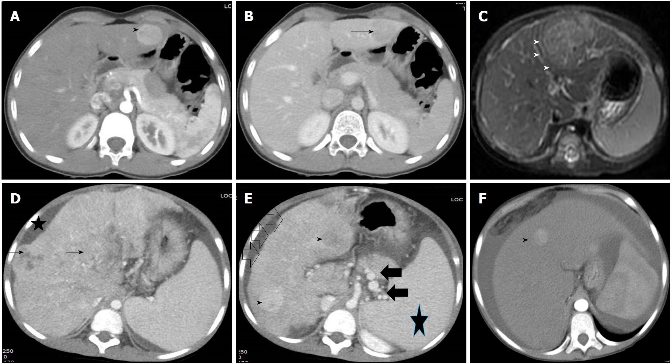
Figure 3 Radiological images in children with hepatocellular carcinoma.
A and B: 14 and half years old boy with chronic hepatitis-B (Immunoclearance phase), HBeAg+, DNA 5 log, AFP = 3 ng/mL (normal), with a segment 3 lesion (2.5 cm × 2.4 cm), BCLC stage A. Contrast-enhanced CT (CECT) image showing enhancement of the lesion (arrow) in the arterial phase (A) and washout in delayed venous phase (B) in a background of non-cirrhotic liver; C: 10 mo old boy with infantile cholestasis, bile salt export pump (BSEP) deficiency on immunostaining, incidentally found to have lesion in segment II and IVa abutting the capsule, BCLC stage B. The lesion was T2 hyperintense with enhancement in the arterial phase (arrows) and washout in the delayed phase; D and E: 8-1/2 yr old boy with decompensated chronic liver disease, chronic hepatitis-B (Immunoclearance phase), HBeAg+, DNA 7 log, AFP = 250000 ng/mL, with multifocal HCC, BCLC stage D, Child-Pugh C. Contrast-enhanced CT (CECT) image showing heterogenous lesion (arrows, D and E) involving whole of the right lobe with enhancement in the arterial phase in few lesions (arrows, E) in a background of cirrhotic liver with nodular margins (open black arrows, E), ascites (star, D) and collaterals (black solid arrows, E) and splenomegaly (star, E); F: 14 yr old girl with hepatic venous outflow tract obstruction, ascites, portal hypertension, incidentally found to have a lesion in segment 4 (1 cm × 1 cm), BCLC stage D, Child-Pugh C. Contrast-enhanced CT (CECT) image showing enhancement of lesion in the arterial phase (arrow).
- Citation: Khanna R, Verma SK. Pediatric hepatocellular carcinoma. World J Gastroenterol 2018; 24(35): 3980-3999
- URL: https://www.wjgnet.com/1007-9327/full/v24/i35/3980.htm
- DOI: https://dx.doi.org/10.3748/wjg.v24.i35.3980


