Copyright
©The Author(s) 2017.
World J Gastroenterol. Feb 7, 2017; 23(5): 792-799
Published online Feb 7, 2017. doi: 10.3748/wjg.v23.i5.792
Published online Feb 7, 2017. doi: 10.3748/wjg.v23.i5.792
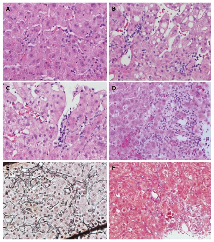
Figure 2 Spectrum of sinusoidal pathology.
A: lymphocytes in sinusoidal spaces, with adhesion to endothelium but without lifting of endothelial cells; B: Grade 1 sinusoidal endotheliitis with subendothelial linear lymphocytic infiltration; C: Grade 2 sinusoidal endotheliitis with subendothelial lymphocyte clusters and partially disrupted endothelium; D: Grade 3 sinusoidal endotheliitis with endothelial damage, fresh hemorrhage, lymphohistiocytic infiltration and adjacent liver cell necrosis; E: Grade 3 sinusoidal endotheliitis with collapsed liver cell plates on reticulin staining; F: Grade 3 sinusoidal endotheliitis with collagen deposition on Masson trichrome staining (hematoxylin-eosin staining, magnification × 400).
- Citation: Shi Y, Dong K, Zhang YG, Michel RP, Marcus V, Wang YY, Chen Y, Gao ZH. Sinusoidal endotheliitis as a histological parameter for diagnosing acute liver allograft rejection. World J Gastroenterol 2017; 23(5): 792-799
- URL: https://www.wjgnet.com/1007-9327/full/v23/i5/792.htm
- DOI: https://dx.doi.org/10.3748/wjg.v23.i5.792


