INTRODUCTION
Ischemia/reperfusion (I/R) injury is a frequent sequel of liver transplantation (LT) and significantly predisposes patients to graft dysfunction, which is related to increased risk of morbidity and mortality[1,2]. It is also a major obstacle to increasing the donor pool using marginal grafts, which are prone to a higher degree of I/R injury[3]. Evidence has shown that I/R induces vascular endothelial dysfunction, which is defined as abolished endothelium-dependent dilation[2,4]. Endothelial nitric oxide synthase (eNOS) protein expression, which is responsible for the basal production of endothelium-derived nitric oxide (NO), is markedly reduced along with the abolished dilation of endothelium[5,6]. It has been suggested that increasing NO availability notably improves the microcirculation and attenuates I/R injury, possibly due to the role of NO in reducing the generation of reactive oxygen species (ROS)/reactive nitrogen species (RNS)[7].
ROS/RNS play a pivotal role in signaling cascades that are essential for I/R injury[8]. ROS, including the superoxide anion (O2-), H2O2 and the hydroxyl radical, are generated with the reintroduction of O2 to ischemic tissues. This is associated with mitochondrial depolarization, which is responsible for a positive feedback loop of ROS-induced ROS release[9-15]. RNS, which include NO· and peroxynitrite (ONOO-), the latter originating from a reaction of O2- with transient initial excessive NO on reperfusion[16], together with ROS are responsible for oxidative/nitrative stress of I/R injury[17,18].
Remote ischemic conditioning (RIC), including remote ischemic preconditioning (RIPreC)[19], remote ischemic postconditioning (RIPostC)[20] and the recently described remote ischemic perconditioning (RIPerC)[21], was first described by Przyklenk et al[22], who demonstrated that short periods of ischemic reperfusion to a distant organ can protect the target organ[23]. To date, only three major studies have mentioned RIPerC in liver I/R injury[24-26]. There is only limited information about the relationship of oxidative/nitrative stress and NO during hepatic I/R, and about RIC vs classic surgical conditioning techniques of IPreC[27] and IPostC[28], which reduce I/R injury in liver[29]. The effects of RIC on liver grafts have not been reported.
We have established an LT model of RIC/RIPerC and validated its protection against I/R injury[30], and here, we further investigate the underlying mechanisms. We postulated that the liver graft protection of RIPerC involved inhibition of oxidative/nitrative stress and up-regulation of the eNOS/NO pathway, and compared it with IPostC, which is also effective in our established LT I/R injury model.
MATERIALS AND METHODS
Animals and experimental design
Adult male Sprague-Dawley rats (250-300 g) were kept at 25-30 °C in a humidity-controlled environment and allowed access to a standard diet and water ad libitum. All experimental protocols were approved by the Institutional Animal Care and Use Committee of the First Affiliated Hospital, Zhejiang University School of Medicine and were conducted in accordance with the ARRIVE (Animal Research: Reporting in vivo Experiments) guidelines (http://www.nc3rs.org/ARRIVE). Thirty-five rats (including 15 donors) were randomly assigned to four groups (n = 5 for each group) which were subjected to the following procedures. The Sham group (Group 1) underwent opening and closure of the abdomen under anesthesia, lasting approximately 75 min, which is the mean total ischemic time of orthotopic liver transplantation (OLT) in our center. The OLT group (Group 2) was subjected to standard OLT as above. The IPostC group (Group 3) underwent OLT with portal vein reperfusion and reocclusion for six 10-s cycles applied immediately at the onset of reperfusion (Figure 1). The RIPerC group (Group 4) underwent OLT with hindlimb ischemia and reperfusion for three 5-min cycles starting at the beginning of the anhepatic phase (Figure 1).
Figure 1 Ischemic postconditioning and remote ischemic perconditioning models.
IPostC was performed by six 10-s cycles of reperfusion and 10 s reocclusion of the portal vein. RIPerC was performed by three 5-min cycles of reperfusion and 5 min reocclusion by tourniquet. IPostC: Ischemic postconditioning; RIPerC: Remote ischemic perconditioning.
Rat OLT, IPostC and RIPerC models
The OLT model has been described previously[30]. The animals were anesthetized with 4% chloral hydrate (Shanghai No. 1 Biochemical and Pharmaceutical Co. Ltd, China). After isolation of the donor liver, the graft was perfused by cold saline containing 25 U/mL heparin through the portal vein, then placed into cold saline (0-4 °C) for approximately 40 min before transplantation. After the completion of anastomosis of the suprahepatic vena cava, followed by inserting the cuffs into the recipient portal vein, the liver was reperfused to end the anhepatic period, which lasted approximately 15 min, with the hepatic artery being ligated. Subsequently, the same cuff procedure was carried out on the infrahepatic vena cava, and the common bile duct with a stent was also reconstructed. The abdominal incision of the recipient was closed. Immediately, 1.5 mL saline was injected through the penile vein. All rats were anesthetized with 4% chloral hydrate at 3 h after OLT for the collection of samples. The rats were killed at 3 h after the portal vein of the recipients was opened. During the operation, we manipulated the rats lightly and softly to ameliorate any suffering. The blood collected from the portal vein was immediately centrifuged to obtain a plasma supernatant and stored at -80 °C until it was assayed for organ function tests. The left and median liver lobes were obtained and stored at -80 °C for further analysis.
The IPostC model with six 10-s cycles of reperfusion and 10 s reocclusion of the portal vein was applied immediately at the onset of reperfusion. The RIPreC model consisted of clamping of the femoral vessels for 5 min followed by 5 min of reperfusion for a total of three cycles applied immediately at the onset of the anhepatic phase to the recipient hindlimb, using a standard tourniquet with 1 kg weight on both sides.
Biochemical assay of blood samples
Blood samples were taken for measurement of alanine aminotransferase (ALT), aspartate aminotransferase (AST), creatinine (Cr) and creatinine kinase-myocardial band (CK-MB) by standard laboratory methods using the Hitachi 7600 automatic analyzer (Tokyo, Japan).
Primary hepatocyte culture
Primary hepatocytes were cultured as described previously[31], with modification. The rat livers were dissected mechanically and disaggregated with collagenase under sterile conditions. The fragmented liver tissues were pipetted 10 times with 10 mL Dulbecco’s modified Eagle’s medium (Gibco, Grand Island, NY, United States) through a nylon mesh. The cell suspension was centrifuged (50 ×g, 5 min, 4 °C) and the supernatant was removed. The precipitate was rinsed with Hank’s solution and centrifuged again (50 ×g, 5 min, 4 °C), and this process was repeated three times. The cell suspension was seeded (6.25 × 10⁴ cells/well) on a 96-well plate. All cells were maintained in a humidified atmosphere of 50 mL/L CO2 at 37 °C and cultured according to standard cell culture techniques.
Mitochondrial membrane potential (ΔΨm), ROS, H2O2 and NOx assays
ΔΨm, ROS, H2O2 and NOx activity were determined using the JC-1 Mitochondrial Membrane Potential Detection Kit (Biotium, Fremont, CA, United States), ROS Assay Kit (Beyotime Institute of Biotechnology, Shanghai, China), H2O2 Assay Kit (Beyotime Institute of Biotechnology) and Total NOx Assay (R and D Systems, Minneapolis, MN, United States), respectively. Fluorescence of JC-1 (5,5,6,6’-tetrachloro-1,1’,3,3’ tetraethylbenzimidazolylcarbocyanine iodide) was measured in the channel FL-1 and FL-2 by flow cytometry (FCM, Cytomics FC500; Beckman Coulter, Miami, FL, United States). In non-apoptotic cells, the mitochondria appeared red following aggregation of the JC-1 reagent, with an emission centered at 590 nm. In apoptotic cells, the dye remained in its monomeric form and appeared green with an emission centered at 530 nm. ROS were determined by measuring the oxidative conversion of cell-permeable 2’,7’dichlorofluorescein (DCF) diacetate to cell-impermeable fluorescent DCF. Then, DCF fluorescence distribution was detected by confocal microscopy (FV1000; Olympus, Tokyo, Japan) analysis at an excitation/emission wavelength of 488/525 nm. H2O2 activity was determined in samples of tissue homogenate. H2O2-oxidized ferrous (Fe2+) ions to ferric (Fe3+) ions reacted with an indicator dye, xylenol orange, to form a purple complex that was detected at 560 nm using a microplate spectrophotometer (Biotek Instruments, Winooski, VT, United States). The amount of H2O2 released was calculated according to the standard curve originating from standard solutions from identical experiments. Total NO in the liver tissue was determined indirectly as the stable end products of nitrate and nitrite by the Griess reaction. NO3- was converted to NO2- with Aspergillus nitrite reductase and the total NO3- plus NO2- (NOx) was measured by measuring NO2- with the Griess reagent. OD540 was determined (wavelength correction at 690 nm).
Detection of mRNA expression by reverse transcription-polymerase chain reaction
Total RNA was isolated from liver tissue homogenate using TRIzol reagent (Thermo Fisher Scientific, Waltham, MA, United States). One microgram of total RNA was isolated and reverse-transcribed to cDNA using a PrimeScript RT Reagent Kit with gDNA Eraser (Takara Bio, Shiga, Japan). The PCR system contained 0.8 μL forward primer (10 μmol/L), 0.8 μL reverse primer (10 μmol/L), 2 μL template cDNA, 0.4 μL ROX Reference Dye or Dye II (50 ×), 10 μL SYBR Premix Ex TaqII (Tli RNaseH Plus) (2 ×) and 6 μL distilled water to a total volume of 20 μL. The target gene expression was quantified using an ABI 7500 Fast Real-Time PCR (Foster City, CA, United States). Homogeneity was detected in each sample in all wells using dissociation curve analysis and the relative quantification was calculated using comparative cycle threshold (CT) method. The eNOS primers used were 5’-GTATTTGATGCTCGGGACTG-3’ (forward) and 5-AGATTGCCTCGGTTTGTTG-3’ (reverse). The amplification conditions for p-eNOS were: 30 s at 95 °C for one cycle and 5 s at 95 °C, followed by 30 s at 60 °C for 40 cycles.
Western blot analysis for eNOS and 3-nitrotyrosine
Protein was isolated from liver tissue after incubation in RIPA Lysis Buffer (Beyotime Institute of Biotechnology) supplemented with protease inhibitor cocktail (Sigma-Aldrich, Dublin, Ireland) for 1 h on ice. After centrifugation (14000 ×g, 4 °C, 15 min), the supernatants were collected for protein concentration measurement using a Bicinchoninic Acid Protein Assay Kit (Thermo Fisher Scientific). Denatured protein was separated by SDS-PAGE (Invitrogen, Carlsbad, CA, United States) and transferred to nitrocellulose membranes. After blocking with 50 mL/L non-fat milk in Tris-buffered saline with Tween (TBST; 10 mmol/L Tris-HCl, 0.5 mL/L Tween 20 and 0.15 mol/L NaCl, pH 7.2) for 3 h, the membranes were incubated with primary antibodies against β-actin (1:1000; Abcam, Cambridge, MA, United States), total eNOS (1:1000; Abcam), p-eNOS (1:500; Abcam), 3-nitrotyrosine (1:1000; Abcam) for 15 h at 4 °C. Blots were washed in TBST and incubated with appropriate horseradish-peroxidase-linked anti-mouse or anti-rabbit secondary antibodies (goat anti-mouse IgA for total eNOS, goat anti-rabbit IgG for p-eNOS and goat anti-mouse IgG2a for 3-nitrotyrosine; all supplied by Abcam) for 1 h at room temperature. Enhanced chemiluminescence was conducted using an ECL kit (Pierce Biotechnology, Rockford, IL, United States).
Statistical analysis
Data were expressed as mean ± SD. For comparison of statistical differences between experimental groups, analysis of one-way analysis of variance was used, followed by the Dunnett’s test or Student’s t-test for the histopathological outcomes. P < 0.05 was considered statistically significant.
RESULTS
Effects of RIPerC treatment on liver graft and other remote organ functions
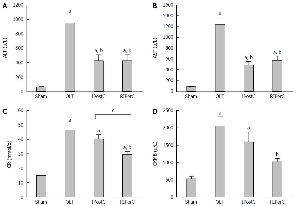
Compared with the Sham group, markers for acute damage of the liver, kidney and heart in all the other groups were elevated (P < 0.05). ALT, AST and parameters for hepatocellular damage were significantly lower in the RIPerC (P < 0.001) group as compared with OLT (Figure 2A and B) group. Serum Cr, a marker of acute kidney injury, was significantly decreased in the RIPerC group as compared with the OLT group (P < 0.05). CK-MB, a marker for acute myocardial damage, was significantly decreased in the RIPerC group as compared with the OLT group (P < 0.05) (Figure 2D).
Figure 2 Effects of remote ischemic perconditioning treatment on liver graft and other organ functions.
A and B: Effects of remote ischemic perconditioning (RIPerC) and ischemic postconditioning (IPostC) on alanine aminotransferase (ALT) and aspartate aminotransferase (AST), parameters for hepatocellular damage; C: Effects of RIPerC and IPostC on creatinine (Cr), a marker of acute kidney injury; D: Effects of RIPerC and IPostC on creatinine kinase-myocardial band (CK-MB), a marker for acute myocardial damage. Data represent mean ± SEM for 5 animals per group. aP < 0.05 vs sham group; bP < 0.05 vs orthotopic liver transplantation (OLT) group; cP < 0.05 RIPerC vs IPostC.
Effects of RIPerC treatment on I/R-induced oxidative stress
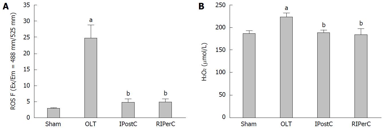
Levels of ROS were significantly elevated by OLT I/R injury (P < 0.001) as compared with the Sham group. RIPerC decreased the ROS level (P < 0.001) (Figure 3A). Levels of H2O2 were also significantly elevated in the OLT group (P < 0.05) as compared with the Sham group. RIPerC reversed this trend (P < 0.05) (Figure 3B).
Figure 3 Effects of remote ischemic perconditioning on ischemia/reperfusion-induced oxidative stress.
A: Effects of remote ischemic perconditioning (RIPerC) and ischemic postconditioning (IPostC) on ROS expression; B: Effects of RIPerC and IPostC on H2O2 expression. Data represent mean ± SEM for 5 animals per group. aP < 0.05 vs sham group; bP < 0.05 vs orthotopic liver transplantation (OLT) group.
Effects of RIPerC treatment on I/R-induced nitrative stress
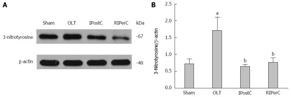
Expression of 3-nitrotyrosine was measured by western blot analysis. 3-nitrotyrosine levels were significantly elevated in reperfused hepatocytes (P < 0.05). I/R-induced 3-nitrotyrosine was significantly reduced in the RIPerC group (P < 0.05) (Figure 4).
Figure 4 Effects of remote ischemic perconditioning on ischemia/reperfusion-induced nitrative stress.
A: Protein expression levels of 3-nitrotyrosine were measured by western blot analysis (the gels were run under the same experimental conditions); B: Statistical analysis of 3-nitrotyrosine in liver tissues is shown. Data represent mean ± SEM for 3 animals per group. aP < 0.05 vs sham group; bP < 0.05 vs orthotopic liver transplantation (OLT) group. RIPerC: Remote ischemic perconditioning.
Effects of RIPerC treatment on ΔΨm
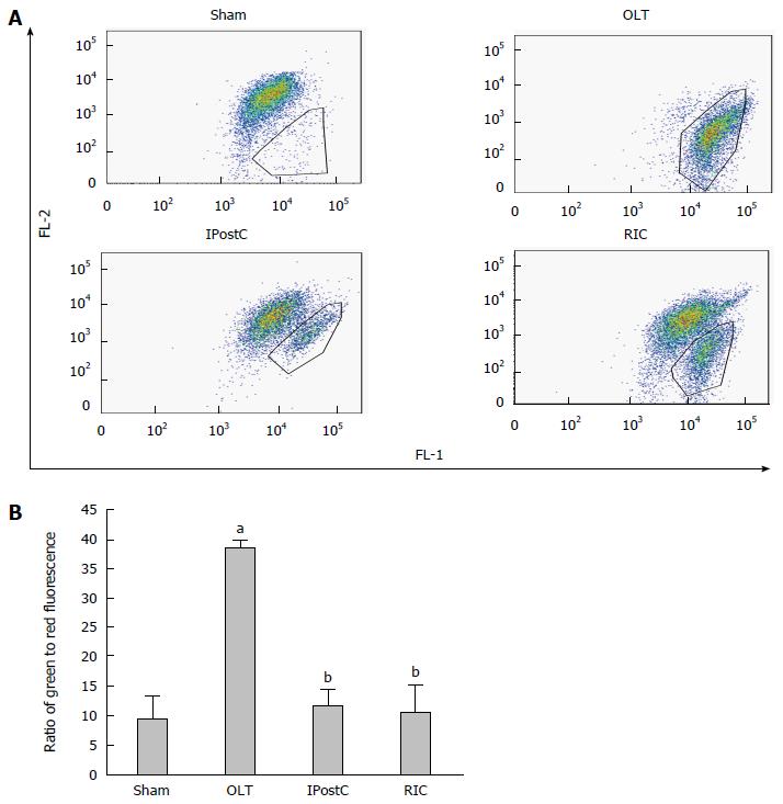
JC-1 fluorescence shifted from red to green, indicating a collapse of ΔΨm. The ratio of green to red fluorescence was significantly increased in the liver graft after I/R injury (P < 0.001) as compared with the Sham group. The fluorescence shift induced by OLT I/R injury was significantly attenuated in the RIPerC group (P < 0.001) (Figure 5).
Figure 5 Effects of remote ischemic perconditioning on ΔΨm.
A: Typical dot plot analysis of JC-1 of Δm. A dot plot of red fluorescence (FL2) vs green fluorescence (FL1) resolved live liver cells with intact ΔΨm (Sham group) from apoptotic and dead cells with lost ΔΨm (OLT group); B: The change of Δm was reported by the ratio of green to red fluorescence. Data represent mean ± SEM for 5 animals per group. aP < 0.05 vs sham group; bP < 0.05 vs OLT group. OLT: Orthotopic liver transplantation.
Effects of RIPerC on eNOS and NO expression
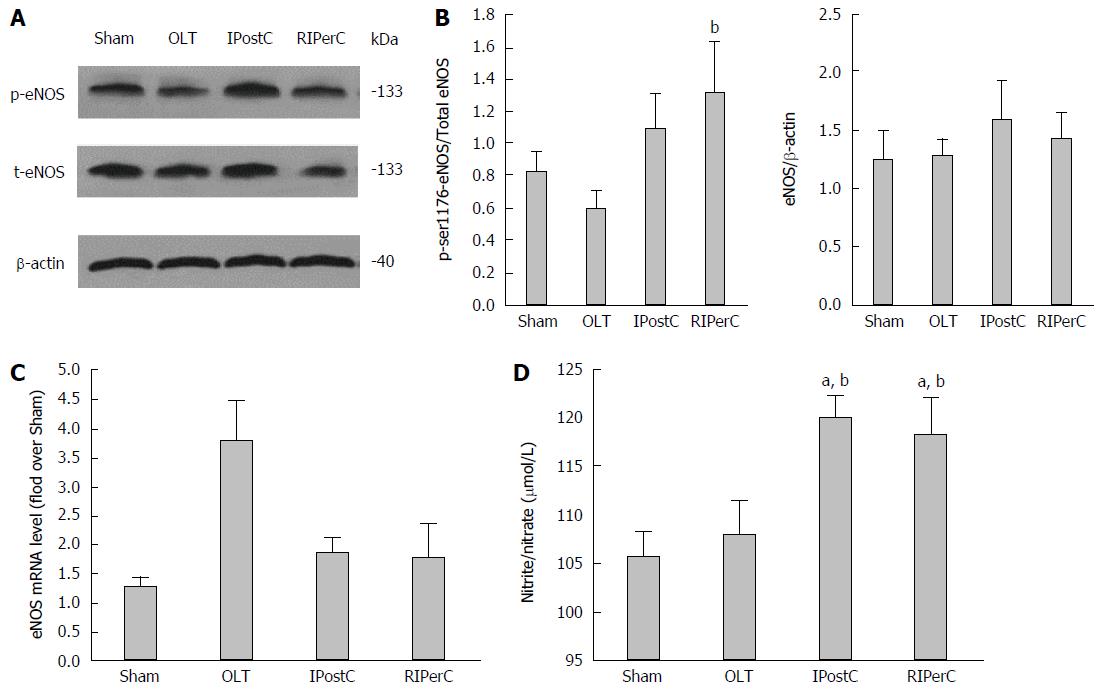
Expression of total eNOS was measured by western blotting and RT-PCR, and no significant alteration was found. There was a marked increase of eNOS phosphorylation on serine-1176 in the liver graft of the RIPerC group (P < 0.05) as compared with the OLT group (Figure 6A-C). NO expression, which was determined indirectly as the stable end products of nitrate and nitrite, was significantly increased in the RIPerC as compared with the OLT group (P < 0.05) (Figure 6D).
Figure 6 Effects of remote ischemic perconditioning on endothelial nitric oxide synthase and nitric oxide expression.
A: Western blots of phospho-ser1176-endothelial nitric oxide synthase (eNOS) [p-eNOS] and total eNOS (t-eNOS) detection in liver tissues (gels run under the same experimental conditions); B: Statistical analysis of phospho-ser1176-eNOS and total eNOS in liver tissues; C: mRNA level of eNOS; D: Total nitrate/nitrite content in the four groups. Data represent mean ± SEM for 5 animals per group. aP < 0.05 vs sham group; bP < 0.05 vs orthotopic liver transplantation (OLT) group.
Effects of RIPerC vs IPostC
Compared with the OLT group, the IPostC group had significantly lower levels of ALT, AST (P < 0.001) (Figure 2A and B), ROS (P < 0.001), H2O2 (P < 0.05) (Figure 3A and B) and 3-nitrotyrosine (P < 0.05), and significantly higher expression of Δm (P < 0.001) (Figure 5) and NO (P < 0.05) (Figure 6D). eNOS phosphorylation of serine-1176 in the IPostC group was increased by 1-8-fold compared with the OLT group, although this difference was not significant (P = 0.106) (Figure 6A-C). No significant differences were observed between the RIPerC and IPostC groups expect for Cr. The Cr level was lower in the RIPerC group than in the IPostC group (P < 0.01) (Figure 2C).
DISCUSSION
We previously established a new rat model of RIC and validated its protection against I/R injury by its anti-inflammatory and antioxidative activities and effects of activating the PI3K/Akt pathway[30]. Currently, we used the same model of hindlimb RIPerC (5 min × 3) to explore further the downstream effector proteins of the PI3K/Akt pathway, and compared it with IPostC under the same treatment conditions. We showed that RIPerC is similar to or better than IPostC, which prevented liver graft dysfunction and apoptosis induced by hepatic I/R injury by inhibiting oxidative/nitrative stress, as assessed by ROS and 3-nitrotyrosine, through the eNOS/NO pathway. We also found additional beneficial effects of RIPerC, such as reduced levels of Cr and CK-MB, compared with the OLT group, which indicates protection of other organs. To the best of our knowledge, this is the first investigation of RIC using a novel model in LT to identify the PI3K/Akt/eNOS/NO axis involved.
The present study provides new information about RIC after I/R injury beyond that in previous studies showing that protection associated with RIC mainly involves the heart. While most of the previous studies have only investigated the effects of RIPreC and RIPostC on organ protection, the present study focused on the novel model of RIPerC protection against hepatocyte I/R injury of LT. We found a significant reduction in plasma aminotransferase levels, which reflected liver function in the RIPerC group vs the I/R group, and exerted a protective effect by attenuating the congestion and hepatocyte necrosis shown in our previous study[31]. Several studies have also reported these protective effects of RIC, using different models from ours, in other organs, including the heart[32], brain[33] and kidneys[34,35]. However, those results contrast with the lack of improved clinical outcomes of RIPreC in patients undergoing elective on-pump coronary artery bypass graft, with or without valve surgery[36] (Figure 2A and B).
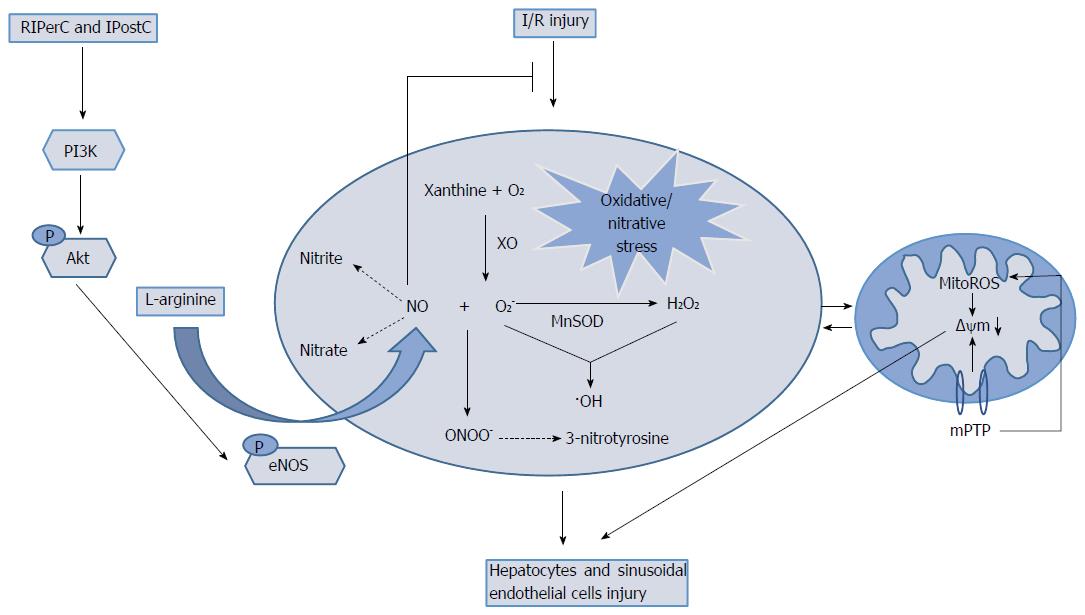
Increased oxidative stress was one of the features observed in the liver and the isolated liver mitochondria during the initial phase of hepatic I/R[37]. We found that elevated production of ROS in the OLT and RIPerC groups could reverse this situation. In particular, H2O2, which is not a free radical, can be formed from O2- and generate hydroxyl radicals. Because H2O2 is very reactive, it is often discussed along with ROS[38,39]. H2O2 triggers release of proinflammatory cytokines and induces apoptosis, leading to tissue oxidative damage[40]. As there is non-specific suppression of ROS in the human body, H2O2-responsive co-polyoxalate containing vanillyl alcohol nanoparticles can serve as I/R-targeted nanotherapeutic agents[41]. Therefore, we also measured the H2O2 level alone, which reflected the same trend (Figure 3). Oxidant stress is an effective trigger of mitochondrial permeability transition pore opening in hepatocytes[42], leading to ΔΨm collapse, which is a hallmark of apoptosis[43]. The deleterious effects of ROS, such as apoptosis, are blocked using various antioxidants during I/R[44]. In the present study, RIPerC attenuated the related ΔΨm decrease induced by liver transplantation I/R injury, which was correlated with an increase in hepatocyte apoptosis (Figure 5). In early reperfusion, increased production of ROS is accompanied by reduced availability of NO[16]. Thus, we attempted to clarify the relationship between RIPerC protection against I/R injury and NO. The proposed mechanism is shown in Figure 7.
Figure 7 Simplified mechanisms of remote ischemic perconditioning protective effects against ischemia/reperfusion injury.
Remote ischemic perconditioning (RIPerC) liver graft protection involves inhibition of oxidative/nitrative stress and up-regulation of the PI3K/Akt/eNOS/NO pathway. •OH: Hydroxyl radical; XO: Xanthine oxidase; MitoROS: Mitochondrial reactive oxygen species. IPostC: Ischemic postconditioning; I/R: Ischemia/reperfusion.
Experimental evidence indicates that eNOS-derived NO is hepatoprotective against liver I/R injury[45-47], and eNOS knockout mice exhibit significantly greater liver injury following I/R compared with wild-type counterparts[2]. It has also been demonstrated that genetic ablation of eNOS abolishes the cardioprotective effect in anesthetized mice and that stimulation of eNOS during RIPerC contributes to cardioprotection[48]. Phosphorylation of eNOS in the myocardium was higher in the RIPerC group compared to the control group, parallel to infarct size reduction[49]. In agreement with the above findings, we found that phosphorylation of eNOS on serine-1176, through which RIPerC mediated protection against liver I/R injury, increased along with eNOS-derived NO, while there was no significant change in total eNOS (Figure 6). However, during early reperfusion, a transient initial excessive release of NO occurred in the presence of O2-via a diffusion-limited reaction to form ONOO-[16], further impairing the mitochondrial[50] and cellular functions and increasing ROS generation[51]. These radicals further promote the post-translational modification of tyrosine to 3-nitrotyrosine[52], a marker for RNS and nitrative stress[37]. Consistent with these findings, our data showed that the liver tissue protein levels of 3-nitrotyrosine were higher after I/R injury compared to the sham-operated rats (Figure 4). Therefore, we propose that the tyrosine-nitrated proteins may experience a loss of function, attenuating the protective roles of eNOS, which contributes to I/R injury. Previous work has also shown that tyrosine nitration inactivates the PI3K/Akt pathway[51]. We determined Akt phosphorylation and observed significant increases in the RIPerC group[30], which indicated an inhibitory effect of tyrosine nitration. Akt activates phosphorylation of eNOS, resulting in increased production of NO in the endothelium[53]. We hypothesized that RIPerC exerts protection against LT I/R injury through the eNOS/NO pathway, as reported by previous studies that Akt is an important upstream regulator of eNOS activation[54-56]. The proposed mechanism is shown in Figure 7.
Compared to the local ischemic conditioning, RIC overcomes the main concern of increasing total ischemic time that may lead to sequence problems[30]. However, almost no work has been conducted comparing the protective effects between them. We found that the effect of RIPerC was similar to or better than that of IPostC in preventing liver graft I/R injury. RIPerC appears to be the most promising technique to avoid I/R injury of LT and is clinically convenient.
We revealed an increase in the levels of CK-MB and Cr 3 h after reperfusion of LT. A few studies have been conducted under similar conditions. A large single-center study showed for the first time that hepatic I/R injury plays a modifiable and critical role in the pathogenesis of acute kidney injury[57]. RIPerC induces multiple beneficial effects following valve replacement surgery, including reduced drainage and myocardial damage, attenuated hyperbilirubinemia, and acute lung injury[58]. Based on these studies, we attempted to demonstrate the hypothesis that RIPerC could mitigate the acute injury to other organs induced by liver I/R injury. Therefore, we measured CK-MB and Cr for acute heart and kidney injury, respectively, and verified our hypothesis. The underlying mechanism deserves further investigation.
We failed to verify the direct relationship between RIPerC-induced hepatic I/R injury protection and Akt/eNOS phosphorylation. Further studies using specific Akt or eNOS inhibitors could help to clarify the protection of this pathway in this setting. Nevertheless, this work provides clear clues for future studies.
In conclusion, our findings suggest that the liver graft protection of RIPerC involves inhibition of oxidative/nitrative stress and up-regulation of the PI3K/Akt/eNOS/NO pathway. The procedure of RIPerC for 5 min × 3 is simple to perform, particularly during LT, and is potentially applicable in clinical practice.
COMMENTS
Background
Remote ischemic conditioning (RIC) includes remote ischemic preconditioning (RIPreC), remote ischemic postconditioning (RIPostC) and the recently described remote ischemic perconditioning (RIPerC). To date, only three major studies have mentioned RIPerC in liver ischemia/reperfusion (I/R) injury. There is limited information available on the relationship between oxidative/nitrative stress and NO during hepatic I/R, and between RIC and classic surgical conditioning techniques of IPreC and ischemic postconditioning (IPostC), which are useful in reducing I/R injury in liver. The effects of RIC on liver grafts have also not been reported.
Research frontiers
Previous studies have demonstrated that the protection of RIC mainly revolves around the heart. Most of these studies have only investigated the organ-protective effects of RIPreC and RIPostC. The protective activity of RIPerC in liver I/R injury and the mechanism of action need to be explored.
Innovations and breakthroughs
This is believed to be the first study to investigate RIC using a novel model in liver transplantation and to identify PI3K/Akt/eNOS/NO axis involvement.
Applications
This study showed that the effect of RIPerC, which overcomes the main concern of increasing total ischemic time, was similar to or better than that of IPostC in preventing liver graft I/R injury. RIPerC appears to be the most promising technique to avoid I/R injury of liver transplantation and is clinically convenient.
Terminology
RIC was originally developed by Przyklenk et al in 1993, who showed that brief ischemia in one organ conferred protection on important distant organs without direct stress on the target organ. Compared to IPostC, RIC can be applied before ischemia of the target organ (RIPreC), after ischemia, and before perfusion (RIPerC) or at onset of reperfusion (remote ischemic postconditioning), without increasing the total ischemic time.
Peer-review
The authors established a new rat model of RIC and validated its protection against I/R injury by the PI3K/Akt/eNOS/NO axis. These results are interesting. The strategy of RIPerC 5 min × 3 is relatively simple to perform, particularly during liver transplantation, and may be considered as potentially applicable in the clinical field.


